3827
Ultra-Wideband Self-Grounded Bow-Tie Antenna Building Block for Thermal Intervention, Diagnostic MRI and MR Thermometry at 7.0 Tesla1Berlin Ultrahigh Field Facility, Max-Delbrück Center for Molecular Medicine in the Helmholtz Association, Berlin, Germany, 2Physikalisch-Technische Bundesanstalt (PTB), Berlin, Germany, 3MRI.TOOLS GmbH, Berlin, Germany, 4Department of Electrical Engineering, Chalmers University of Technology, Gothenburg, Sweden, 5Experimental and Clinical Research Center (ECRC), a joint cooperation between the Charité Medical Faculty and the Max-Delbrück Center for Molecular Medicine, Berlin, Germany
Synopsis
A compact resonator antenna for broadband thermal intervention, diagnostic proton (1H) and fluorine (19F) imaging and MR-Thermometry was developed for operating at 7.0T MRI. The antenna is based on the concept of a self-grounded bow-tie (SGBT) antenna to enable thermal intervention frequencies ranging from 250MHz to 516MHz. The self-grounded bow-tie resonator antenna is smaller and lighter compared to previous dipole-antenna designs, enabling a high-density multi-channel array. The proposed antenna is demonstrated to be suitable for diagnostic imaging, thermal intervention, and MR thermometry.
Purpose
Temperature is a physical parameter with diverse biological implications and crucial clinical relevance 1. Targeted temperature increase can be beneficial for thermal manipulation of inflamed tissues, treatment of cancer by potentiating cells for chemo- and radiotherapy, targeted drug delivery afforded by thermo-responsive nano-carriers and increased blood-brain barrier permeability 2,3. Focal and targeted temperature manipulation facilitated by radiofrequency applicators is highly dependent on the operating frequency, which ranges between 50 and 1000MHz for published implementations 2,4,5. Recent reports on self-grounded bow-tie SGBT antennae use a broadband approach, enabling frequency and focal temperature adjustment 6,7. Here we report on the development of a compact ultra-wideband self-grounded bow-tie resonator antenna that supports targeted thermal intervention, diagnostic proton (1H) MRI, 1H MR temperature mapping and fluorine (19F) MRI in a single device on a 7.0T MRI system.Methods
CST
Microwave Studio 2018 (CST–Computer Simulation
Technology
GmbH, Darmstadt, Germany) was used for electromagnetic field (EMF) and
temperature simulations. The antenna and resonator geometry optimization was performed on a rectangular phantom (240x240x150mm³) with
muscle tissue mimicking material 8. For obtaining the
resonator and antenna dimensions, a genetic algorithm was used for
optimization, pursuing
the reflection coefficient S11<-13dB for the targeted operating
bandwidth. Based on the findings, an antenna was manufactured and placed
in an
additive manufactured housing filled with deuterium oxide (99.9% D2O,
Sigma Aldrich GmbH, Munich, Germany). An exponential tapered strip-line
balanced-unbalanced-transformer with an overall size of 39.3x26.5mm² was employed to enable
wideband response 9,10. A phantom with muscle tissue mimicking material
was constructed for MR imaging and radiofrequency
(RF) heating experiments. The dielectric properties of
the
phantom were measured for correct reproduction in the EMF and
temperature
simulations. Phantom density (1230.89 g/l), heat capacity (2.9635 J/g/K)
and
thermal conductivity (0.4355 W/m/K) estimations were based on 11,12.
Transmission (B1+)
fields at f=280 MHz (19F) and f=298 MHz (1H) were
examined numerically and validated with pre-saturation based B1+
mapping 13. Temperature and SAR simulations based on IEEE/IEC
standard 62704-1 were performed at f=300MHz, f=400MHz, and f=500MHz.
Magnetic
resonance thermometry was conducted using a 7.0T whole-body MRI system
(MAGNETOM, Siemens Healthineers, Erlangen,
Germany). For this purpose proton
resonance frequency shift (PRFS) thermometry based on double echo method
(TR=102ms, TE1=2.26ms and TE2=6.34ms at a special resolution of
1.5x1.5x4.0mm³)
was combined with fiberoptic probe
measurements (Neoptix, Québec, Canada) 14.Results
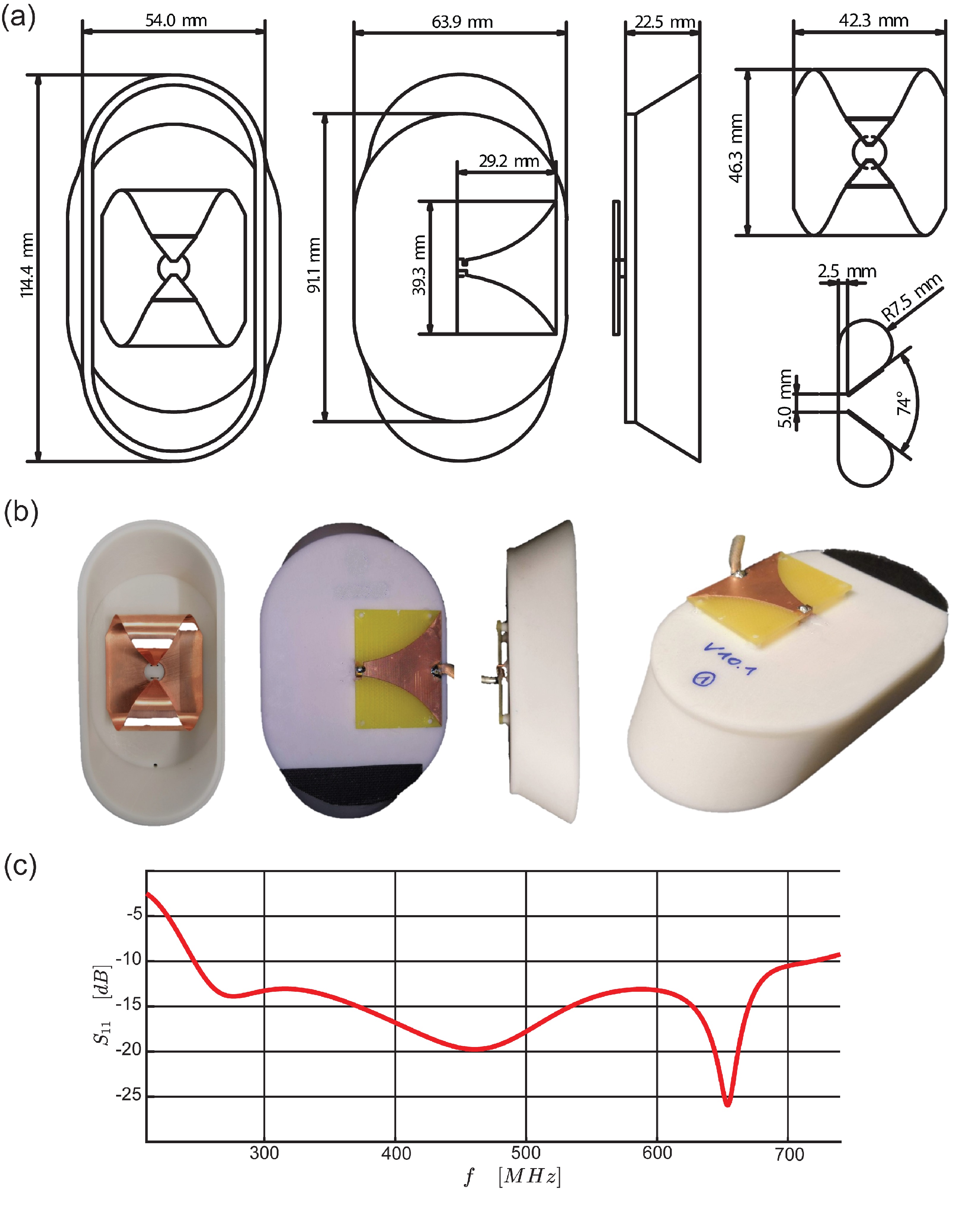
Figure 1 shows the antenna building block design and the
reflection coefficient S11 for a bandwidth of
467MHz on muscle tissue. The dielectric
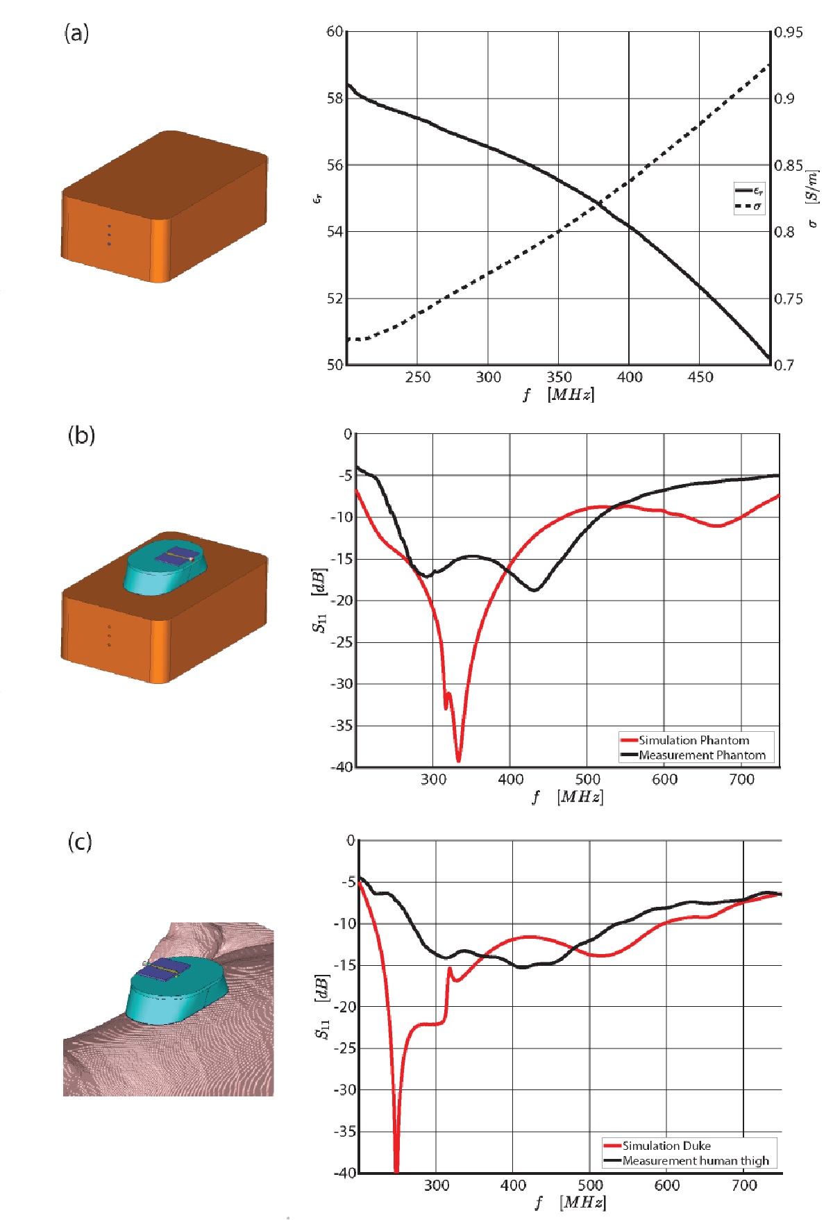
parameters of the phantom are shown in Figure 2 (a).
Figure 2 (b-c)
shows the reflection coefficient S11
of the resonator antenna when placed on a phantom and on the thigh of the human
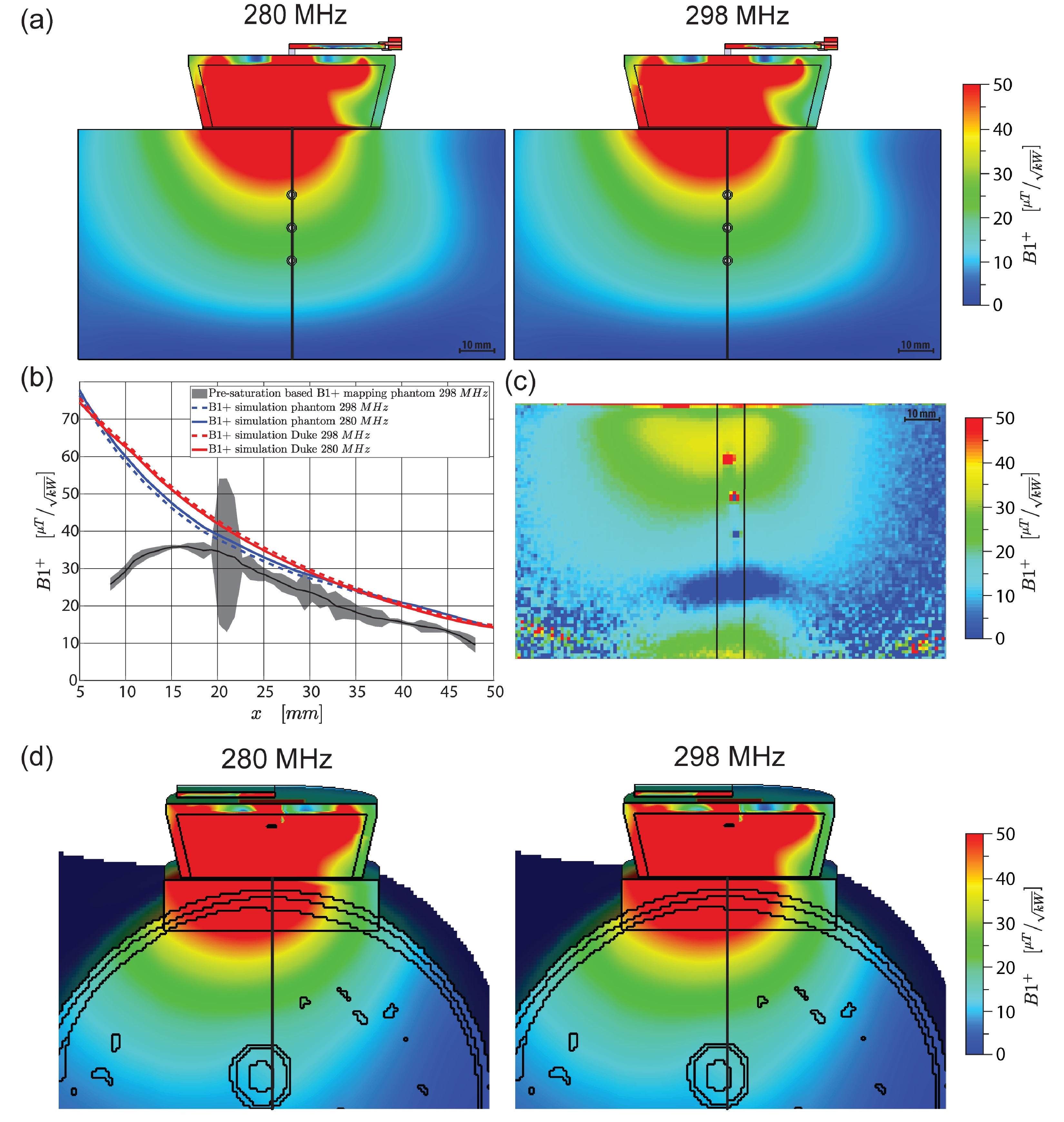
voxel model Duke 15. Results of EMF simulations for B1+
at f=280MHz and f=298MHz together with B1+ mapping at f=298MHz
of the phantom are demonstrated in Figure 3. B1+
simulations and measurements show a difference of approximately 8% offering transmission
fields of >15μT/√kW at 40mm distance to the antenna which is very well
suitable for 1H MRI and MR themometry.
The losses of the imaging signal chain of -2.12dB at f=298MHz were considered in
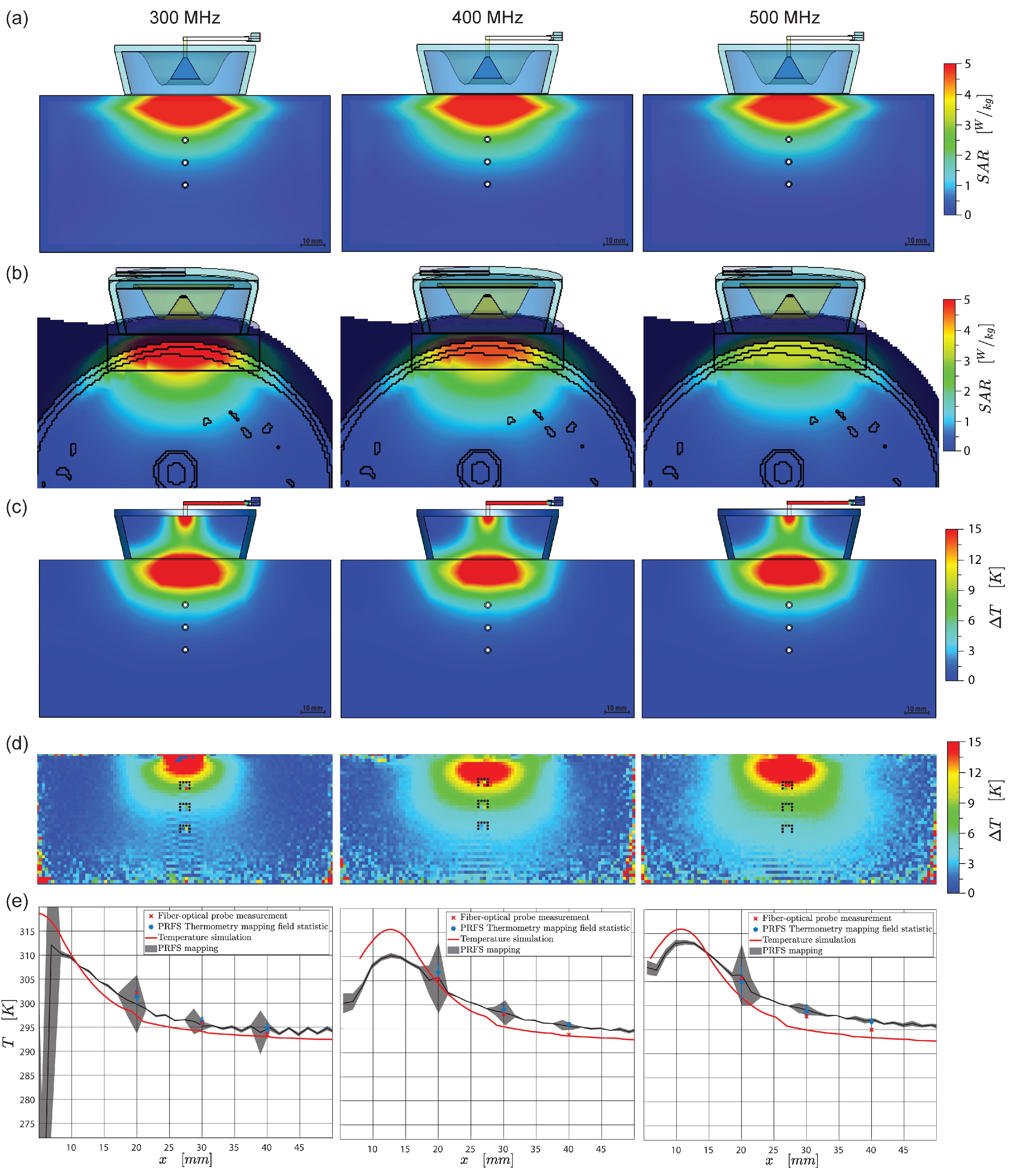
the simulations. SAR and temperature simulation results obtained for f=300MHz, f=400MHz,
and f=500MHz are shown in Figure 4 (a-c).
The heating transmit chain exhibited frequency-dependent losses of -1.95dB,
-2.26dB and -2.59dB for the 3 thermal intervention frequencies. The
experimental results obtained from MR thermometry matched the temperature
simulations qualitatively and quantitatively. Figure 4 (d-e)
highlights the results deduced from MR thermometry mapping and fiberoptic probe temperature reference measurements
for f=300MHz, f=400MHz, and f=500MHz. Considering
the match of MR thermometry and the fiberoptic probes, a difference of <1K
for the first two measurement positions was achieved.Discussion and Conclusion
The presented compact SGBT resonator broadband antenna
supports thermal intervention, diagnostic imaging, and temperature mapping at 7.0T, while offering a frequency
bandwidth ranging from 250MHz to 516MHz. The proposed SGBT building block pushes
the boundaries of antenna design by affording 54.9% or 72.2% reduced antenna
volume than previously reported for SGBT (107x78x31mm³) or bow-tie
(150x70x40mm³) antenna configurations 4,7. In addition to 19F
and 1H imaging, this antenna supports RF-induced heating for a broad frequency range. Temperature
simulations, MR thermometry and temperature measurement showed good agreement
between simulations and measurements. To take this project to the next level,
we envision a high-density array of SGBT
building blocks customized for 1H and 19F MRI, broadband thermal interventions, and MR-thermometry all being integrated into a 7.0T MRI scanner.Acknowledgements
This project was funded in part by an advanced ERC grant (EU project ThermalMR: 743077).References
1. Lamb G M Gedroyc W M Interventional magnetic resonance imaging. Br J Radiol. 1997;70:81-88.
2. Wust P, et al. Hyperthermia in combined treatment of cancer. Lancet Oncol. 2002;3(8):487-497.
3. Issels R, et al. Effect of Neoadjuvant Chemotherapy Plus Regional Hyperthermia on Long-term Outcomes Among Patients With Localized High-Risk Soft Tissue Sarcoma. JAMA Oncol. 2018;4(4):483
4. Winter L, et al. Design and Evaluation of a Hybrid Radiofrequency Applicator for Magnetic Resonance Imaging and RF Induced Hyperthermia: Electromagnetic Field Simulations up to 14.0 Tesla and Proof-of-Concept at 7.0 Tesla. PLoS One. 2013;8(4):e61661.
5. Guérin B, et al. Computation of ultimate SAR amplification factors for radiofrequency hyperthermia in non-uniform body models: impact of frequency and tumour location. Int J Hyperth. 2018;34(1):87-100.
6. Takook P, et al. Compact self-grounded Bow-Tie antenna design for an UWB phased-array hyperthermia applicator. Int J Hyperth. 2017;33(4):387-400.
7.Winter L, et al. Ultrahighfield, One for all: Ultra-wideband (279-500MHz) self-grounded bow-tie antenna for MR. ISMRM-ESMRMB. 2018:#4281.
8. IT’IS Foundation. Tissue Properties Database V4.0. 2018.
9. Yang J, Kishk A. A Novel Low-Profile Compact Directional Ultra-Wideband Antenna: The Self-Grounded Bow-Tie Antenna. IEEE Trans Antennas Propag. 2012;60(3):1214-1220.
10. Kazemipour A, Begaud X. Calculable Dipole Antenna for EMC Measurements with Low-Loss Wide-Band Balun from 30 MHz to 2 GHz. Electromagnetics. 2005;25(3):187-202.
11. Asadi M. Beet-Sugar Handbook. John Wiley & Sons; 2007
12. Buchanan EJ. Economic Design and Operation of Process Heat Exchange Equipment. Proc S Afr Sug Technol Ass. 1966;44:89-101
13. Yarnykh VL. Actual flip-angle imaging in the pulsed steady state: A method for rapid three-dimensional mapping of the transmitted radiofrequency field. Magn Reson Med. 2007;57(1):192-200.
14. Rieke V, Pauly KB. MR thermometry. J Magn Reson Imaging. 2008;27(2):376-390.
15. Christ A, et al. The Virtual Family - Development of surface-based anatomical models of two adults and two children for dosimetric simulations. Phys Med Biol. 2010;55(2)
Figures