3819
Development of 2D Cartesian, radial and UTE MP2RAGE sequences for fast T1 mapping: application to MR thermometry1CNRS - Univ. Bordeaux, CRMSB UMR 5536, Bordeaux Cedex, France
Synopsis
A surrogate strategy to the Proton Resonance Frequency technique that fails when short T2* tissue are present is the measurement of the longitudinal relaxation times T1. In order to monitor temperature in real-time, a 2D version of the Magnetization Prepared 2 Gradient Echo (MP2RAGE) sequence has been developed, and the cartesian encoding has been replaced by radial ones to obtain an ultra-short Echo Time (UTE). This enabled to measure T1 of short T2*-phantoms and to perform MR thermometry every 8s over a wide range of temperatures and consequently of T1.
INTRODUCTION
Thermal ablations (either by hyperthermia or by cryo-ablation) are getting more used to treat a variety of pathologies. The Proton Resonance Frequency (PRF) technique is the standard technique to measure temperature in real-time, due to its accuracy and its rapidity. Nevertheless, this technique fails when adipose tissue or short T2* tissue (lungs, bones, tissue close to a metallic implant) are present. There is a growing interest in measuring temperature in these conditions, as more patients get implants, and tumors/metastases developing in the lungs for exemple, are very frequent in many types of cancer. A surrogate strategy is to measure relaxation times, especially the longitudinal one (T1), that had been shown to be linear with temperature1. Nevertheless, due to its high sensitivity to slice excitation profiles, the actual fastest 2D T1 mapping technique (Variable Flip Angle VFA) only generated approximate values of T12. The Magnetization Prepared 2 Gradient Echo (MP2RAGE) sequence was developed for 3D T1 mapping in Neuro-imaging, requiring 10 minutes acquisition using parallel imaging. This sequence is thus too long at this state to monitor temperature during thermo-ablation. Consequently, the goal of this study was to develop 2D MP2RAGE sequences in order to fasten acquisition duration. In order to apply this sequence for MR thermometry on tissues affected by respiration or having short T2*, non-Cartesian encodings were also implemented and an ultra-short echo time (UTE) was reached. The influence of the slice profile on the robustness of the T1 measurements was simulated. Then, demonstration of the applicability of the new sequences to MR thermometry was performed on phantoms over a wide range of temperatures.MATERIALS and METHODS
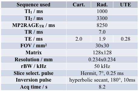
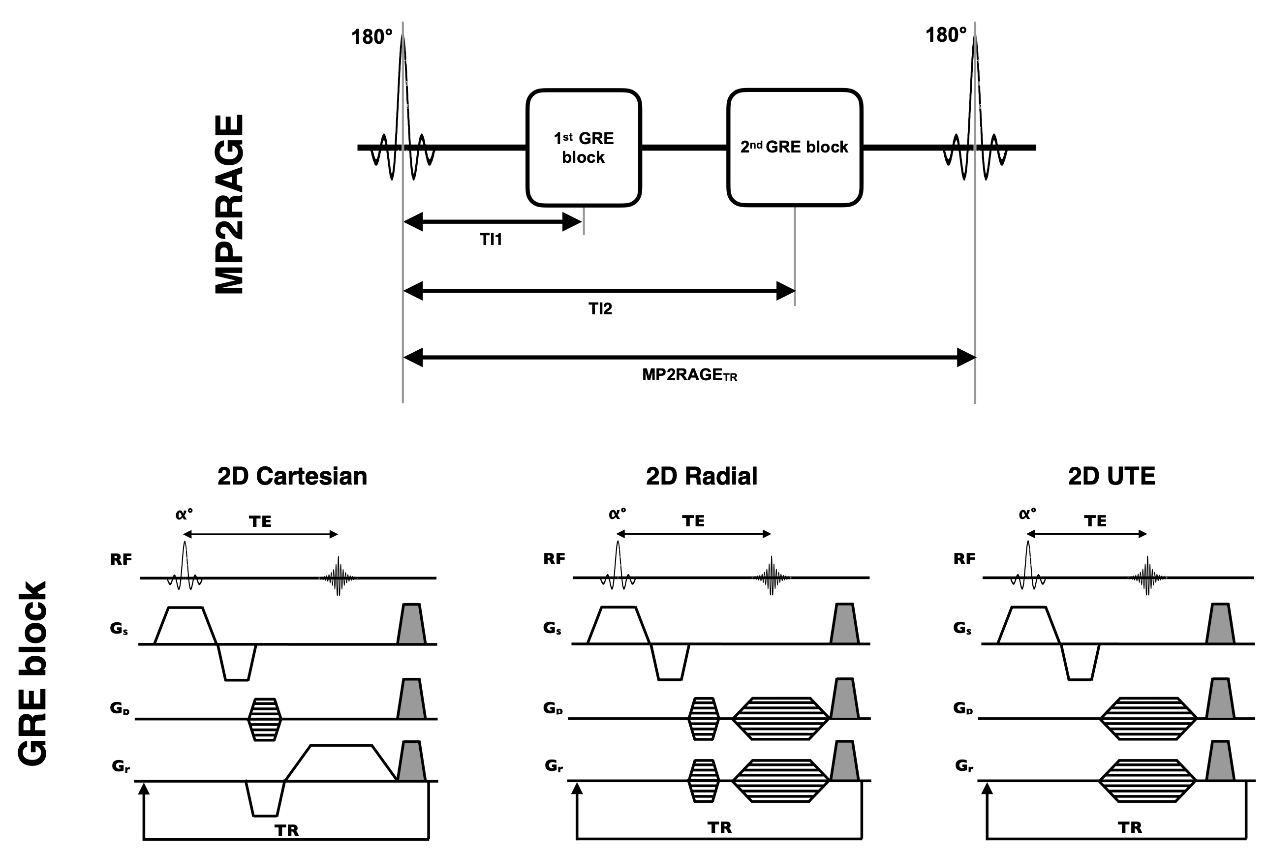
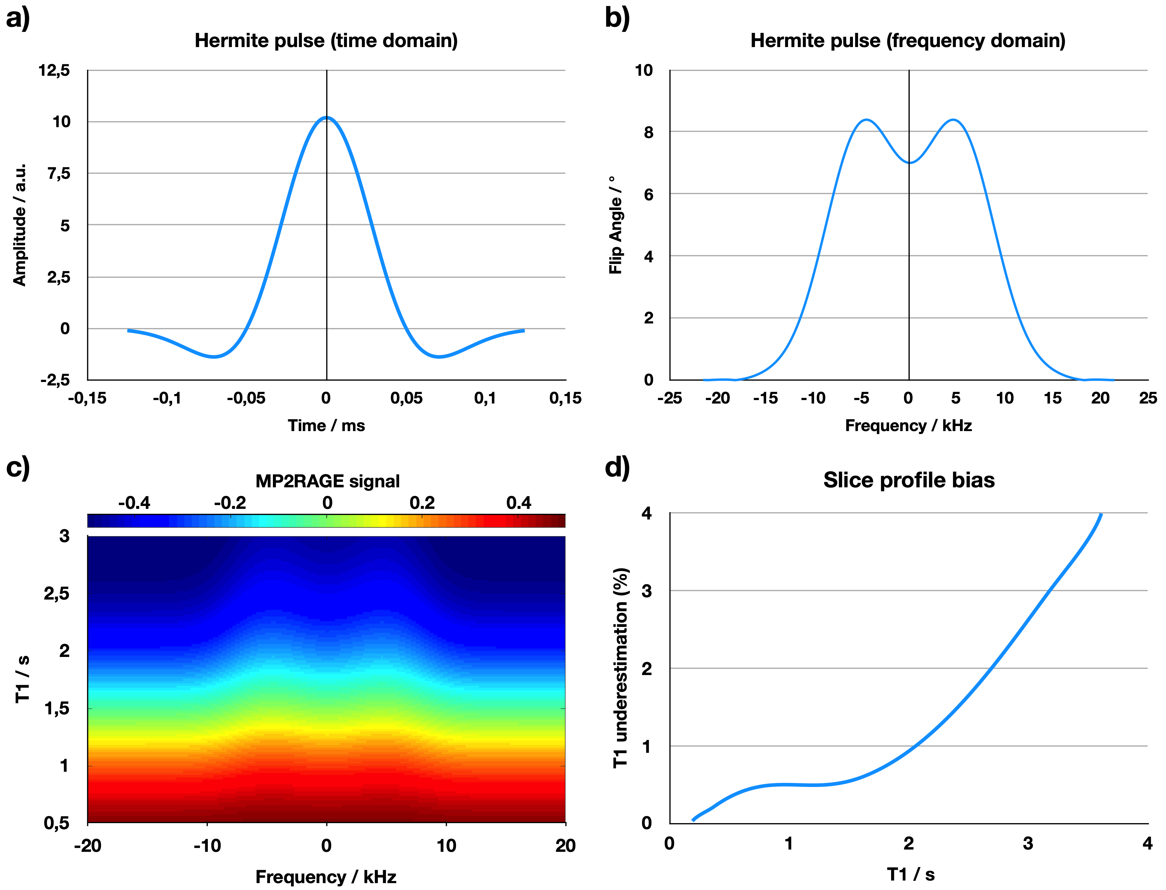
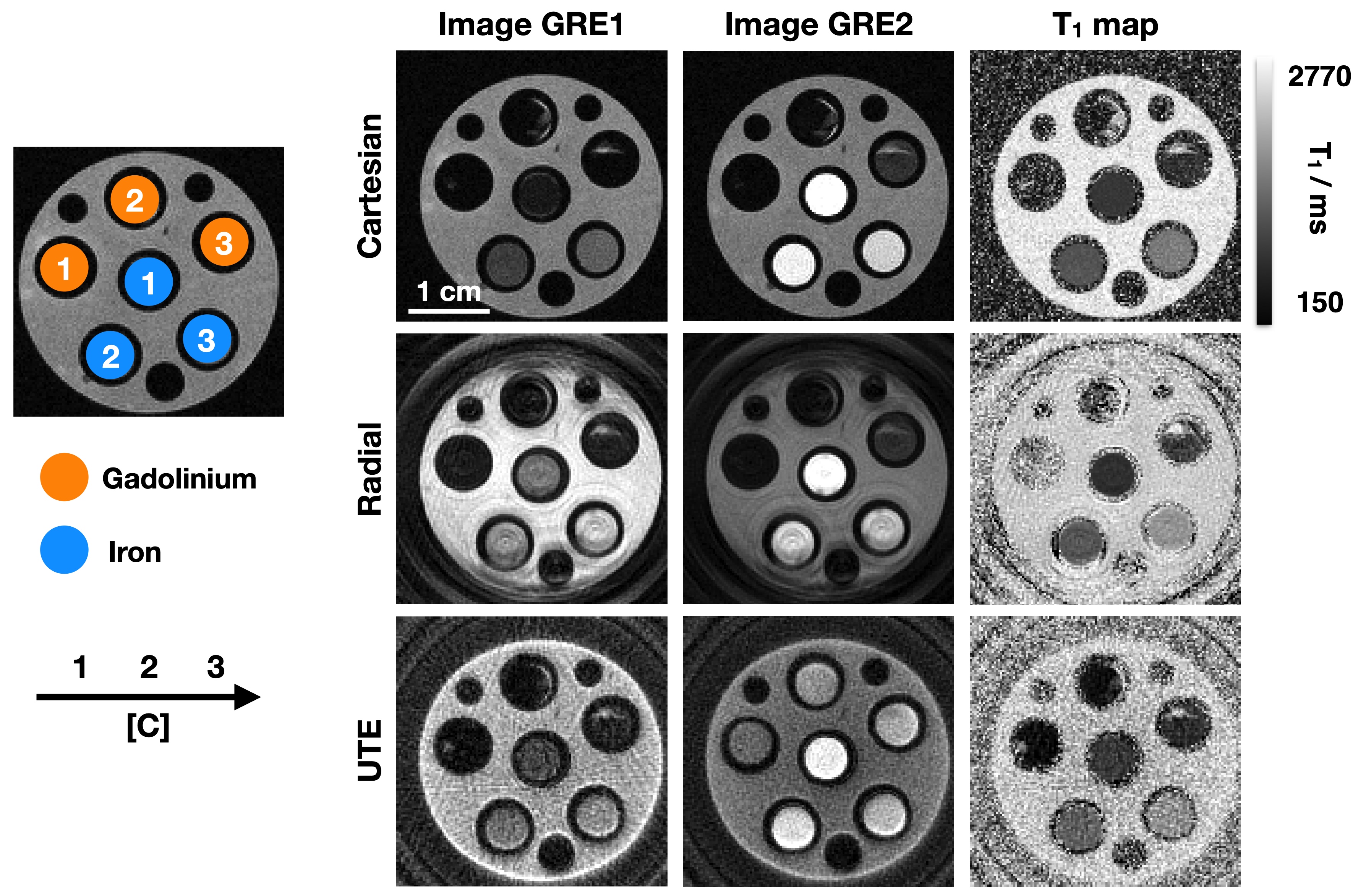
A 2D MP2RAGE sequence was developed using a slice-selection within the GRE block (Figure 1). Influence of this slice-selection on the T1 measurements was simulated using Hermit pulses (Figure 2). Then, a radial encoding was implemented, necessitating to take into account all the projections to simulate the corresponding lookup-table. To prevent that some regions receive less signal than others during relaxation, the projections were separated by a golden angle. This encoding was further modified to start the spokes at the center of the k-space and enable to shorten echo time. These 3 sequences are called « Cartesian », « Radial » and « UTE », thereafter. The other parameters of the sequences are summed up in Table 1. Experiments were performed on a 7T Bruker BioSpec system. Tubes containing gadolinium (75, 100, 135, 215 and 300 μM) or Micron sized Particles of Iron Oxide (MPIO, 1.2, 2.3 and 4.7 mM) were prepared in order to mimic different tissues of the body. Finally, MR thermometry was performed to measure T1 of three Gd-phantoms, inserted within a home-made holder where heated water (from 10°C to 60°C) was circulating.RESULTS
Using a Hermit pulse (Figure 2a), the flip angle varies within the slice depending on the frequency (Figure 2b). Consequently, the MP2RAGE signal varies along the position in the slice (Figure 2c). This imperfection in the slice profile does not significantly alter the T1 measurements (Figure 2d).
A phantom containing Gd and MPIO phantoms were scanned using the 2D Cartesian, Radial and UTE sequences (Figure 3). All three sequences enabled to measure T1 of the Gd tubes. Nevertheless, only the UTE sequence enabled to measure T1 when MPIO are present (T2*<1ms).
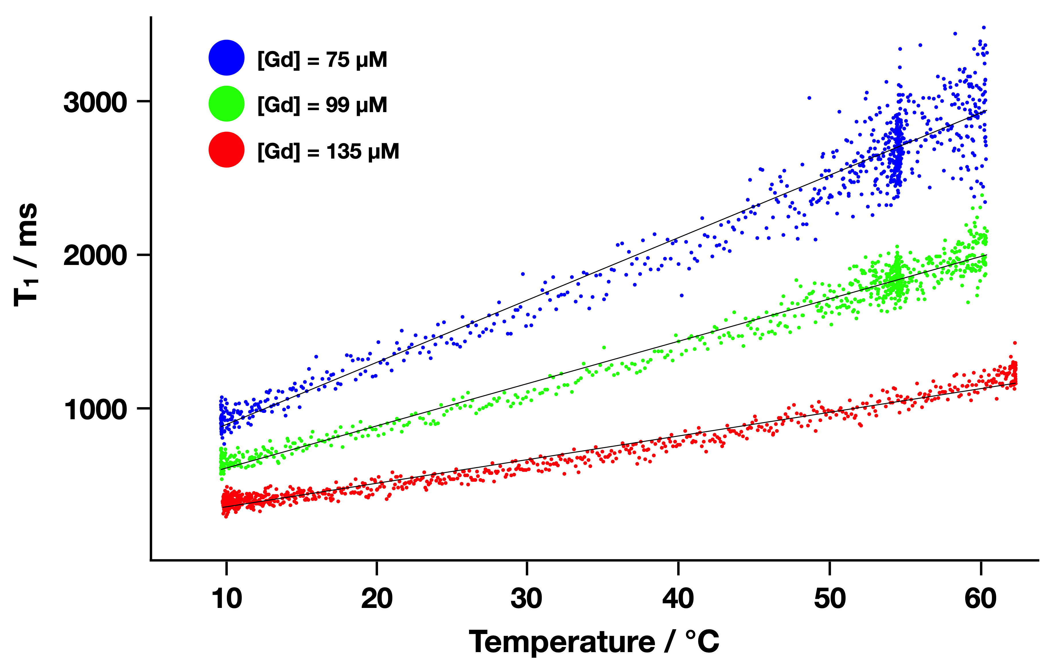
T1 maps acquired in only 8s were obtained over a large scale of temperatures on three tubes containing increasing concentrations of Gd. Figure 4 shows a linear evolution of T1 with the temperature. The slope decreased with the Gd concentration. The uncertainty of the measurements increased with T1 and consequently with temperature. Overall, a temperature uncertainty of 3-4 °C was measured.
DISCUSSION
Using a 2D implementation, the MP2RAGE sequence can be used for T1 mapping with a time resolution below 10s. The slice selection has a small influence on the T1 measurements, which shows the robustness of this T1 mapping sequence compared to other conventional ones like the Variable Flip Angle. The radial encoding has the potential to decrease the sensitivity to motion. The ability to drastically reduce TE enabled to measure T1 of short T2* phantoms.CONCLUSION
This study demonstrate the versatility and the rapidity of 2D versions of the MP2RAGE sequence in vitro. These sequences could be surrogates when the Proton Resonance Frequency (PRF) method fails. They could also be translated to rapidly measure T1 on the whole-body of animals and then on patients.Acknowledgements
No acknowledgement found.References
[1] Quesson B, de Zwart JA, Moonen CTW. Magnetic Resonance Temperature Imaging for Guidance of Thermotherapy. J Magn Reson Imaging 2000;12:525–533
[2] Svedin BT and Parker DL. Technical Note: The effect of 2D excitation profile on T1 measurement accuracy using the variable flip angle method with an average flip angle assumption. Med. Phys. 2017;44 (11):5930-5937
Figures