3817
Fast 3D MR thermometry using echo-shifted sequence with parallel acquisition acceleration1Paul C. Lauterbur Research Center for Biomedical Imaging, Shenzhen Institutes of Advanced Technology, CAS, Shenzhen, China
Synopsis
A high-precision, fast MR thermometry is always preferred for HIFU monitoring. However, the coverage of MRT is also important for the safety concern as the unwanted burn usually takes place in the tissue interface which might be far away from the focal area. In this work, we combined 3D echo-shifted GRE sequence with 2D parallel imaging acceleration to implement a fast 3D MRT.
Introduction
High-intensity focused ultrasound (HIFU) is a useful noninvasive thermal ablation therapy. To ensure its safety and effectiveness, a high-precision, fast thermometry is needed to monitor the treatment. MR thermometry based on proton resonance frequency shift (PRFS) has good linearity and high temperature sensitivity, and is often used for HIFU monitoring[1]. However in practical applications, the greatest potential danger of HIFU is the unwanted skin burns, which is usually far away from the focal area. As a result, the coverage of MRT is also important. In this work, a fast 3D MR thermometry was proposed based on echo-shifted GRE sequence with 2D parallel imaging acceleration.Materials and Methods
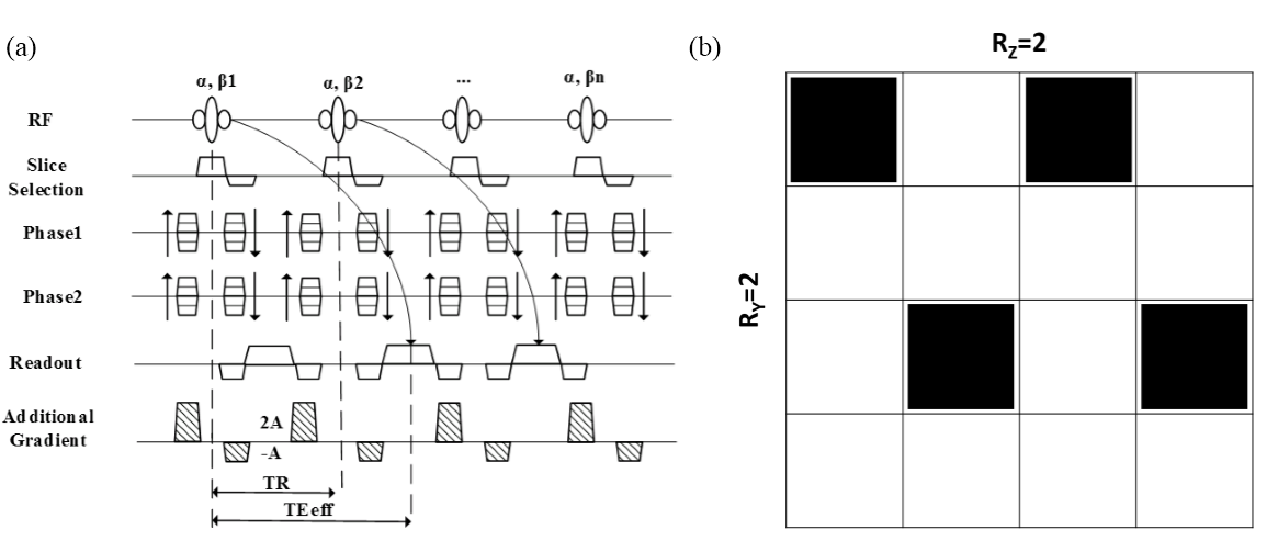
The echo-shifted GRE sequence (cf. Fig. 1(a)) adds a pair of opposite crusher gradients in each of the TRs in both the slice selection direction and the readout direction, which can shift the echo to the next TR. Thus the effective TE becomes TR+TE. Compared to traditional GRE sequences, echo-shifted GRE sequence increases the temperature sensitivity while reducing scanning time[2]. Figure 1(b) shows the under-sampling pattern used by the 2D parallel imaging to accelerate in the two phase encodings directions ky and kz respectively[3].
The algorithm used in this paper is based on SENSE reconstruction algorithm, the coil sensitivity was acquired and estimated ahead of successive measurements[4] then the reconstructed image can be solved from the aliasing image.
All experiments were conducted in a Siemens TIM Trio 3T system. The basic protocol of the sequence: TR/TE=9/5ms, slices per slab = 12, slice thickness = 5 mm. Imaging resolution was 1.5mm in phantom experiment while 2mm in in-vivo experiment.
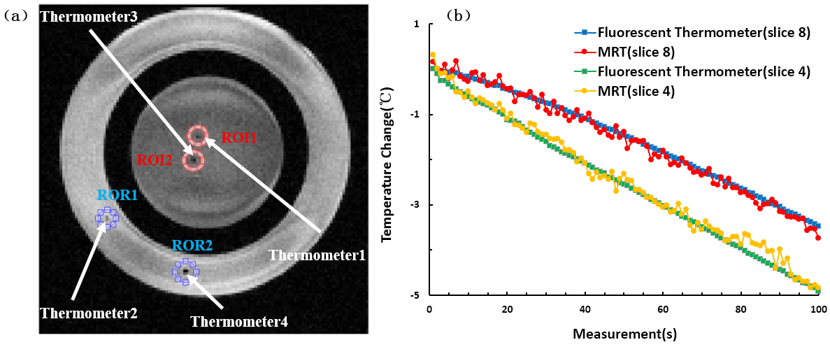
Phantom experiment was conducted to calibrate the temperature measurement accuracy compared to fiber optical thermometer (INDIGO PRECISION, FOTS-DINA-1000-S) which was inserted in the phantom .Thermometer 1 and 2 was in the 4th slice while thermometer 3 and 4 was in the 8th slice (cf. Fig.2(a)). Room experiment was done to compare the temperature precision before and after parallel imaging acceleration. Finally, the echo-shifted sequence (R=4, TAacc=2.6s) with CAIPI was used to monitor a 3D volume during HIFU sonication in rabbit thigh and compare the temperature accuracy before and after parallel imaging acceleration.
Results
Phantom experiment: Figure 2(a) shows an echo-shifted GRE image of a phantom. Figure 2(b) shows the temperature change curve using echo-shifted sequence compared with fiber thermometers. The temperature error in the 4th slice is -0.08±0.14℃and is 0.04±0.11℃ in the 8th slice.
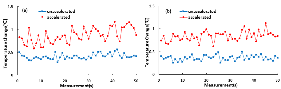
Room temperature experiment: Taking the 6th and 7th slices as an example, Fig. 3 shows the standard deviation of temperature change in an ex-vivo porcine muscle at room temperature. The averaged standard deviation is about 0.40℃ and 0.87℃ before and after acceleration in the 6th slice, is about 0.36℃ and 0.83 in the 7th slice.
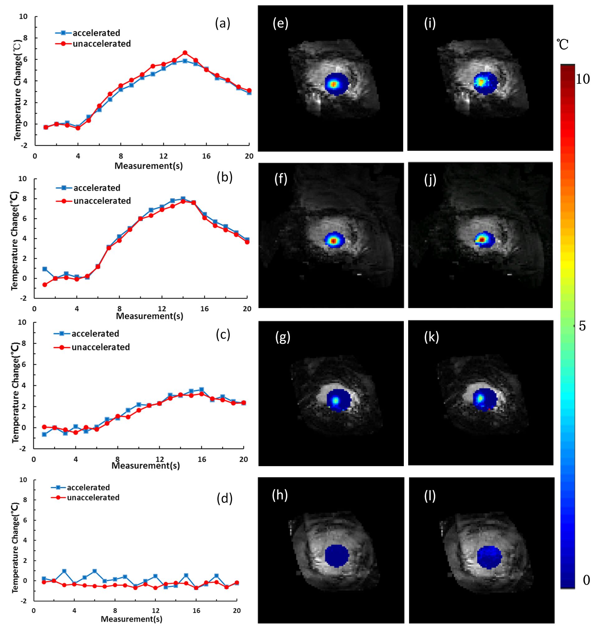
HIFU heating experiment: The mean error of temperature change (cf. Fig. 4) before and after acceleration of the 5th, 6th, 7th and 9th (the skin layer close to ultrasonic transducer) slice is 0.22°C, 0.29°C, 0.15°C and 0.27°C, respectively.
Discussion & conclusions
The 3D MR thermometry based on echo-shifted GRE sequence combined with 2D acceleration has sufficiently large coverage with high temporal resolution and accuracy. The proposed method can perform a temperature measurement with large coverage within 3s after parallel imaging acceleration.
It can be seen from the room temperature experiment that the temperature uncertainty increases by 2.2 and 2.3 times in the 6th and 7th slice after four times acceleration using the custom built 12-channel rabbit coil. After four times acceleration, the temperature uncertainty will increase by two times without considering coil g-factor. Therefore, there is still room for improvement in coil design to further reduce noise amplification caused by g-factor.
Currently, the acceleration is done by retrospective reconstruction, the online real-time reconstruction and visualization are expected in the future work.
Acknowledgements
This work was supported by the Key Laboratory for Magnetic Resonance and Multimodality Imaging of Guang-dong Province (No. 2014B030301013), the National Natural Science Foundation (Nos. 81327801, 81527901,11504401)References
[1] Mougenot, C. , Quesson, B. , de Senneville, B. D., de Oliveira, P. L., Sprinkhuizen, S. , Palussière, J. , Grenier, N. and Moonen, C. T. (2009), Three‐dimensional spatial and temporal temperature control with MR thermometry‐guided focused ultrasound (MRgHIFU). Magn. Reson. Med., 61: 603-614.
[2] Chung, Y. and Duerk, J. L. (1999), Signal formation in echo‐shifted sequences. Magn. Reson. Med., 42: 864-875.
[3] Breuer, F. A., Blaimer, M. , Mueller, M. F., Seiberlich, N. , Heidemann, R. M., Griswold, M. A. and Jakob, P. M. (2006), Controlled aliasing in volumetric parallel imaging (2D CAIPIRINHA). Magn. Reson. Med., 55: 549-556.
[4] Yuhong Peng, Chao Zou, Yangzi Qiao, Changjun Tie, Qian Wan, Rui Jiang, Chuanli Cheng, Dong Liang, Faqi Li, Xin Liu, Hairong Zheng. Fast MR thermometry using an echo-shifted sequence with simultaneous multi-slice imaging. Magnetic Resonance Materials in Physics, Biology and Medicine, 2018, DOI: 10334-018-0692-x
Figures