3812
Detecting T1-based Signal Reduction in Focused Ultrasound Heating of Bone using a 3D Spiral Ultra-Short Echo Time Sequence1Biomedical Engineering, University of Virginia, Charlottesville, VA, United States, 2Autism and Developmental Medicine Institute, Geisinger Health System, Danville, PA, United States, 3Radiology and Medical Imaging, University of Virginia, Charlottesville, VA, United States
Synopsis
MR-guided Focused Ultrasound (MRgFUS) is used transcranially to ablate brain tissue for the treatment of neurological diseases. Temperature monitoring of the skull is desired for increasing treatment safety and efficacy. Proton resonance frequency shift MR fails to detect heating in the cortical bone of the skull. T1-based MR thermometry uses T1 mapping to observe a linear increase in T1 with temperature but requires long acquisitions. We demonstrate a new thermometry method dependent on the linear relationship between signal magnitude from a T1-weighted 3D Spiral Ultra-short Echo Time sequence and test it across three scanners in multiple trials.
Introduction
MR-guided focused ultrasound (MRgFUS) provides a minimally invasive therapy for neurological diseases by employing focused sound waves to heat surgical targets to temperatures greater than 60°C. However, as the skull is highly absorptive to acoustic energy, MRgFUS has been shown to cause unintended skull bone marrow injury to patients treated for movement disorders1. There is currently no direct monitoring of skull heating during clinical treatment because the short T2* constant of bone (<1ms) precludes traditional proton resonance shift thermometry2.
Several groups have performed bone thermometry in ex-vivo bovine femurs2,3,4 by using an Ultrashort Echo Time (UTE) sequence to exploit the temperature dependence of T1 relaxation in cortical bone. A remaining challenge is to balance tradeoffs between time constraints (~1min to detect heat dissipation), signal to noise, and a large field of view. The T1-mapping method by Han et al. requires 8 min. of scan time per temperature point2. In pursuit of a shorter acquisition time, we report on our development and testing of a T1-weighted 3D Spiral Ultra-short Echo Time sequence.
Methods
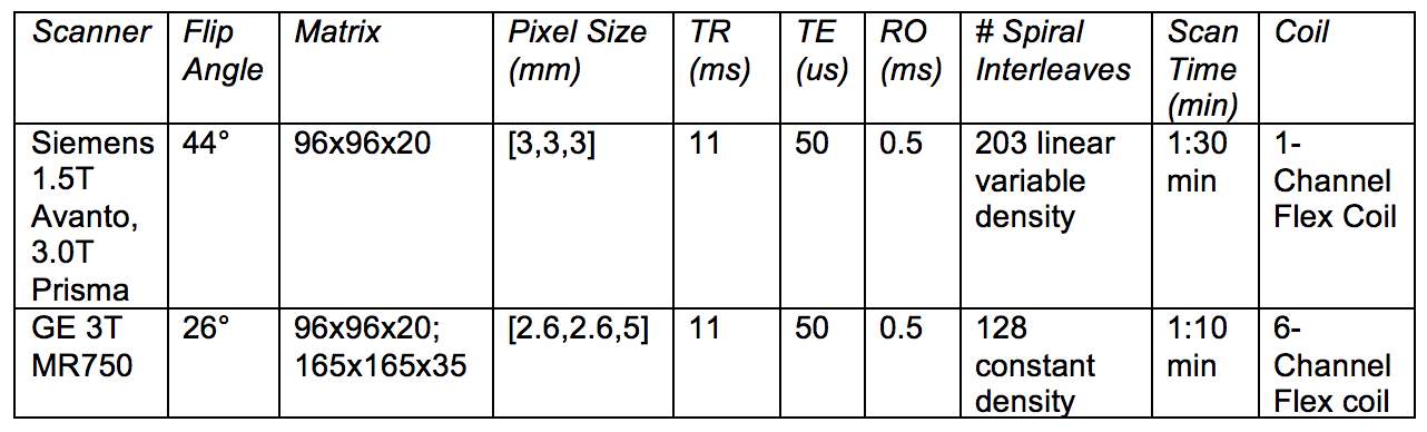
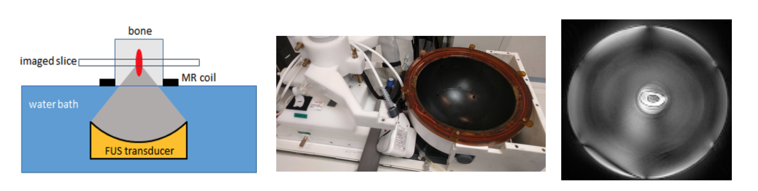
Nine trials were conducted on ex-vivo bovine femurs obtained from the butcher and stored in refrigeration before experimentation. For each trial, a fiberoptic thermocouple was placed in a burr hole drilled into the cortical layer of each sample. Five of the experiments consisted of heating the bone in 70°C water for 10 minutes and then imaging during cooling (after every 5°C of temperature loss). Four of the experiments consisted of heating the bones with FUS. The bones were placed on an ultrasound transparent film above a water tank of 1.1 MHz single element small animal transducer (FUS Instruments Inc.) (Fig. 2). The bones were targeted (1mm3 focus) and ablated with a 45W continuous sonication six times for 135 seconds to temperatures between 30-70°C. Imaging was performed either after each sonication (trial 1) or during sonication to maintain bone temperature (trials 2-4) and performed using a prototype 3D spiral UTE pulse sequence on 1.5T (Siemens Avanto) and 3T scanners (Siemens Prisma and GE MR750). See Fig. 1 for experimental parameters.Results
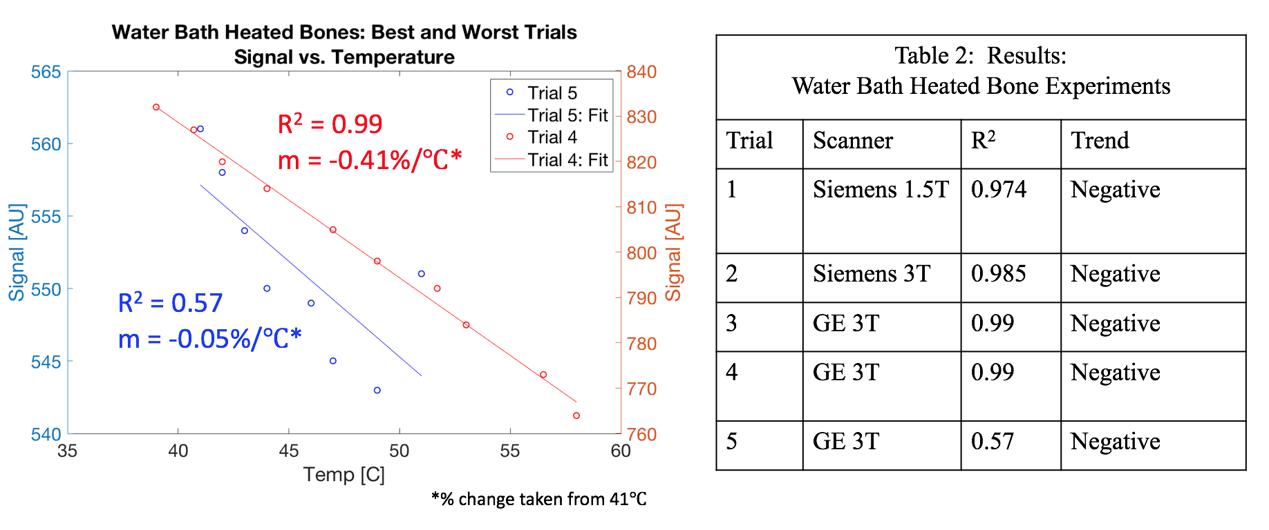
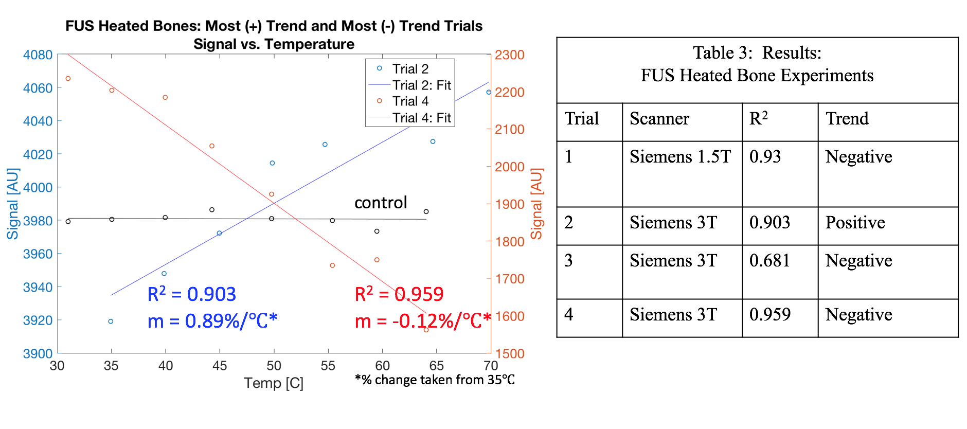
From MR thermometry theory, we expected the T1 weighted signal to linearly decrease with temperature as T1 increases with temperature5. All trials in which bones were heated by a water bath (n=5) showed a strong negative linear dependence between signal and temperature (R2ave = 0.90) (Fig. 4). However, we saw conflicting results when we heated the bones with the small animal FUS transducer. While three trials showed the expected negative trend (R2ave = 0.86), one trial showed an opposite (positive) trend (R2 = 0.90) (Fig. 5).Discussion
We have observed a strong negative trend between signal and temperature in all bones heated by water baths in accordance with our hypothesis while under the constraint of <2 min scan time. However, when bones were heated by the small animal FUS transducer, we observed both positive and negative correlations between the MR signal and temperature. These conflicted observations are corroborated in previous reports of bone thermometry. For example, Ramsay et Al. observed a positive trend using a short echo-time RF-spoiled GRE6. Odeen et Al. also observed a positive trend of measured T1 values (from variable flip angle T1 mapping) decreasing with temperature using a short-echo time GRE3.
There are many possible explanations for this observation. The 1.1MHz transducer may have heated only a very small portion of the bone. The magnetization properties of bone may change depending on how it is stored and whether coagulation occurred due to heating. Furthermore, bone is composed of bound and pore water, which may respond differently to temperature6. As we have used some of the same bones multiple times, their properties could have been altered to no longer show the expected negative trend. Bone processing should be kept consistent between experiments.
To avoid this source of uncertainty, we have implemented the 3D spiral UTE sequence on the clinical 650 kHz, 1024-element Insightec ExAblate (Model 4000) transducer paired with a 3T GE scanner and are currently planning FUS heated temperature experiments on bovine bone. Additionally, the adaption of this sequence to the clinical transducer system will allow us to test our sequence in animals and in humans during essential tremor treatments.
Conclusion
Our preliminary results show the possibility of rapid whole-brain T1-based thermometry using spiral UTE imaging. However, determining the T1 response of bone to temperature will require future study.Acknowledgements
This research was partly supported by Siemens Medical Solutions. The authors acknowledge Josef Pfeuffer and Berthold Kiefer for their help with this project.References
1. Schwartz, Michael L., et al. “Skull Bone Marrow Injury Caused by MR-Guided Focused Ultrasound for Cerebral Functional Procedures.” Journal of Neurosurgery, 2018, pp. 1–5., doi:10.3171/2017.11.jns17968.
2. Han, Misung, et al. “Assessing Temperature Changes in Cortical Bone Using Variable Flip-Angle Ultrashort Echo-Time MRI.” Magn Res Med. 2015 Dec; 74(6): 1548–1555.
3. Odeen O., Bolster B., Jeong E. Parker D. “Investigation of temperature dependent changes in signal intensity, T1 and T2* in cortical bone”. Abstract. In: ISMRM 2016.
4. Miller GW. “MR bone Imaging.” J Ther Ultrasound. 2015; 3(Suppl 1): O37.
5. Odéen, Henrik, and Dennis L. Parker. “Non-Invasive Thermometry with Magnetic Resonance Imaging.” Theory and Applications of Heat Transfer in Humans, 2018, pp. 267–299., doi:10.1002/9781119127420.ch15.
6. Ramsay, Elizabeth, et al. “Temperature-Dependent MR Signals in Cortical Bone: Potential for Monitoring Temperature Changes during High-Intensity Focused Ultrasound Treatment in Bone.” Magnetic Res Med, vol. 74, no. 4, 2014, pp. 1095–1102., doi:10.1002/mrm.25492.
7. Fielden S. et al. ISMRM 2015;23: p3867.
Figures