3811
Novel Acoustic Coupling Design to Improve MR Imaging Guidance for Focused Ultrasound Surgery1Department of Biomedical Engineering, University of Virginia, Charlottesville, VA, United States, 2Department of Mechanical Engineering, Virginia Polytechnic Institute and State School, Blacksburg, VA, United States, 3Department of Biomedical Engineering, Virginia Polytechnic Institute and State School, Blacksburg, VA, United States, 4Graduate Program in Translational Biology, Medicine, and Health, Virginia Polytechnic Institute and State School, Blacksburg, VA, United States, 5Department of Chemical Engineering, Virginia Polytechnic Institute and State School, Blacksburg, VA, United States, 6Department of Radiology, University of Virginia, Charlottesville, VA, United States
Synopsis
We explore the feasibility of an acoustic coupling medium that is invisible to both T2-weighted anatomical scans and MR thermometry scans during MR-guided focused ultrasound surgery and yet still preserves the cooling and acoustic coupling functions normally provided by water.
Introduction
The water bath present during MR-guided, focused ultrasound surgery (FUS) provides crucial cooling and acoustic coupling functions. However, this bath can also artificially expand the MR imaging field of view, introduce motion artifacts, and skew pre-scan calibrations1. The resulting degradations in thermometry and anatomical imaging can hinder the safety and efficacy of a procedure. Minimizing the received signal from the water bath would mitigate these problems.
We propose suspending iron oxide nanoparticles (SPIOs) in the water bath to render it invisible to MR guidance scans. Anatomical and thermometry acquisitions employ long echo times. Dilute suspensions that accelerate the transverse relaxation process should suppress the water bath signal. However, the SPIOs may also seed cavitation nuclei in the water bath, which may falsely signal dangerous cavitation activity in the patient’s vasculature2. Well-designed SPIOs should both suppress the water bath and not seed cavitation. Below, we examine the feasibility of using SPIOs to suppress the water bath’s MR signal.
Methods
Water baths with SPIOs (Stock# US7568, US-Nano-Research, Houston, TX) of concentrations 0 and 0.25 mM were respectively examined using dynamic light scattering and subsequently used as a coupling agent while sonicating a gel target. The 0.25mM sample was continuously circulated. Sonication was accomplished using a 30 cm diameter, 650 kHz hemispherical transducer (ExAblate Neuro 4000, Haifa, Israel) situated in the bore of a 3T scanner (MR750, GE, Waukesha, WI). Anatomical imaging consisted of a 2D, T2-weigheted, turbo spin-echo sequence: (TR: 2.5 s, TE: 81 ms, ETL: 24, FOV: 23 cm, resolution: 1.1 mm). Proton resonance shift thermometry, simultaneous to sonication, used a 2D, gradient-echo sequence: (TR: 28 ms, TE: 12.9 ms, FOV: 28 cm, resolution: 1.25 mm). Sonications consisted of 10 s bursts with transmitted acoustic power ranging from 50 to 600 W. This experiment was repeated three times.
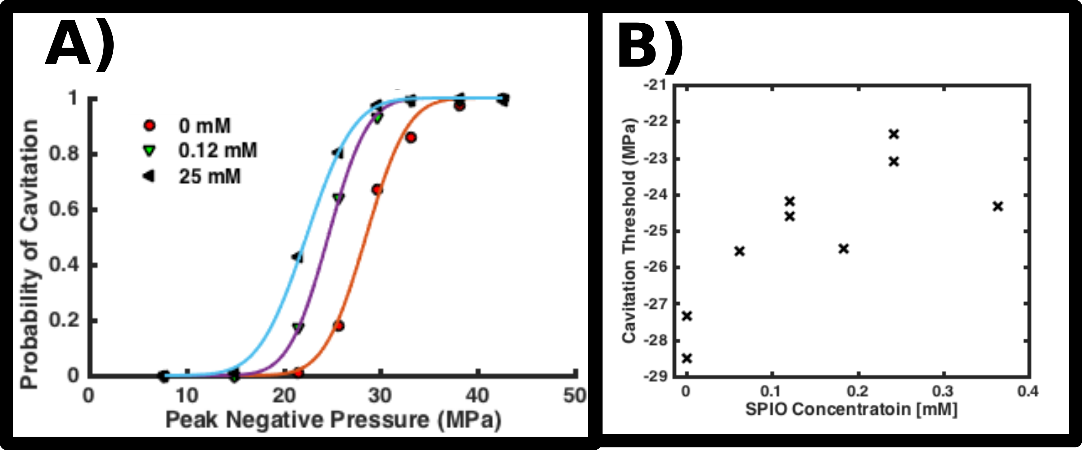
The cavitation threshold for various water bath solutions was estimated by depositing 100 highly focused, shocked, 5 cycle, 700 kHz acoustic pulses at a rate of 1 Hz directly into various SPIO concentrations (0 to 0.36 mM) using a custom transducer (Histosonics, Ann Arbor, MI) in a water tank. Negative acoustic pressures were incremented from -8 to -42 MPa. To prevent settling, the SPIOs were periodically and gently agitated. Cavitation activity was detected with both an optical camera and a passive detection element. Cavitation probability curves were estimated by computing the relative frequency of cavitation events at each negative pressure and then fitting a Gaussian cumulative distribution function3,4. Experimental setups are shown in Figures 1A,B.
Results
Dynamic light scattering revealed that the SPIOs aggregate into about ~240 nm globules. The particles demonstrated a 526 s-1mM-1 transverse relaxivity at 3T. See Figure 1C.
Example magnitude anatomical and thermometry images for both SPIO concentrations are shown in Figure 2. The 0.25 mM SPIO mixture suppressed the water bath signal by 98 and 90% for the spin-echo and gradient-echo scans, respectively. Figure 2 also plots the temperatures achieved during sonication (averaged over 9 voxels) as a function of transmitted acoustic power, with each curve artificially translated by $$$\pm$$$3 W to improve legibility. The presence of SPIOs correlates with a systematic ~2 oC decrease in average temperature for acoustic powers above 200 W.
Figure 3A displays example cavitation probability curves obtained after sonicating varying concentrations of SPIOs. The SPIOs increase the chance lower amplitude pulses will stimulate cavitation events. Figure 3B plots, as a function of SPIO concentration, the 0.5 probability threshold pressure derived from the fitted curves. The SPIOs decrease the magnitude of this threshold by 13 $$$\pm$$$4% from -28 Mpa to -24 $$$\pm$$$1 MPa. Noise in this figure may indicate particle settling or gas absorbing into the SPIO mixture.
Discussion
Our results indicate that the SPIOs can effectively suppress the water bath signal during MR-guided FUS procedures. The systematic ~2 oC decrease in focal temperature suggests that the SPIOs weakly attenuate the acoustic waves. Meanwhile, the observed decrease in the estimated 0.5 probability threshold pressure indicates that, at large enough acoustic amplitudes, the SPIOs seed cavitation.
While a 13% decrease in threshold pressure is non-trivial, acoustic amplitudes steeply attenuate outside the transducer focus and are unlikely to achieve -20 MPa within the coupling water bath. However, continuous wave sonications may stimulate cavitation through rectified diffusion5—an effect not studied here. Future studies should both account for pre-focal pressures as well as characterize cavitation behavior under continuous wave conditions.
Conclusion
Aqueous SPIO solutions effectively suppress the water bath signal during MR-guided FUS procedures and only weakly suppress thermal deposition in gel targets. The SPIOs also decrease the cavitation threshold pressure.Acknowledgements
The authors would like to thank Justin Howell and Dave Moore for their respective assistance with experimental setup.References
1. Odéen, Henrik; Patil, Sunil; Bolster, Bradley; Bhat Himanshu; Parker DL. Evaluation and tradeoffs of 2D and 3D Cartesian MR temperature imaging (MRTI) for brain applications. In: 6th International Symposium on Focused Ultrasound. Reston, VA; 2018. p. BR-46.
2. Hynynen K, Chung AH, Colucci V, Jolesz FA. Potential adverse effects of high-intensity focused ultrasound exposure on blood vessels in vivo. Ultrasound Med. Biol. 1996;22:193–201.
3. Maxwell AD, Wang TY, Cain CA, Fowlkes JB, Sapozhnikov OA, Bailey MR, Xu Z. Cavitation clouds created by shock scattering from bubbles during histotripsy. J. Acoust. Soc. Am. 2011;130:1888–1898.
4. Maxwell AD, Cain CA, Hall TL, Fowlkes JB, Xu Z. Probability of cavitation for single ultrasound pulses applied to tissues and tissue-mimicking materials. Ultrasound Med. Biol. 2013;39:449–465.
5. Leighton, T., The acoustic bubble. 2012. Academic press.
Figures