3810
Enhancement of the HIFU thermal effect in ex-vivo kidneys using a new class of endovascular sono-sensitizers1Image Guided Interventions Laboratory, Faculty of Medicine, University of Geneva, Geneva, Switzerland, Geneva, Switzerland, 2Radiology Department, University Hospitals of Geneva, Geneva, Switzerland, Geneva, Switzerland, 3University of Avignon, CBSA-IBMM (UMR5247), Avignon, France, Avignon, Switzerland, 4Department of Radiology and Medical Informatics, University of Geneva, Switzerland, Geneva, Switzerland, 5Research and Development Laboratory, Visceral and Transplantation Service, University Hospital Geneva, Geneva, Switzerland., Geneva, Switzerland, 6Sorbonne Universités, UPMC Univ Paris 06, CNRS, INSERM, Laboratoire d’Imagerie Biomédicale (LIB), F-75006, Paris, France, Paris, Switzerland
Synopsis
Magnetic Resonance guided High Intensity Focused Ultrasound (MRgHIFU) is a promising approach for the non-invasive ablation of localized tumors. Ablation of highly perfused tumors is challenging due to the heat sink effect. We developed a new concept of endovascular liquid core micro-droplets, used as sono-sensitizers for the enhanced absorption of the HIFU beam. We demonstrated the improvement of the HIFU thermal effect after adjunction of sono-activable micro-droplets in the perfusion fluid of freshly excised viable pig kidneys. Temperature maps were computed using the PRFS method to prove the enhancement of thermal contrast.
Introduction
During the last decades, Magnetic Resonance guided High Intensity Focused Ultrasound (MRgHIFU) has been widely accepted and used for thermal ablation of tumors. Ablation of highly perfused tumors is challenging due to the heat sink effect. We developed a new concept of endovascular liquid-core micro-droplets, used as sono-sensitizers for the enhanced absorption of the HIFU beam. They comprise PFOB liquid-core and are stabilized with biocompatible perfluorinated surfactants [1]. We demonstrated good capillary circulation and efficiency of these micro-droplets to enhance the temperature rise at the focal point in perfused pig kidneys under MR guidance.Method
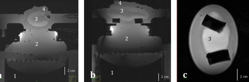
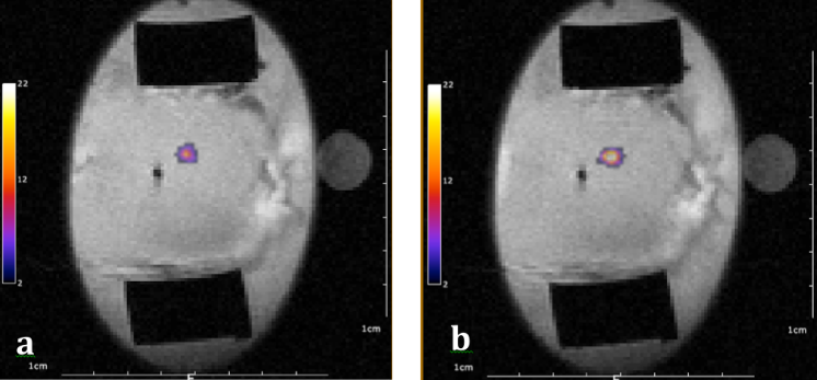
Two pig kidneys were perfused with the MR-compatible perfusion machine described by Buchs et al. [2] delivering oxygen and perfusate to the kidneys. Focused ultrasound was generated by an MR compatible phased array transducer operating at 1 MHz and powered by a 256-channel beam former. Microemulsions of micro-droplets with 2.35µm average diameter were prepared with 2% v/v of PFOB. Two doses of 50mL of the emulsion were successively injected at 30 min interval in 750mL of perfusate and two sonication protocols were conducted to verify the effect of micro-droplet injections on temperature rise. The first protocol consisted in sonications at the fixed focal point with 60W or 135W of acoustic power during 10s or 30s. In the second protocol the energy was delivered in a 4mm-diameter circle, described by 16 points, at 90W for a total duration of 220s. Experimental data were acquired in a whole-body Siemens 3T MRI scanner with a segmented GRE-EPI sequence in the coronal plane through the focal point using a 11 cm diameter receive loop coil. Relative localization of the HIFU transducer and kidney was performed with a high-resolution T1-weighted 3D sequence (Fig 1). The temperature rise was computed by the PRFS sensitive method (voxel size 1x1x3mm3). MR images were reconstructed in real-time and magnitude images were merged with temperature maps as illustrated in Fig 2. The intrarenal microcirculation was verified by injection of gadolinium chelate at the end of the experiment monitored with a 2D saturation-prepared turbo flash sequence (DCE) according to the study by Buchs et al. [3].Results
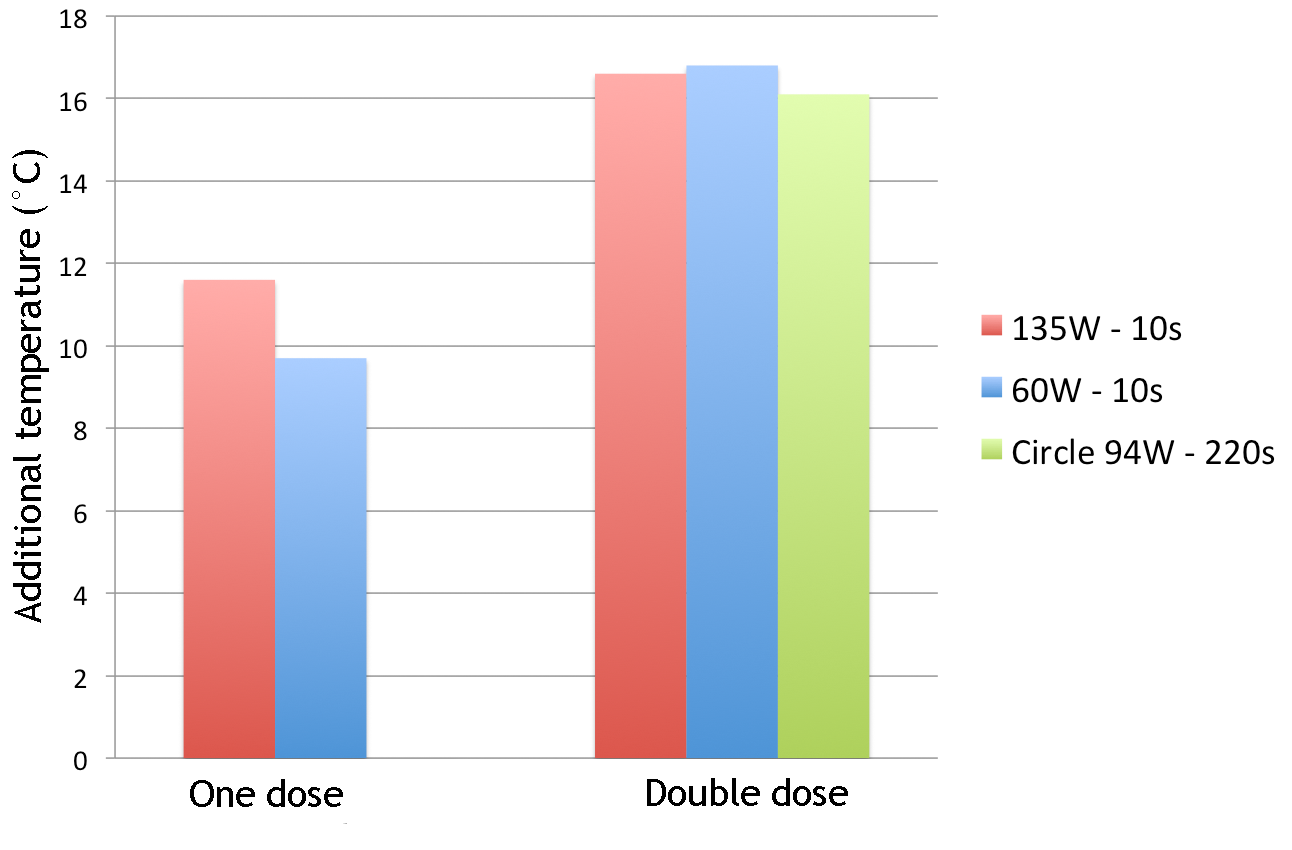
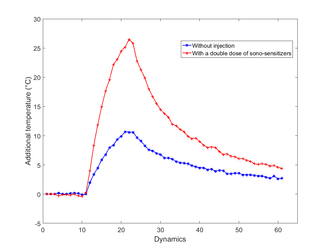
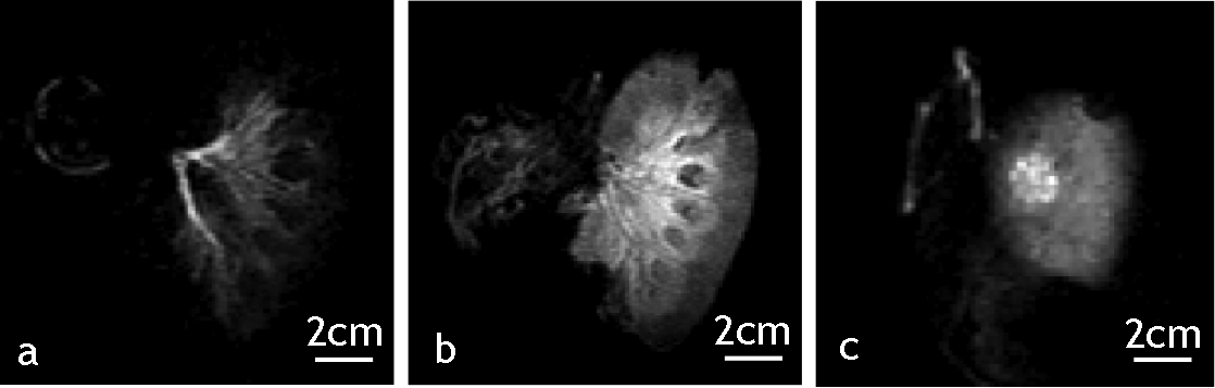
The temperature elevation maps clearly demonstrated an enhanced increase in the achieved temperature upon adjunction of micro-droplets. Figure 3 and 4 shows that the additional temperature elevation achieved with one dose of sono-sensitizer for a 10s single focus sonication was 9.7°C at 60W and 11.6°C at 135W, and the additional temperature elevation achieved after the second dose was 16.8°C at 60W and 16.6°C at 135W. Adjunction of sono-sensitizer coupled to 30s sonication yielded above 100°C tissue temperature and produced a boiling core. For the circular pattern of volumetric sonication, the additional temperature elevation due to sono-sensitizer was 16.1°C. The perfusion controller did not measure overpressure values indicating that the micro-droplets freely circulated into the capillaries without embolism. Repeated sonication at the same location yielded reproducible results demonstrating that the pool of circulating droplets is stable at least within the hour following the injection. DCE MR images showed T1 hyperintense signal at the site of ablation. Outside the target region, DCE imaging demonstrated a good intrarenal circulation and demonstrated that the kidney was viable at the end of the experiment (Fig 5)Discussion
The fluid circulation in the kidney allowed a renewal of fresh micro-droplets at the focal spot, which enabled an optimal efficiency of the method. Experimental data were accurate and demonstrated the feasibility of the method by injection of micro-droplets in perfused ex-vivo tissues. The kidney is a good model for perfused tissue ablation given that 20% of the cardiac flow is driven into it. In our study, HIFU could be applied one minute after the iv injection, unlike the phase shift nanoemulsions which require six hours between injection and HIFU [4].Conclusion
Injections of micro-droplets in the endovascular system demonstrated a dramatic enhancement in the heating efficiency of the MRgHIFU. The use of micro-droplets allowed reaching high temperature with less HIFU power, less technological constraints and less risk for near- or far- field heating.Acknowledgements
No acknowledgement found.References
[1] Astafyeva K, Somaglino , Desgranges D, Berti R, Patinote C, Langevin D, Lazeyras F, Salomir R, Polidori A, Contino-Pépin C, Urbachij W, Taulier N (2015) Perfluorocarbon nanodroplets stabilized by fluorinated surfactants: characterization and potentiality as theranostic agents. J Mater Chem B 3:2892-2907.
[2] Buchs JB, Bühler L, Morel P. A new disposable perfusion machine, nuclear magnetic resonance compatible, to test the marginal organs and the kidneys from non-heart-beating donors before transplantation (2007) Interactive CardioVascular and Thoracic Surgery 6 (2007) 421–424.
[3] Buchs JB, Buehler L, Moll S, Ruttimann R, Nastasi A, Kasten J, Morel P, Lazeyras F. DCD Pigs’ kidneys analyzed by MRI to assess ex vivo their viability (2014) Transplantation Journal. 97(2):148–153.
[4] Kopechek JA, Park E, Mei CS, McDannold NJ, Porter TM. Accumulation of Phase-Shift Nanoemulsions to Enhance MR-Guided Ultrasound-Mediated Tumor Ablation In Vivo (2013) J Healthc Eng. 4(1): 109–126.
Figures