3809
Rapid MR-guided-HIFU using Convolution-based Reconstruction and Parallel Imaging (CORE-PI)1Department of Biomedical Engineering, Technion - Israel Institute of Technology, Haifa, Israel
Synopsis
A novel reconstruction method for accelerated Magnetic Resonance guided High Intensity Focused Ultrasound (MRgHIFU) thermometry is presented. This method utilizes multi-coil acquisition, k-space undersampling and the recently introduced Convolution-based Reconstruction for Parallel Imaging (CORE-PI) technique. The proposed method utilizes data sparsity in the Stationary Wavelet Transform (SWT) domain. It is a parameter-free, non-iterative and calibrationless method. Retrospective experiments with in-vivo data from clinical human prostate ablation treatments show that the proposed method produces accurate temperature maps from two-fold and three-fold subsampled k-space data. The method is therefore suitable for real time application.
Background
MR thermometry is a safe, non-invasive efficient modality for guiding High Intensity Focused Ultrasound (HIFU) treatments1,2. MR-guided-HIFU (MRgHIFU) is clinically used for treating prostate3,4, brain5,6, breast7,8 liver9–11 and heart11. However, common MRgHIFU has a very limited spatial coverage, which currently includes only several few 2D slices12,13. To improve the temperature monitoring, MRgHIFU can be accelerated by k-space undersampling.
Purpose
A novel method for accelerated MRgHIFU from subsampled k-space is proposed. This method utilizes the recently introduced Convolution-based Reconstruction for Parallel MRI (CORE-PI)17 technique. In contrast to previous methods, CORE-PI utilizes parallel MRI acquisition, exploits data sparsity and offers simple non-iterative computations. The proposed method is also parameter-free, i.e. does not require tuning of any parameters, and operates in a calibrationless manner.Theory
The proposed method assumes the existence of one fully sampled baseline dataset. This dataset is acquired at $$$t=0$$$ , prior to heating onset, by an array of $$$N_c$$$ coils. Sensitivity maps of the coils are estimated from that data, and a complex-valued baseline image $$$f_0(x,y)$$$ is computed from the data using Roemer’s optimal method18.
During heating $$$(t>0)$$$, subsampled k-space data are acquired. The method’s goal is to reconstruct $$$f_t(x,y)$$$ (the unknown complex-valued MR image) from the subsampled data using the CORE-PI method. Then, the temperature change can be obtained using the well-established Proton Resonance Frequency (PRF) shift thermometry1.
In contrast to most PI methods, which reconstruct $$$f_t(x,y)$$$ either in the image domain or the Fourier domain (or their hybrid domain), CORE-PI reconstructs the image representation in the Stationary Wavelet Transform (SWT) domain. Since SWT-domain data are highly sparse and redundant, CORE-PI obtains the full SWT of $$$f_t(x,y)$$$ from the subsampled k-space data, using only estimated sensitivity maps.
The proposed method steps are:
- Applying CORE-PI17 for computation of $$$\Psi f_t(x,y)$$$, which is the SWT of $$$f_t(x,y)$$$, directly from the subsampled k-space data. This step is performed in a two-channel process, which includes a low-pass and a high-pass channel, according to the wavelet filter bank method19. This step produces the full set of SWT coefficients representing $$$f_t(x,y)$$$ in the $$$\Psi$$$ domain.
- Reconstruction of $$$f_t(x,y)$$$ from the recovered coefficeints using $$$\Psi^{-1}$$$, which is the Inverse SWT.
- Reconstruction of the temperature rise from the temporal phase change using the well-established PRF shift method1, with $$$\Delta \angle\phi_t = \angle f_t(x,y) - \angle f_0(x,y)$$$ .
Methods
Imaging. The method was validated using MR data acquired in two clinical in-vivo human prostate treatments using 8-coils, a 3Tesla MR scanner (GE Healthcare, WI) and ExAblate 2100 prostate array (InSightec, Israel). Data was provided by InSightech. The data was fully sampled in 2D Cartesian k-space, deidentified and retrospectively subsampled offline with a regular subsampling scheme.
Reconstruction. CORE-PI was implemented using a Daubechies-2 SWT. Coils sensitivity maps were estimated from baseline data using a Sum Of Squares (SOS). The temperatures reconstructed from the subsampled data were compared to those obtained from the full k-space data using the Normalized Root Mean Square Error (NRMSE). Computations were performed in Matlab™ on a personal computer.
Results & Discussion
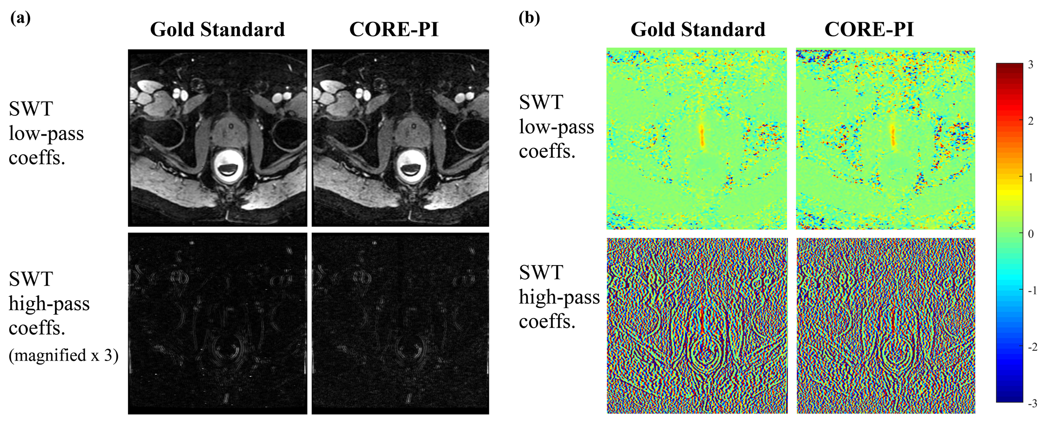
CORE-PI was implemented to the in-vivo data which was subsampled with a reduction factor of R=2. Figure 1 shows the SWT coefficients that were computed by CORE-PI, i.e. the $$$\Psi f_t(x,y)$$$ representation, and the coefficients obtained form the fully sampled data. Clearly, CORE-PI produced a highly accurate reconstruction of the SWT coefficients, both in magnitude and in phase, in both the low-pass and high-pass channels. The CORE-PI images include all the anatomical structures and HIFU-induced phase modifications that are present in the gold standard images, without discernible artifacts.
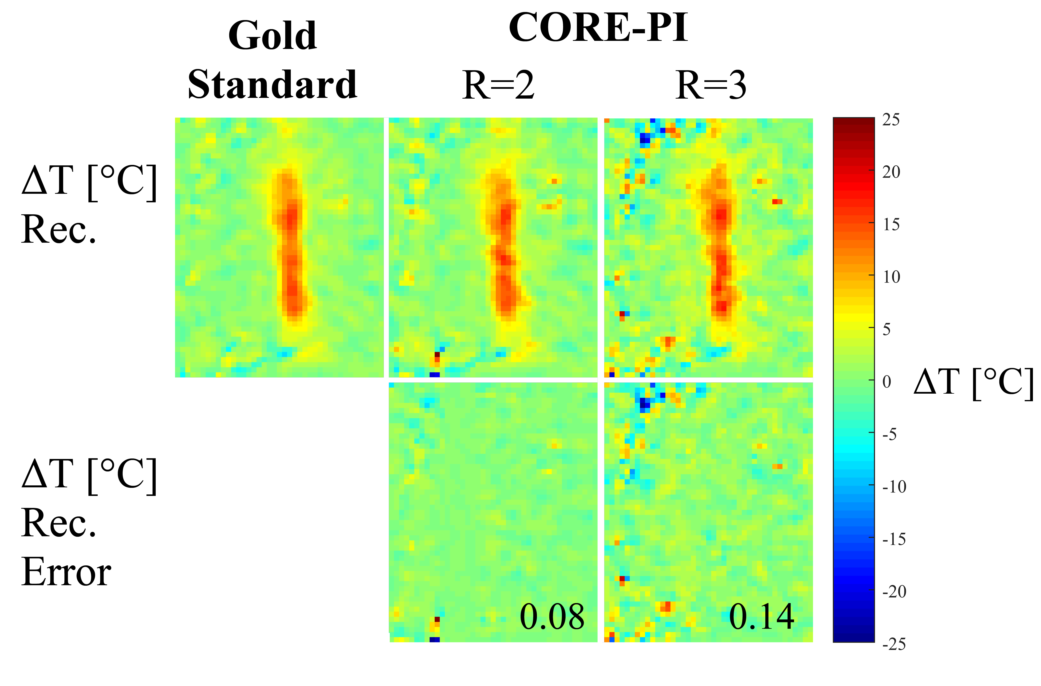
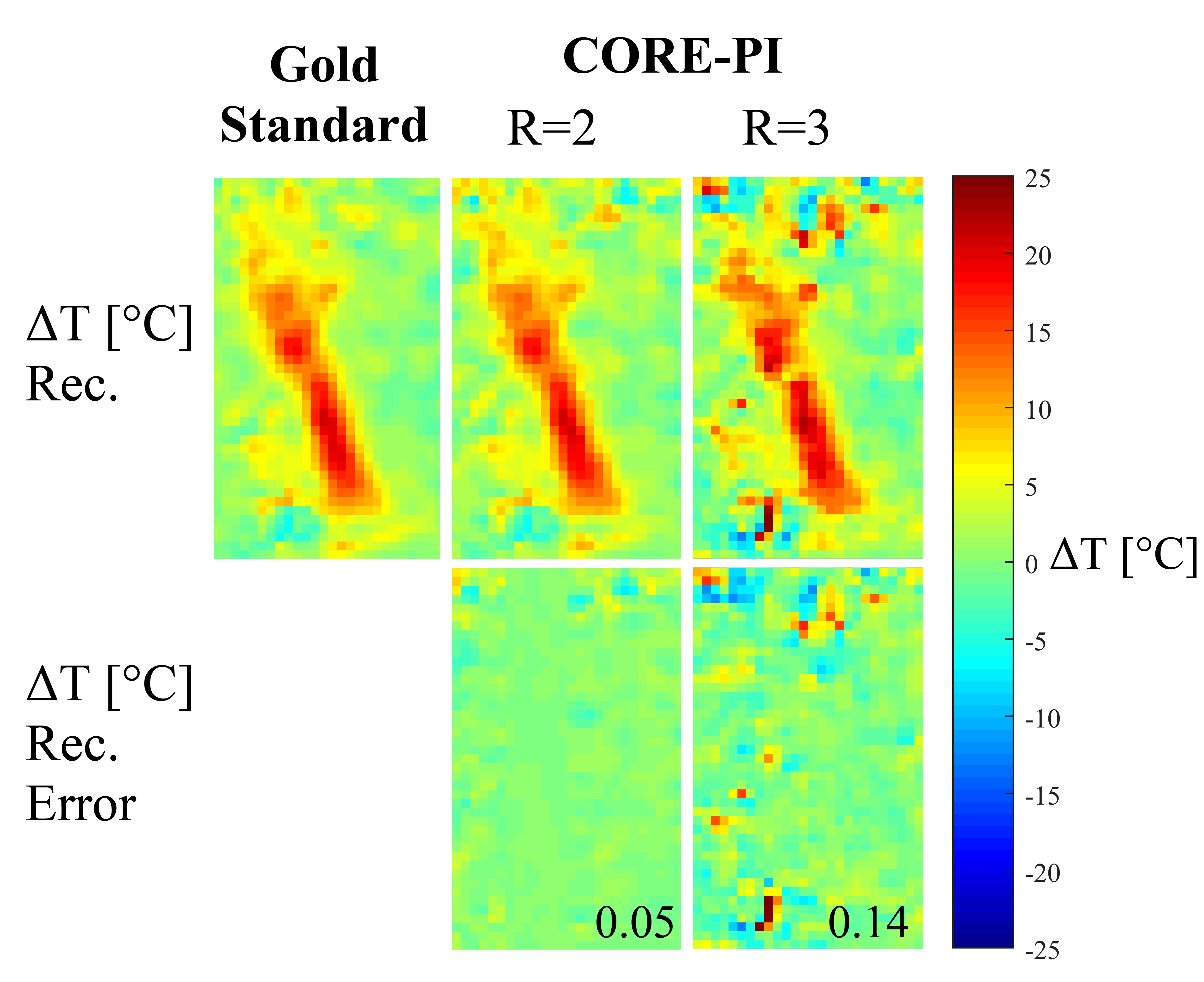
Figure 2 shows the results of the temperature changes reconstructed by CORE-PI from the SWT decomposition, for both R=2 and R=3. Evidently, the CORE-PI reconstructions of the HIFU-induced temperature rise are similar both in value and shape to the gold standard. Similar results are shown in Figure 3, which shows CORE-PI reconstructions for data of a different patient. Markedly, in both Figure 2 and Figure 3, there are no severe errors of temperature reconstruction with in the HIFU-heated zone. This high accuracy of CORE-PI is reflected by the low NRMSE values (0.05-1.4).
Conclusion
This work proposes the implementation of CORE-PI for accelerated MRgHIFU. CORE-PI exploits data sparsity, offers simple non-iterative computations, and is a parameter-free calibrationless method. The in-vivo results show that: (1) CORE-PI reconstructs $$$\Psi f_t(x,y)$$$ accurately and directly from the multicoil subsampled k-space data, and (2) it produces highly accurate temperature rise maps. It is therefore highly suitable for accelerating MRgHIFU.Acknowledgements
The authors thank InSightech for providing the in-vivo data, and Dr. Yoav Levy from InSightech for useful discussions.References
1. Rieke, V. & Butts Pauly, K. MR thermometry. J. Magn. Reson. Imaging 27, 376–390 (2008).
2. Kuroda, K. MR techniques for guiding high-intensity focused ultrasound (HIFU) treatments. J. Magn. Reson. Imaging 47, 316–331 (2018).
3. Chopra, R., Burtnyk, M., Haider, M. A. & Bronskill, M. J. Method for MRI-guided conformal thermal therapy of prostate with planar transurethral ultrasound heating applicators. Phys. Med. Biol. 50, 4957–4975 (2005).
4. Pauly, K. B. et al. Magnetic Resonance-Guided High-Intensity Ultrasound Ablation of the Prostate. Top. Magn. Reson. Imaging 17, 195–207 (2006).
5. Wang, T. R., Dallapiazza, R. & Elias, W. J. Neurological applications of transcranial high intensity focused ultrasound. Int. J. Hyperth. 31, 285–291 (2015).
6. Ghanouni, P. et al. Transcranial MRI-Guided Focused Ultrasound: A Review of the Technologic and Neurologic Applications. Am. J. Roentgenol. 205, 150–159 (2015).
7. Minalga, E. et al. An 11-channel radio frequency phased array coil for magnetic resonance guided high-intensity focused ultrasound of the breast. Magn. Reson. Med. 69, 295–302 (2013).
8. Deckers, R. et al. Performance analysis of a dedicated breast MR-HIFU system for tumor ablation in breast cancer patients. Phys. Med. Biol. 60, 5527–5542 (2015).
9. Weidensteiner, C. et al. Stability of real-time MR temperature mapping in healthy and diseased human liver. J. Magn. Reson. Imaging 19, 438–446 (2004).
10. de Senneville, B. D., Moonen, C. & Ries, M. MRI-Guided HIFU Methods for the Ablation of Liver and Renal Cancers. in 43–63 (Springer, Cham, 2016).
11. Grissom, W. A. et al. Hybrid referenceless and multibaseline subtraction MR thermometry for monitoring thermal therapies in moving organs. Med. Phys. 37, 5014–5026 (2010).
12. Svedin, B. T., Payne, A., Bolster, B. D. & Parker, D. L. Multiecho pseudo-golden angle stack of stars thermometry with high spatial and temporal resolution using k-space weighted image contrast. Magn. Reson. Med. 79, 1407–1419 (2018).
13. Gaur, P. & Grissom, W. A. Accelerated MRI thermometry by direct estimation of temperature from undersampled k-space data. Magn. Reson. Med. 73, 1914–1925 (2015).
14. Guo, J.-Y., Kholmovski, E. G., Zhang, L., Jeong, E.-K. & Parker, D. L. K-space Inherited Parallel Acquisition (KIPA): application on dynamic magnetic resonance imaging thermometry. Magn. Reson. Imaging 24, 903–915 (2006).
15. Todd, N., Adluru, G., Payne, A., DiBella, E. V. R. & Parker, D. Temporally constrained reconstruction applied to MRI temperature data. Magn. Reson. Med. 62, 406–419 (2009).
16. Cao, Z. et al. Complex difference constrained compressed sensing reconstruction for accelerated PRF thermometry with application to MRI-induced RF heating. Magn. Reson. Med. 73, 1420–1431 (2015).
17. Shimron, E., Webb G., A. & Azhari, H. CORE-PI: Non-iterative Convolution-based Reconstruction for Parallel MRI in the Wavelet Domain. Med. Phys. (2018). DOI:10.1002/MP.13260
18. Roemer, P. B., Edelstein, W. A., Hayes, C. E., Souza, S. P. & Mueller, O. M. The NMR phased array. Magn. Reson. Med. 16, 192–225 (1990).
19. Mallat, S. G. A wavelet tour of signal processing. (Academic Press, 1999).
Figures