3801
Altered connectivity following olfaction fMRI in Patients with Parkinson’s Disease: A comparative study using two different odors1Department of NMR & MRI Facility, All India Institute of Medical Sciences, New Delhi, India, 2Neurology, All India Institute of Medical Sciences, New Delhi, India, 3National Drug Dependence Treatment Centre, All India Institute of Medical Sciences, New Delhi, India
Synopsis
Olfactory impairments are one of the cardinal non-motor symptoms in Parkinson’s Disease (PD). Functional Connectivity analysis was carried out for olfaction task with floral fragrance (smell 1) and citrus fragrance (smell 2) on 10 PD patients (9M/1F, mean age ± SD=57.7±5.6 years). Main effects depicted heterogeneous connectivity and differential lateralization for both the smells. Amygdala and the thalamus were affected for both the smells in patients with PD and may be attributed to the pathology. Olfaction specificity for hippocampal connectivity in floral smell and for orbitofrontal, insula and anterior cingulate in citrus smell suggest differential recruitment of cognition linked areas.
Introduction
Methodology
Results
Discussion
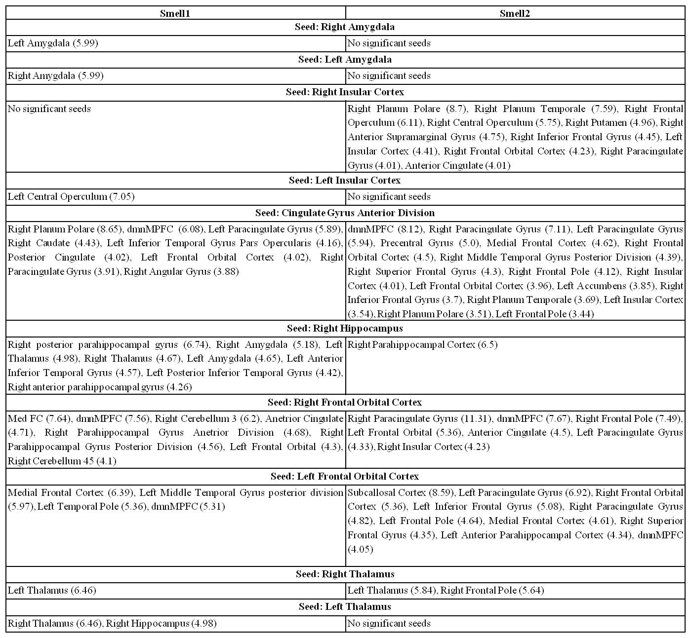
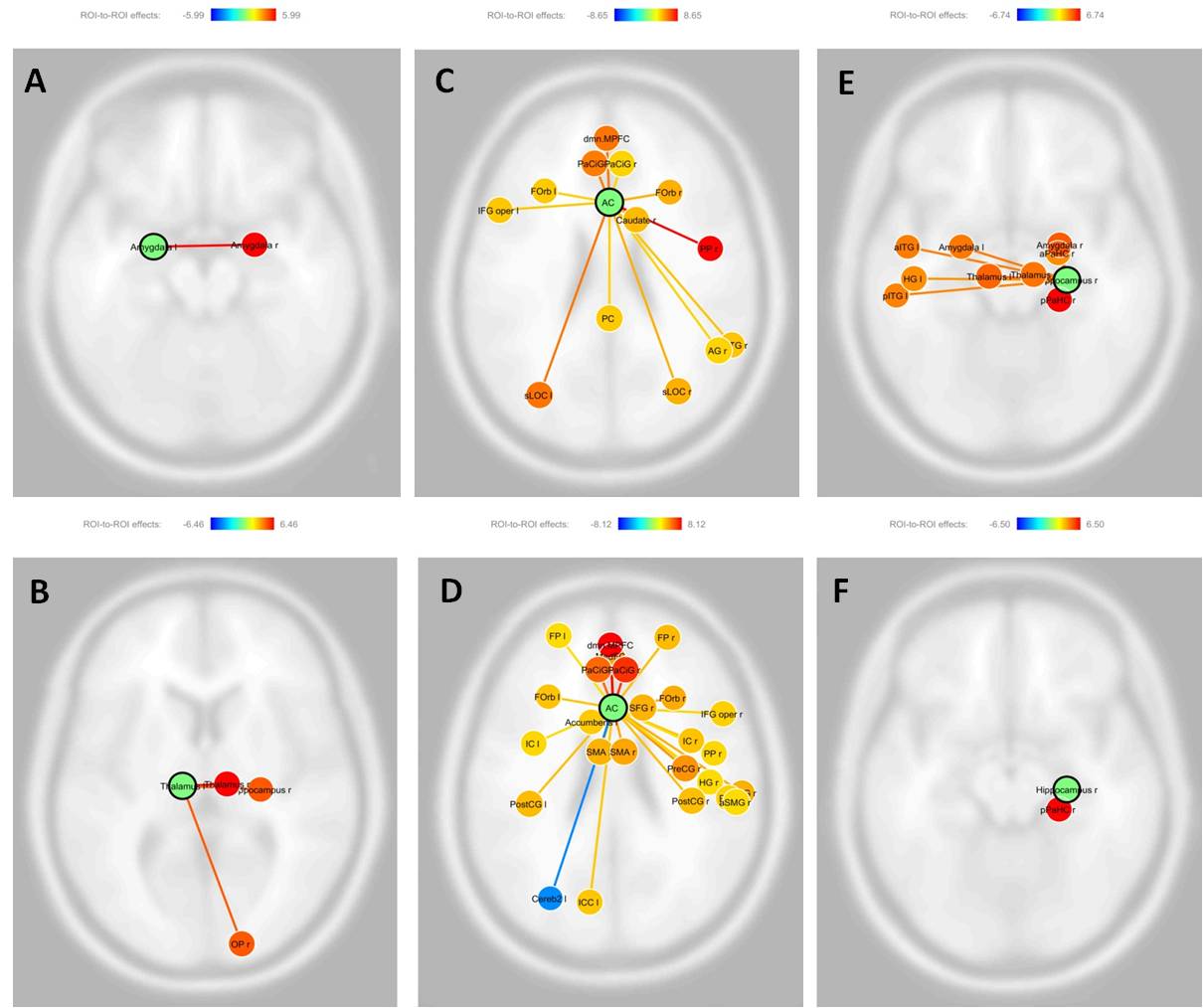
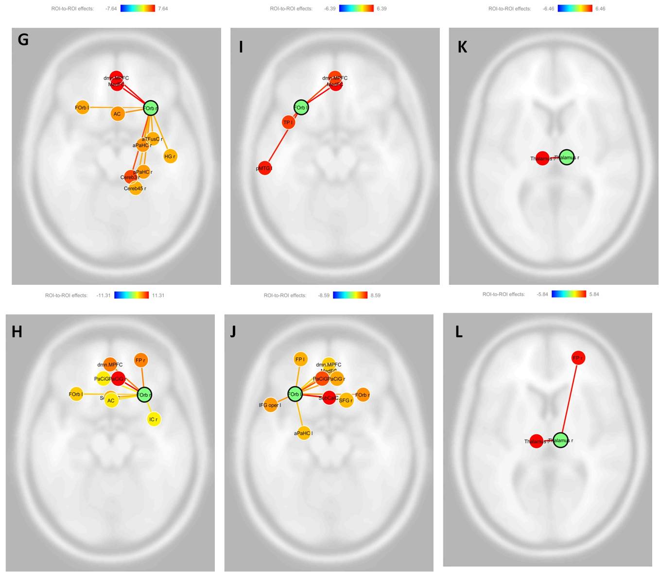
Connectivity between bilateral amygdala for smell 1, but not for smell 2 suggests emotional response to the pleasantness of smell 1 (rose) as the odor intensity was similar between the two smells2. Left central operculum (with no clear role in olfaction3) connectivity for left hemisphere and absence of connections for right insular cortex infer impaired insular connectivity corresponding to smell 1. The connectivity for the anterior cingulate gyrus was similar during both the smells except for the absence of some of the olfaction linked frontal, temporal and mid brain regions for smell 1, indicating inability to elicit more of the cognition linked path of olfaction. The right insular cortex connections with frontal orbital4, cingulate5, putamen6 and other frontal/parietal operculum7,8 having direct or indirect olfaction link, suggest optimal activation of olfactory circuit in smell 2, and right hemispheric lateralization may be attributed to odor stimulated gustatory response for the presented citrus smell9.
Major olfaction connections associated with right hippocampus10 were preserved for smell 1, while smell 2 exhibited only right hippocampus - parahippocampus gyrus link in spite of better memory responses to smell 2. More specific olfaction regions of the orbital frontal cortex11 connectivity were observed for smell 2. This suggests a differential recruitment of memory regions with respect to smell category.
Except for the absence of smell 2 connectivity for left thalamus, not much difference was observed for both the smells suggesting impairment of thalamus relay circuit of odor perception in PD, affecting the link between olfactory cortex to orbitofrontal associative cortex12.
In summary, we observed heterogeneous connectivity and differential lateralization for both the smells. The primary relay structure, amygdala and the upstream structure thalamus were affected for both the smells and may be attributed to PD associated pathology. Olfaction specificity for hippocampal connectivity in smell 1 and for orbitofrontal, insula and anterior cingulate in smell 2 suggest differential recruitment of cognition associated areas which might be functionally compensating each other while perceiving the two categories of smells.
Conclusion
Missing connectivity (regions) is associated with altered olfaction in patients with Parkinson’s disease and the pattern differs with the nature of odor used.Acknowledgements
Funding from SERB (for fellowship of first author) and DST (for the project), Govt. India is acknowledged.References
1. Hummel T et al. Olfactory FMRI in patients with Parkinson's disease. Front Integr Neurosci. 2010; 4:125.
2. Soudry Y et al. Olfactory system and emotion: common substrates. Eur Ann Otorhinolaryngol Head Neck Dis. 2011; 1:18-23.
3. Angela D. Friederici. The brain basis of language processing: from structure to function. Physiology Review. 2011; 91:1357-1392.
4. Savage R et al. The role of temporal lobe and orbitofrontal cortices in olfactory memory function. Arch Clin Neuropsychol. 2002; 4:305-18.
5. Soudry Y et al. Olfactory system and emotion: common substrates. Eur Ann Otorhinolaryngol Head Neck Dis. 2011.128:18-23.
6. Larsson M et al. Age-related loss of olfactory sensitivity: association to dopamine transporter binding in putamen. Neuroscience. 2009; 161:422-6.
7. Veldhuizen MG et al. Identification of human gustatory cortex by activation likelihood estimation. Hum Brain Mapp. 2011; 32:2256-66.
8. Doty RL, Bromley SM. Effects of drugs on olfaction and taste. Otolaryngol Clin North Am. 2004; 37:1229-54.
9. Stevenson RJ, Miller LA, McGrillen K. Perception of odor-induced tastes following insular cortex lesion. Neurocase. 2015; 1: 33-43.
10. Deshmukh SS, Bhalla US. Representation of odor habituation and timing in the hippocampus. J Neurosci. 2003; 23:1903-15.
11. Adina Michael-Titus, Patricia Revest, Peter Shortland. Organization of the nervous system. The Nervous System (SECOND EDITION) 2010, 1-30.
12. Emmanuelle C. and Donald A. The olfactory thalamus: unanswered questions about the role of the mediodorsal thalamic nucleus in olfaction. Front Neural Circuits. 2015; 9: 49.
13. Purves D, Augustine GJ, Fitzpatrick D, et al.,
editors. Neuroscience. 2nd edition. Sunderland (MA): Sinauer Associates; 2001.
Figures