3800
Graph theoretic analysis of language sub-networks in Arabic-English bilingual patients undergoing fMRI for pre-surgical planning1Johns Hopkins University School of Medicine, Baltimore, MD, United States, 2Kennedy Krieger Institute, Baltimore, MD, United States
Synopsis
Task fMRI is routinely performed on patients for pre-surgical planning to localize critical components of the language network and assess hemispheric language dominance. However, the cortical representation of language in bilingual patients (those who are fluent in more than one language) is more complex than in monolinguals, and may involve overlapping but distinct sub-networks. The focus of our current study is to assess the functional connectivity strength of language networks derived from task fMRI activation maps obtained using paradigms performed in both primary native (L1) and secondary (L2) languages in a group of Arabic-English bilingual patients.
Purpose
Task-based fMRI is routinely performed at many centers for presurgical language cortical localization and hemispheric language lateralization. Recent American Society of Functional Neuroradiology (ASFNR) guidelines have recommended the sentence completion (SC) task as the most effective task for both purposes1,2. Since primary (L1) and secondary (L2) language networks can differ in bilinguals, our aim is to use graph theoretic metrics to assess differences between L1 and L2 in Arabic-English bilingual patients undergoing presurgical mapping with task fMRI.Methods
From an overall study pool of 227 patients undergoing clinical presurgical fMRI mapping from year 2011 to year 2017, a total of five patients (age range 18-35 years, 3F, 2M, all right-handed) performed the SC task both in primary (Arabic) and secondary (English) languages. These patients had left hemispheric lesions, including one with left mesial temporal sclerosis, one with a left hemispheric cavernous malformation and 3 with low grade tumors. The age of English acquisition varied across subjects. Patient 1 had lack of overall familiarity with English. Patients 2 & 3 were early acquisition bilinguals (L2 acquired below the age of 7) while patients 4 & 5 were late acquisition bilinguals.
Imaging was performed on a 3.0 T Siemens Trio MRI with a 12-channel head matrix coil using 2D GE-EPI T2* weighted BOLD sequences for functional MR imaging (TR 2000ms/TE 30ms/64x64x33 matrix). The SC task is a covert block design paradigm involving alternating control and active blocks lasting 20 seconds each for a total task duration of 4 min. SPM12 was used for preprocessing of fMRI data (slice timing correction, realignment, normalization to MNI space at 2mm voxel resolution, and spatial smoothing using a 6 mm FWHM Gaussian kernel). T maps were obtained from the general linear model (GLM) analysis using the standard SPM canonical HRF (reflecting activation vs. rest).
Lateralization Index (LI) was computed as (L-R)/(L+R) where L refers to number of left and R number of right hemispheric suprathreshold activated voxels (T>3.0). LI ≥ 0.2 represents left hemispheric dominance, LI ≤ −0.2 represents right hemispheric dominance and (-0.2<LI<0.2) designates bilateral dominance3.
Graph-theory based network analysis was performed on task fMRI data to explore the connectivity strength and topology of activated language clusters in L1 & L2. Network nodes were defined by parcellating the language network into 34 regions of interest (ROIs) (17 for the left hemisphere and 17 for the right hemisphere) according to the Anatomical Automatic Labeling atlas (AAL)4. A representative mean time series for each region was extracted by averaging the time series of all voxels within that region of the thresholded task fMRI (T>3.0) activation map. The Pearson's correlation coefficients (r) were calculated between each possible pair of nodes’ time series to obtain the correlation matrix. The correlation matrix was further thresholded for graph density (r>0.4) and further analysis was performed on the weighted network of the undirected graph G (N, K) with N nodes and K edges. The following network measures of brain connectivity were investigated for both L1 and L2 language networks: average degree, small world properties5, characteristic path length, global clustering coefficient, global network efficiency6, and betweenness centrality.
Results
Table 1 (column 3) includes LI values from L1 and L2 task fMRI activation maps. Each patient demonstrated left hemispheric dominance (LI ≥ 0.2) with greater lateralization of L1 than L2 (see Figure 1). L2 displays higher average degree compared to L1 (Table 1, column 6).
In patients 2 & 3 (early acquisition bilinguals) the L2 language network displays higher global network efficiency as well as more pronounced small-worldness (i.e. short average path lengths and high clustering) structure compared to L1. In late acquisition bilinguals 4 & 5, L1 displays greater global efficiency and small-worldness compared to L2.
Patient No 1 had overall lack of familiarity with L2, which is reflected in very low betweenness centrality of L2 with respect to L1.
Discussion
We found that although language lateralization is higher in the primary language (L1), L2 displays higher average degree. In early acquisition bilinguals, the L2 network displays more pronounced characteristics of small-worldness and for late acquisition bilinguals, the L1 network displays more pronounced characteristics of small-worldness.Conclusion
The topological properties of the L1 and L2 language networks in Arabic-English bilingual patients are quite distinct, as assessed using graph theoretic metrics, suggesting fundamental differences in language representation that need to be considered in presurgical planning and other clinical applications of fMRI in bilingual patients.Acknowledgements
Partly supported by NIH grant R42 CA173976-02 (NCI)References
- Black DF, et al. American Society of Functional Neuroradiology-Recommended fMRI paradigm algorithms for presurgical language assessment. AJNR 2017; 38(10):E65–E73
- Agarwal S, et al. Repeatability of language fMRI lateralization and localization metrics in brain tumor patients. Hum Brain Mapp 2018 Aug 4; doi: 10.1002/hbm.24318
- Gaillard WD, et al. Language dominance in partial epilepsy patients identified with an fMRI reading task. Neurology 2002;59(2):256–265
- Tzourio-Mazoyer N et al. Hum Brain Mapp 2002;17:143–55
- Watts DJ and Strogatz SH. Collective dynamics of “small-world” networks. Nature 1998; 393:440–442
- Latora V and Marchiori M. Efficient behavior of small-world networks. Physics Review Letters 2001;87:198701
Figures
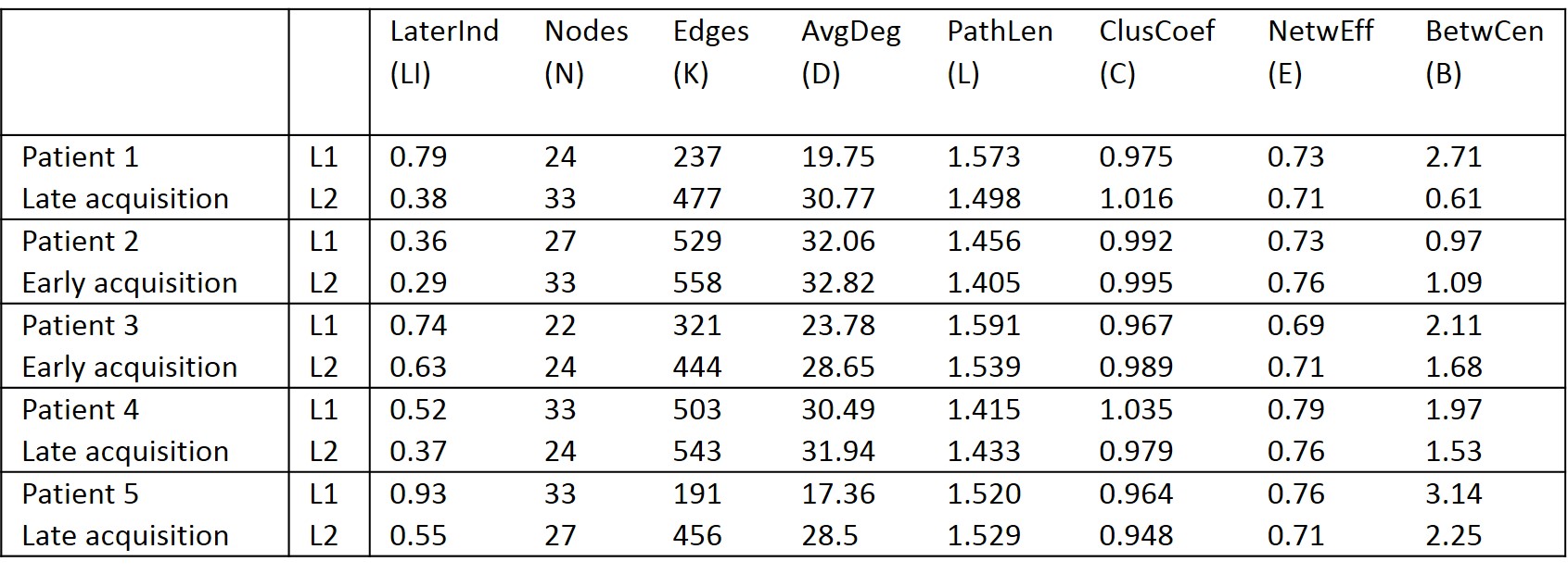
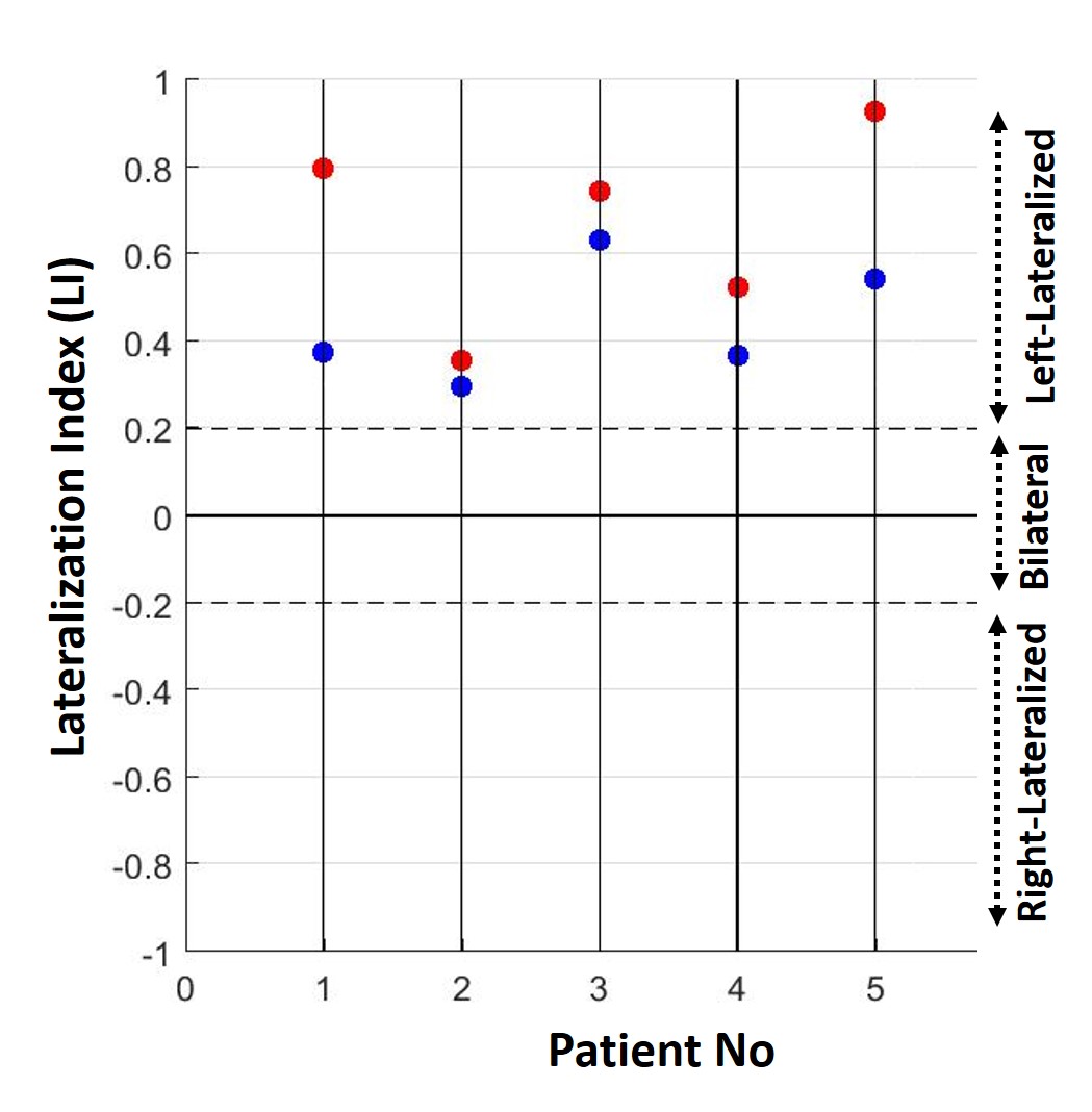
Figure1: Lateralization index LI = (L − H)/(L + H) where L refers to number of suprathreshold activated voxels in left hemisphere and R refers to those in right hemisphere (T>3.0) of task-based fMRI activation map. LI ≥ 0.2 represents left hemispheric dominance, LI ≤ −0.2 represents right hemispheric dominance, (-0.2<LI<0.2) designates bilateral dominance. Red dots in figure represents LI of L1 and blue dots represents LI of L2. Note that, although each patient demonstrates left hemispheric language dominance (LI ≥ 0.2) for both in L1 as well as in L2, L1 demonstrates more lateralized language network than in L2.