3797
Dynamic up- and down-regulation of large-scale cortical networks during task-on and task-off periods.1Department of Biological and Medical Psychology, University of Bergen, Bergen, Norway, 2NORMENT Centre of Excellence, University of Oslo, Oslo, Norway, 3Department of Education, The Arctic University of Norway, Tromsø, Norway, 4Mohn Medical Imaging and Visualization Centre, Haukeland University Hospital, Bergen, Norway, 5Institute of Psychology, Univeristy of Oslo, Oslo, Norway, 6Department of Radiology, Haukeland University Hospital, Bergen, Norway, 7Department of Physics and Technology, University of Bergen, Bergen, Norway, 8Department of Clinical Engineering, Haukeland University Hospital, Bergen, Norway, 9Division of Psychiatry, Haukeland University Hospital, Bergen, Norway
Synopsis
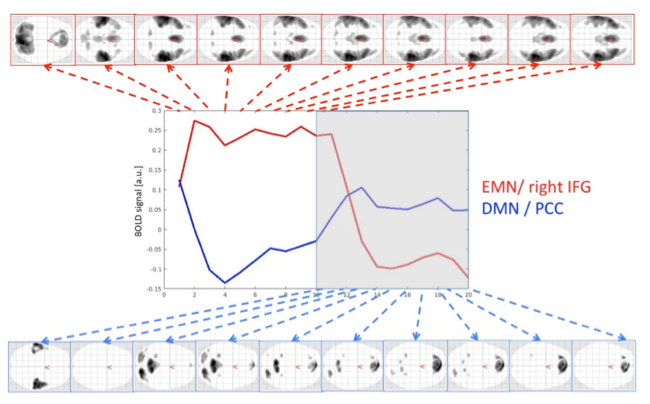
This study was intended to capture the dynamics of networks up- and down-regulation over time, with focus on the extrinsic mode network (EMN) and the default mode network (DMN), using functional magnetic resonance imaging (fMRI). Finite impulse response function (FIR) featured in the SPM software package was used to estimate the BOLD response. The results featured initial rapid activation of EMN in task-periods, while DMN began to drift towards up-regulation already by the end of the task-period.
Introduction
Building on the studies of the default mode network (DMN)1, Hugdahl et al. proposed the existence of a generalized task-positive cortical network that is up-regulated whenever the task to be performed requires the allocation of generalized non-specific cognitive resources.2 It was labelled extrinsic mode network (EMN). The aim of this study was to investigate up- and down- regulations of the DMN and EMN cortical networks while subjects alternated between task-on and taks-off periods.Methods
The participants were 104 healthy adults, with mean age of 29.3 years +/- 8.3 years with an equal distribution of gender. The MR scanner used was a 3T GE SignaHDx scanner. The study was following the protocol used in the study of Kompus et al3.3It included the initial anatomical scanning, a Fast Spoiled Gradient Recall sequence (FSPGR), TE = 14 ms, TR = 400 ms, TI = 500 ms), acquiring 188 consecutive sagittal slices (1 mm thick, no gap, scan matrix: 256 x 256; field of view 256 mm) was applied to acquire T1-weighted 3D images of the whole brain. Afterwards, 10 EPI-volume scans were acquired, during each of the task-present and task-absent blocks, each of 55 sec. Total session time for the EPI-imaging part was thus (9 x 55) x 2 = 16.5 min, with alternating task-present and task-absent blocks. The cognitive task was an auditory speech perception task, with repeated dichotic presentations of two different consonant-vowel syllables presented on each trial, one in the right ear and the other at the same time in the left ear4. The data was analyzed with the SPM12 software package5. The preprocessing steps of EPI data included realignment, unwrapping, and normalization to the MNI template. Low-pass filter set to 480s. The first level analysis was performed by using finite-impulse response function (FIR) as basis function. This setting allowed for modelling each scan per taks-present and task-absent block separately. Resulting beta-images were used for the next 2ndlevel analysis, which was defined as a 3x20 repeated measure ANOVA model. To look at the dynamics between task-positive and resting-state networks, separate contrasts were specified for each time-bin. Results were explored, using family-wise error (FWE)-corrected significance level of p < .05 in the analyses, to protect against Type-I errors and at least 10 voxel per cluster.Results
The approach of modelling taks-on and task-off periods separately showed an initial rapid activation of the extrinsic mode network (EMN), which remained throughout the task period and faded away during the first scan of the resting period. The switch between the EMN and the DMN was observed with an initial time-lag of about 3 seconds, where neither of the networks were active. Afterwards the default mode network was up-regulated and was fading away towards the end of the resting period. The time-courses for the two networks also differed in their dynamics, where the EMN remained stable for the entire task period, while the up-regulation of the DMN began already during the second half of the task-period. The task positive activations started with visual areas, followed by the activation in the auditory and the EMN network.Discussion
The main findings included two orthogonal, non-overlapping activation patterns corresponding to task-processing versus resting, here labelled the EMN and DMN functional networks, respectively. The task-on period activations revealed more lateral and anterior activation pattern, with anterior cingulate as dominant. The task-off blocks resulted in activations within medial and posterior areas, especially in the precuneus and the parietal lobules as the dominant lateral activation. The drift towards the upregulation for the default mode network already during the second half of the task-period is possibly reflecting the anticipatory shift of attention focus.Conclusion
The analysis of fMRI-BOLD data presented two non-overlapping networks that share spatial distribution with the DMN and EMN. Alternating task-on and task-off periods might be more realistic situation to study further task-positive and negative network interactions. It might also be hypothesized that the dynamic interaction between task-positive (EMN) and task-negative (DMN) networks may be disrupted in certain psychiatric and neurological disorders, extending what previously has been suggested for DMN abnormality6,7.Acknowledgements
The present study was supported by grant to Kenneth Hugdahl from the European Research Council (ERC).References
1. Raichle ME, MacLeod AM, Snyder AZ, Powers WJ, Gusnard DA, Shulman GL, A default mode of brain function. Proc Natl Acad Sci. 2001, 98:676–682.
2. Hugdahl K, Raichle ME, Mitra A, Specht K. On existence of generalized non-specific task-dependent network. Front Hum Neurosci. 2015, 9:430.
3. Kompus K, Specht K, Ersland L, Juvodden HT, van Wageningen H, Hugdahl K, Weterhausen R. A forced-attention dichotic listening fMRI study on 113 subjects. Brain an Language 2012; 121:240-247
4. Hugdahl K. Dichotic listening studies of brain asymmetry. In Squire, L.R. (Ed.) Encyclopedia of Neuroscience 2009, Vol. 3: 517-522.
5. SPM12 (6906) software package, Wellcome Department of Cognitive Neurology, London, UK, http://www.fil.ion.ucl.ac.uk/spm/
6. Manoliu, A., Riedl, V., Zherdin, A., Mühlau, M., Schwerthöffer, D., Scherr. M., Peters, H., Zimmer, C., Först, H., Bäuml, J., Wohlschläger, A.M., Sorg, C. Aberrant dependence of default mode/central executive network interactions on anterior insular salience network activity in schizophrenia. Schizophrenia Bulletin 2014; 40:428-437.
7. Northoff G, Qin P, How can the brain's resting state activity generate hallucinations? A 'resting state hypothesis' of auditory verbal hallucinations. Schizophrenia Research 2011; 127:202-214.
Figures