3796
Influence of smoking cessation treatment by varenicline to brain—a resting-fMRI study1Radiology, Beijing Chao-Yang Hospital, Beijing, China, 2School of biomedical engineering, Capital medical university, Beijing, China, 3Clinical research centre, Beijing Chao-Yang Hospital, Beijing, China, 4HC NEA DI MR Siemens Healthcare Ltd, Beijing, China
Synopsis
This study investigated the activation changes of brain regions in smokers before and after cessation treatment using a resting state fMRI. The results demonstrated that treated smokers will have improved functions of brain regions related to emotion and memory after using varenicline. This improvement is not only the direct benefit of quitting smoking, but also helps quitters cope with the discomfort and negative emotions of quitting. All subjects showed a decrease in smoking addiction and withdrawal response, suggesting that varenicline can help quitters maintain their withdrawal status.
Introduction
Smoking is an addictive disease that can easily relapse. Even if the physiological addiction has been lifted, psychological addiction may still exist and influence for a long time. Several previous studies have examined smokers’ brain response to smoking-related clues in different status, such as in nicotine abstinence status and in resting status after cessation1,2. However, little is known about the changes in brain responses caused by smoking cessation drugs. Understanding the effects of cessation drugs on brain activity, especially the changes of brain activity in resting state, will help us to comprehend and evaluate the effects of medical cessation treatment more comprehensively and provide other targeted non-drug adjuvant therapy. Finally, reduce relapse rate and increase smoking cessation success rate. Therefore, we performed the current study aiming to evaluate the changes of resting-state brain activity before and after smoking cessation treatment by using varenicline, a nicotinic receptor blocker.Methods
Data were collected using a MAGNETOM Prisma 3T MR scanner with a 64-channel head coil from a total of 23 smokers (18 males and 5 females). They aged 23-40 (mean 30.7±4.2) years. All subjects underwent resting-fMRI data acquisition before cessation and 1.5 months after cessation treatment by varenicline. The resting state functional images were acquired using an EPI sequence with the following parameters: TR=3000 ms, TE=30 ms, field of view=220 mm× 220 mm, flip angle=90°, image matrix=64 × 64, thickness=3 mm, gap=1 mm, 36 axial slices, 180 volumes. The EPI data were preprocessed with the Data Processing Assistant for Resting State fMRI (http://www. restfmri.net/forum/DPARSF) that works with SPM12 on the Matlab 7.5 platform. The first ten volumes of the scanning sessions were removed to allow for scanner calibration and participants’ adaptation to the scanning environment. All of the subjects’ head movements were less than 1 mm maximum displacement in any direction and less than 1˚ in any angular dimension. After realignment, all of the data were normalized to Montreal Neurological Institute space, resampled with 3 mm × 3 mm ×3 mm resolution and smoothed with a Gaussian kernel of 6 mm full width. fALFF and ReHo analyses were performed. One-sample T-test was performed to compare the activation changes before and after cessation treatment (p<0.01 was considered significant). We also did Fagerström Test for Nicotine Dependence (FTND) before and after treatment to evaluate smokers' changes in smoking habits.Results
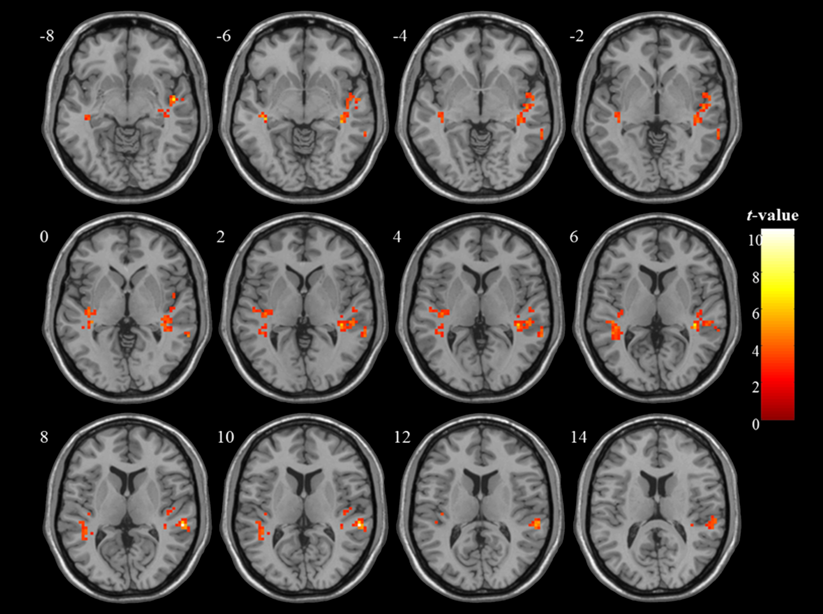
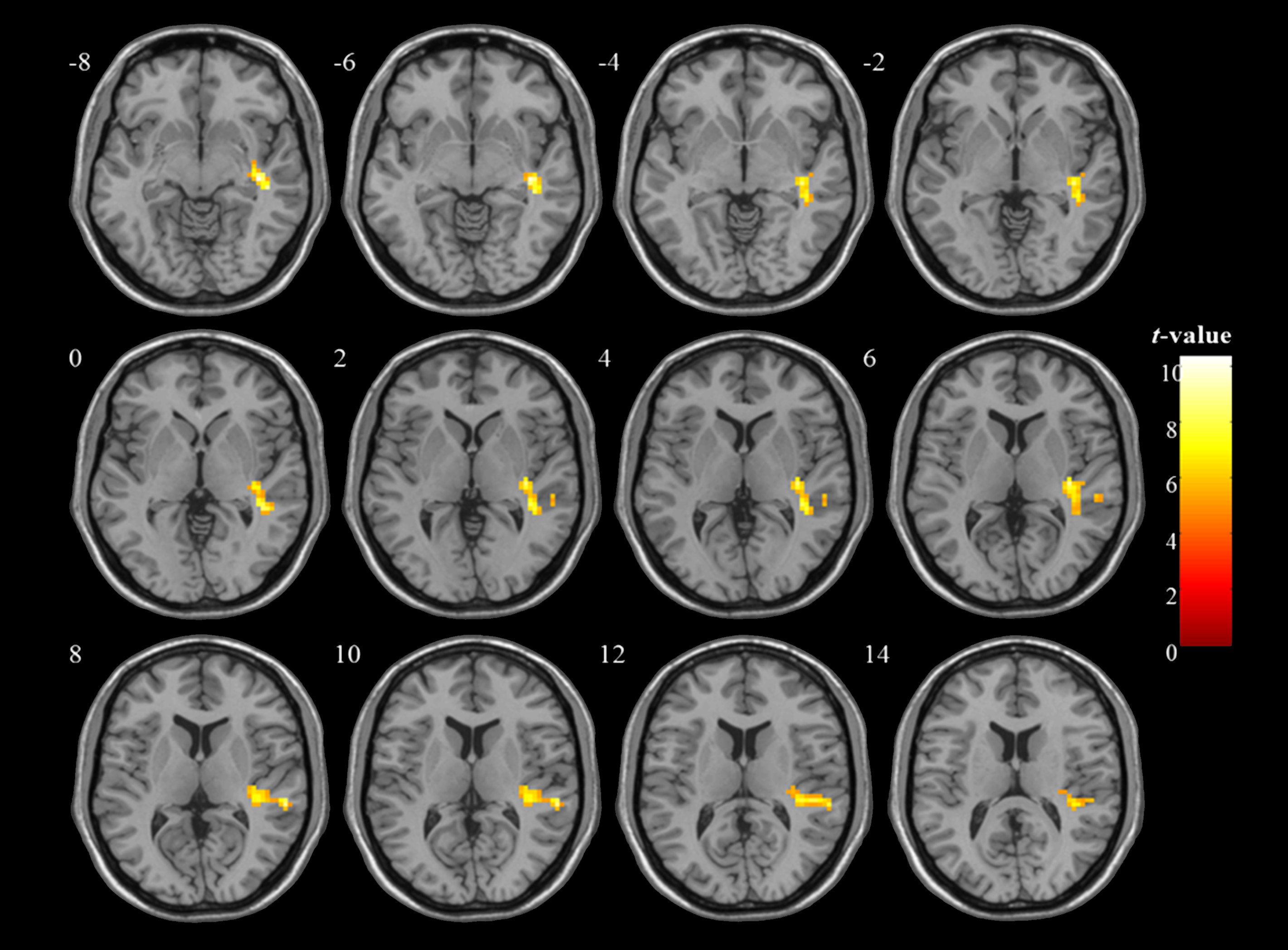
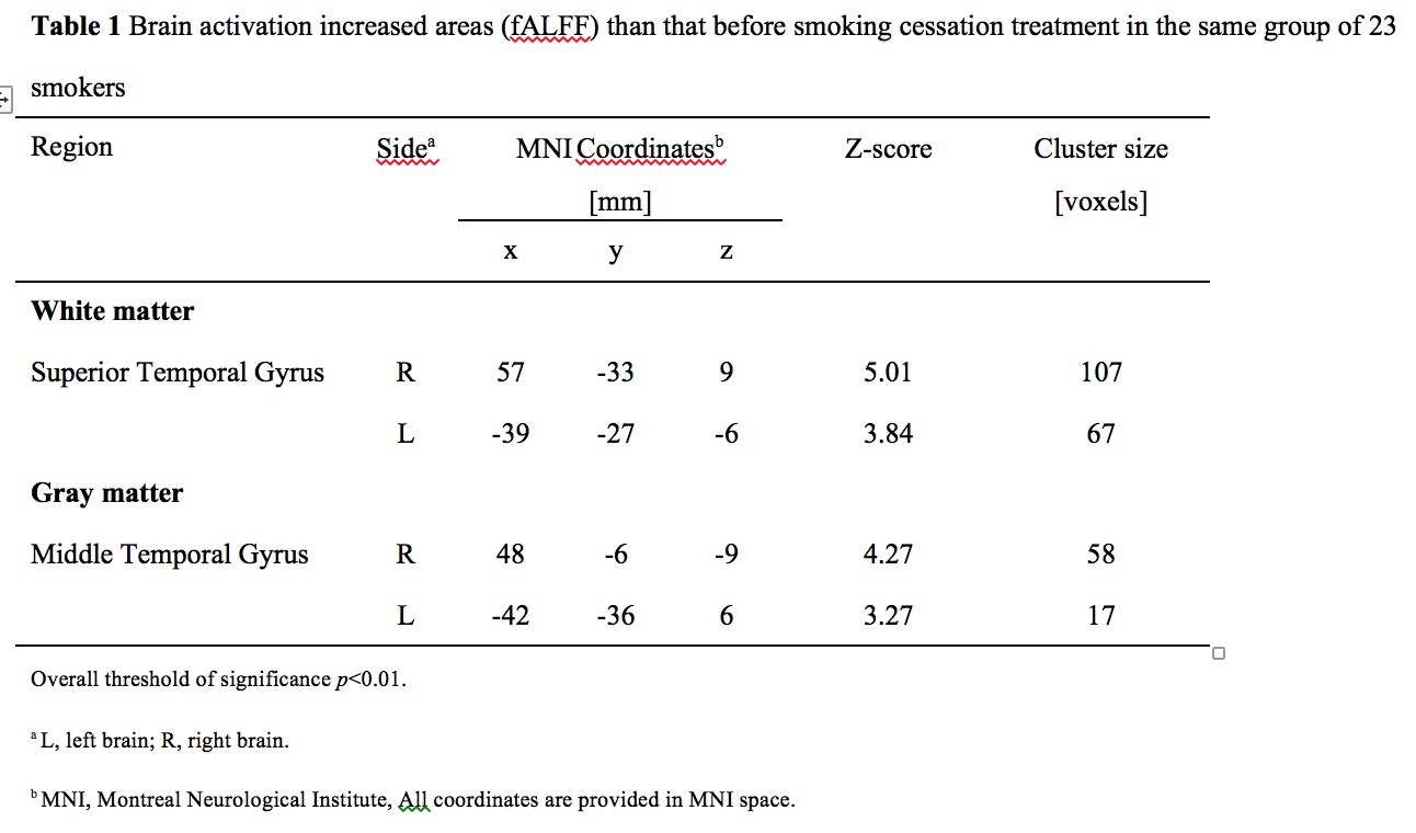
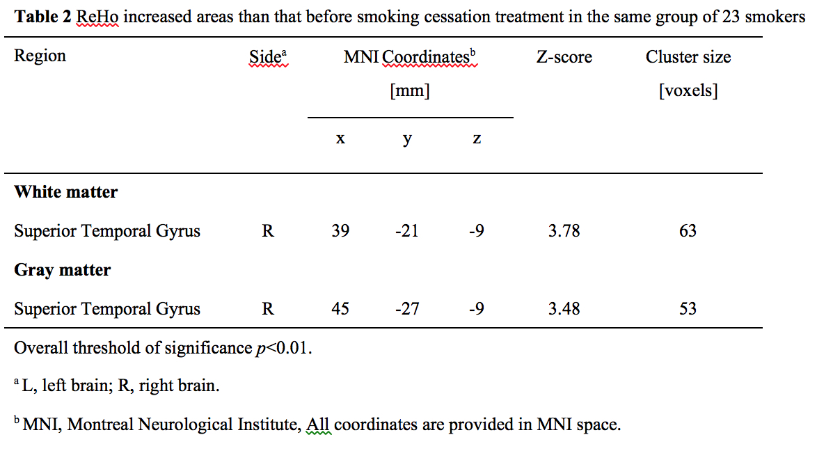
We found that fALFF which reflecting brain activation
in smokers increased after cessation treatment. No decreased fALFF areas were
found. The increased brain activation regions were concentrated in bilateral
temporal lobe. ReHo also increased after treatment and the increased areas were
concentrated on the right temporal lobe. Smokers generally had a lower degree of smoking
dependence, and their satisfaction after smoking decreased significantly.Discussion
Previous findings suggested that brain activation of
smokers in resting state was decreased in temporal gyrus than non-smokers. And severe
nicotine dependent smokers also showed decreased fALFF in temporal gyrus than
mild-moderate addicts3. The temporal lobe is olfactory
and auditory centre and relates to memory and emotion4,5.
Changes in temporal lobe activity explained previous studies’ finding that smoking
reduces people’s attention and working memory, and reduces people's response to
natural rewards6,7. Visual
and emotional brain activation in smokers who quit smoking were reduced after
treatment with varenicline by visual stimulation with smoking-inducing cues8. These findings confirmed that smokers,
especially severe nicotine dependent smokers, have impaired function in the brain
regions responsible for emotion and memory9,10. Varenicline can effectively reduce the effects
of smoking cues on visual and memory stimuli in ex-smokers and reduce relapse
risk11. Our study found that the emotional and memory
functions of treated smokers in resting state without smoking clue stimulation
were higher than those before treated, suggesting that medical treatment is also
helpful to recover resting state brain function. We analysed both fALFF and
ReHo and obtained similar results. It
suggests that the improvement of brain function is not only simple increase of activation
but also change of activity sate12.
However, activity sate changes were lagging behind simple activation changes,
so ReHo only found changes in the right temporal lobe, but neither fALFF nor
ReHo found hypofunctional brain areas than pretherapy. The FTND test also
showed that the dependence level of treated smokers decreased and withdrawal
reaction reduced significantly, which may also be the effects of improved
emotional function13. The
expansion of the subjects will further confirm our findings.Conclusion
Using varenicline can increase the activation of
olfactory and auditory related brain regions and improve the emotional and
memory functions of treated smokers just in a short period of treatment. That
is very important to help the smokers to maintain the state of quitting smoking.Acknowledgements
No acknowledgement found.References
1. Chao W, Zhujing S, Peiyu H, et al. Altered spontaneous activity of posterior cingulate cortex and superior temporal gyrus are associated with a smoking cessation treatment outcome using varenicline revealedby regional homogeneity. Brain Imaging Behav. 2017;11(3):611-618.
2. Janes AC, Frederick Bd, Richardt S, et al. Brain fMRI responses to smoking-related images prior to and during extended smoking abstinence. Exp Clin Psychopharmacol. 2009;17(6):365-373.
3. S Chu, D Xiao, S Wang, et al. Spontaneous brain activity in chronic smokers revealed by fractional amplitude of low frequency fluctuation analysis: a resting state functional magnetic resonance imaging study. Chin Med J.2014;127(8):1504-1509.
4. Fichtenholtz HM, Dean HL, Dillon DG, et al. Emotion-attention network interactions during a visual oddball task. Brain Research Cognitive Brain Research. 2004;20(1):67–80.
5. Keightley ML, Winocur G, Graham SJ, et al. An fMRI study investigating cognitive modulation of brain regions associated with emotional processing of visual stimuli. Neuropsychologia. 2003;41(5):585–596.
6. Rose EJ, Ross TJ, Salmeron BJ, et al. Chronic exposure to nicotine is associated with reduced reward-related activity in the striatum but not the midbrain. Biol Psychiatry. 2012;71(3)206-213.
7. Rubinstein ML, Luks TL, Moscicki AB, et a1. Smoking-cue induced brain activation in adolescent light smokers. J Adolesc Health. 2011;48(1): 7-12.
8. Loughead J, Ray R, Wileyto EP, et al. Effects of the alpha4beta2 partial agonist varenicline on brain activity and working memory in abstinent smokers. Biol Psychiatry.2010;67(8):715-721.
9. Lawrence NS, Ross TJ, Stein EA. Cognitive mechanisms of nicotine on visual attention. Neuron. 2002;36:539-548.
10. Myers CS, Taylor RC, Moolchan ET, et al. Dose-related enhancement of mood and cognition in smokers administered nicotine nasal spray. Neuropsycho-pharmacology. 2008;33:588-598.
11. Loughead J, Ray R, Wileyto EP, et al. Brain activity and emotional processing in smokers treated with varencline. Addict Biol. 2013;18(4):732-738.
12. Zang Y, Jiang T, Lu Y, et al. Regional homogeneity approach to fMRI data analysis. Neuroimage. 2004; 22(1):394-400.
13. Menossi HS, Goudriaan AE, de Azevedo-Marques Pecrico C, et al. Neural bases of pharmacological treatment of nicotine dependence - insights from functional brain imaging: a systematic review. CNS Drugs.2013;27(11):921-941.
Figures