3795
Detecting CBF to Explore Verbal Memory Impairment Mechanisms in Subcortical Stroke Patients1Department of MRI, The First Affiliated Hospital of Zhengzhou University, Zhengzhou, China, 2The First Affiliated Hospital of Zhengzhou University, Zhengzhou, China, 3GE Healthcare MR Research, Beijing, China
Synopsis
In order to explore the neural substrates underlying verbal memory (VM) impairment in subcortical stroke patients, we recruited sixty patients with chronic subcortical stroke and sixty normal controls. 3D-ASL imaging was used to measure the resting-state values of voxel-wise cerebral blood flow (CBF) and the alterations of functional covariance network were detected. In this study, the different CBF levels in the stroke patients and the normal controls, as well as the close correlation between the CBF values and the VM scores, indicate that the VM impairment in stroke patients may be associated with the disconnection of frontal-lobe network.
Purpose
This study is based on perfusion imaging 3D-ASLto explore mechanisms of memory deficit in patients with a subcortical infarction involving the motor pathway.Method
Sixty right-handed patients with ischemic stroke involving subcortical motor pathway, and sixty age-matched healthy controls were recruited for the study. All patients were first-onset stroke patients who showed motor deficits in both the upper and the lower extremities. MR images were acquired on a 3.0 Tesla GE Discovery 750 MR scanner. Perfusion images were obtained by using the pcASL sequence1 with 3D spiral acquisition and background suppression (TR = 5025 ms, TE = 11.1 ms, post-label delay = 2025 ms, FA = 111°, FOV = 240 mm × 240 mm, reconstruction matrix = 128 × 512, slice thickness = 3 mm, no gap, 48 axial slices, number of excitations = 3, and 1.9 mm × 1.9 mm in-plane resolution). The Rey Auditory Verbal Learning Test was used to evaluate the VM function2. The averaged cerebral blood flow (CBF) values of the two groups in each seeding region were extracted from each participant as a regressor in the general-linear model in SPM8 to produce functional network t-maps. A multiple regression model-based linear-interaction analysis was subsequently used to detect the functional network alteration in the patient group with reference to those in the normal controls. We corrected multiple comparisons using the height level FWE method with a corrected threshold of p < 0.05.Result and Discussion
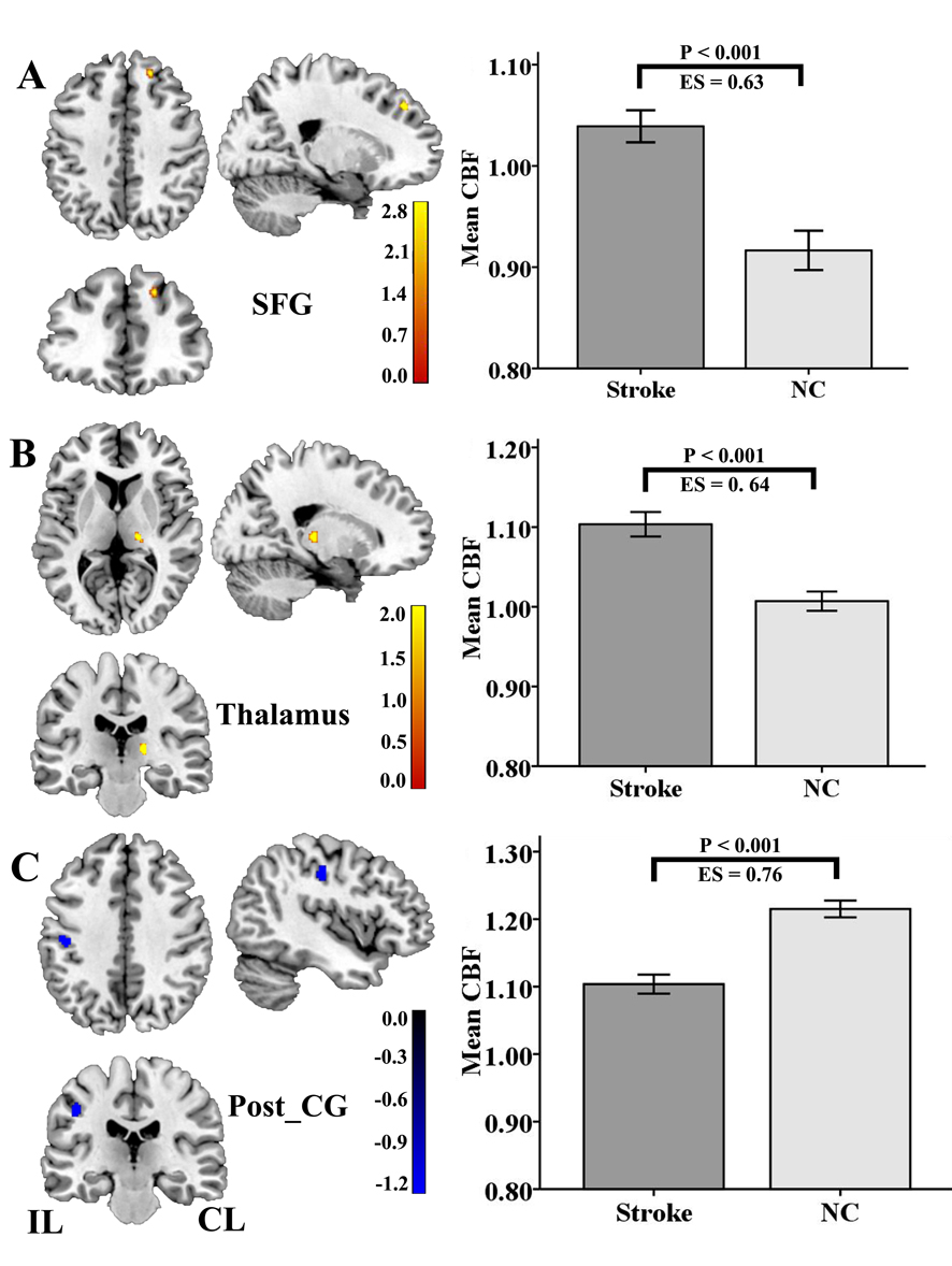
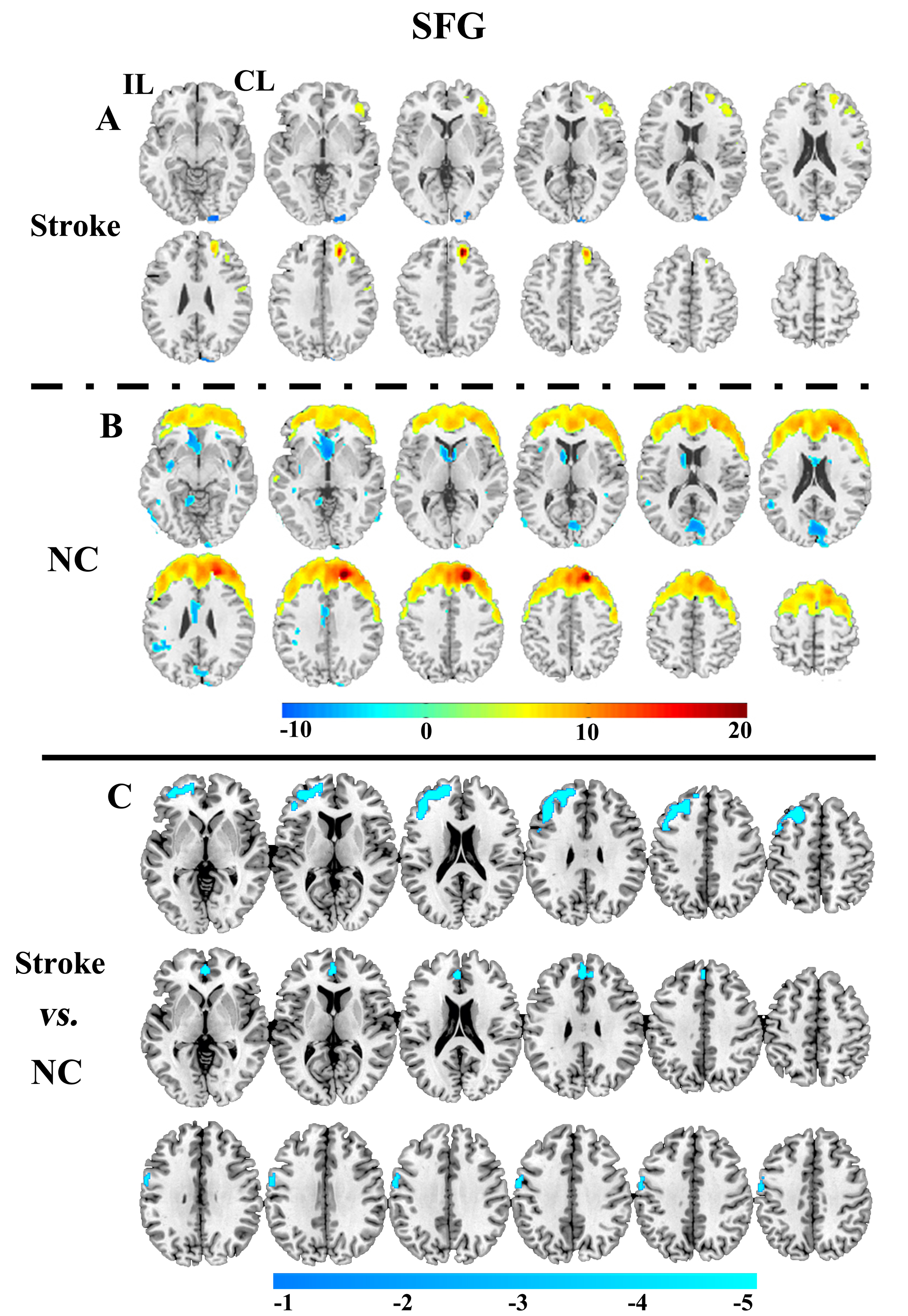
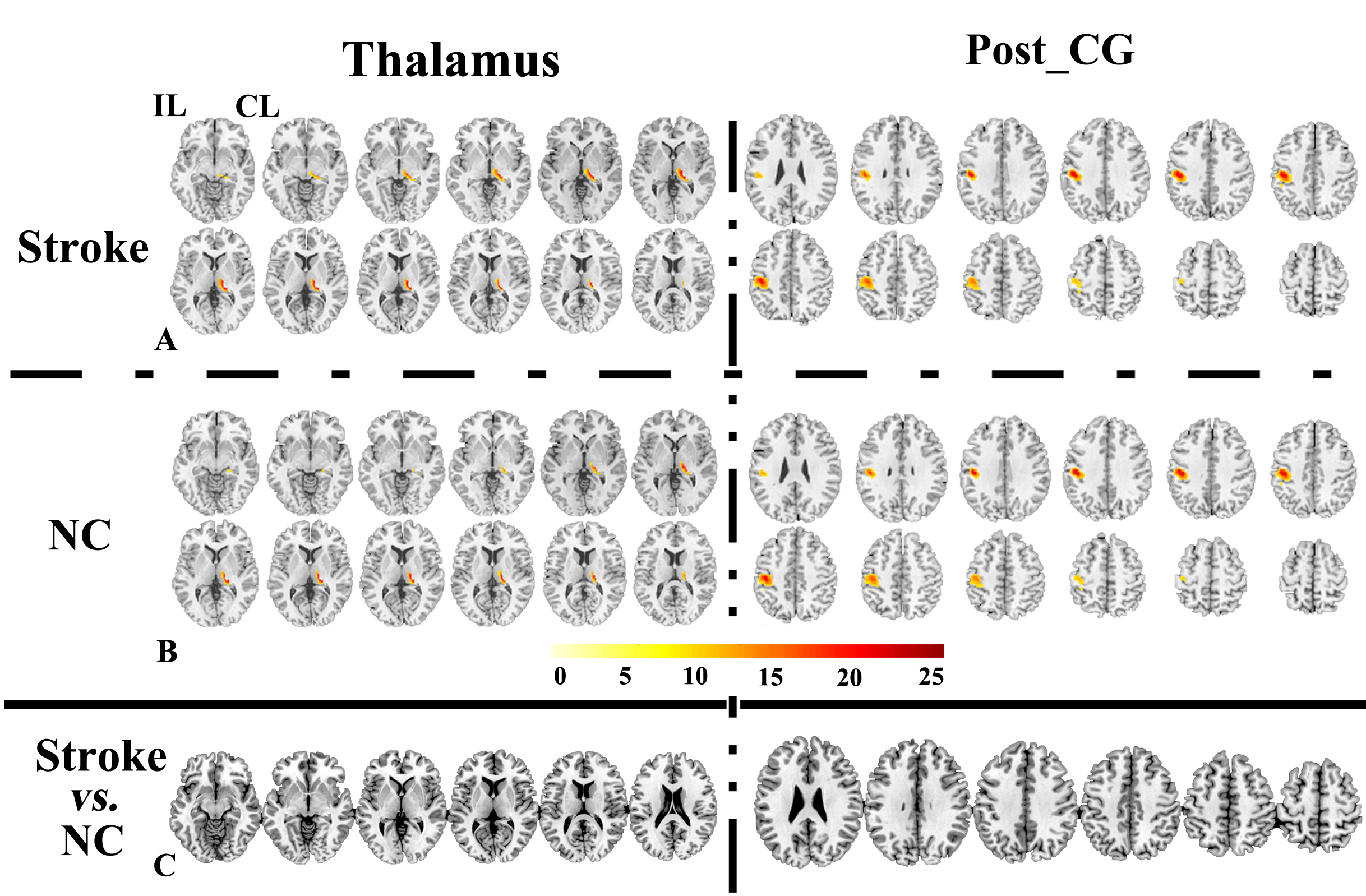
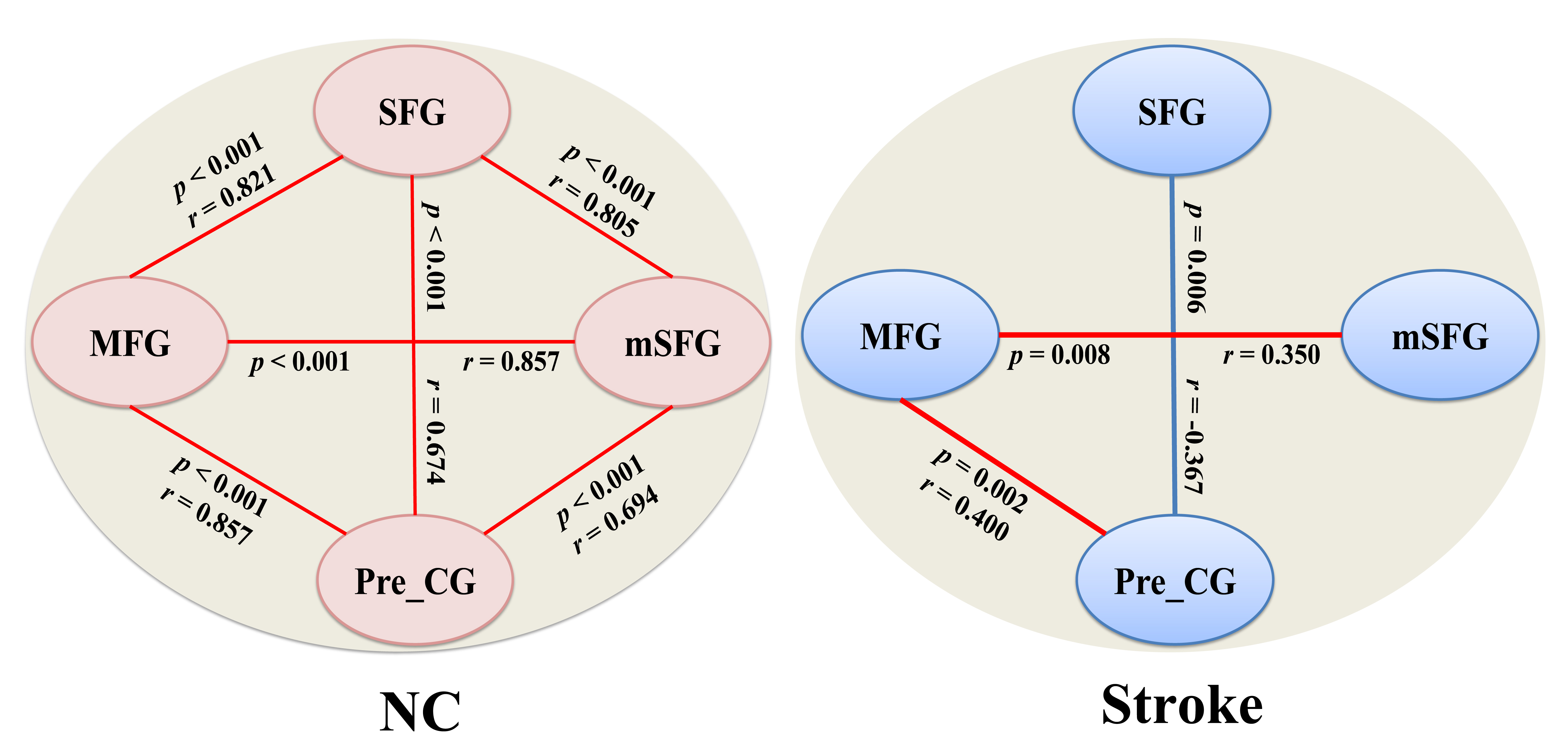
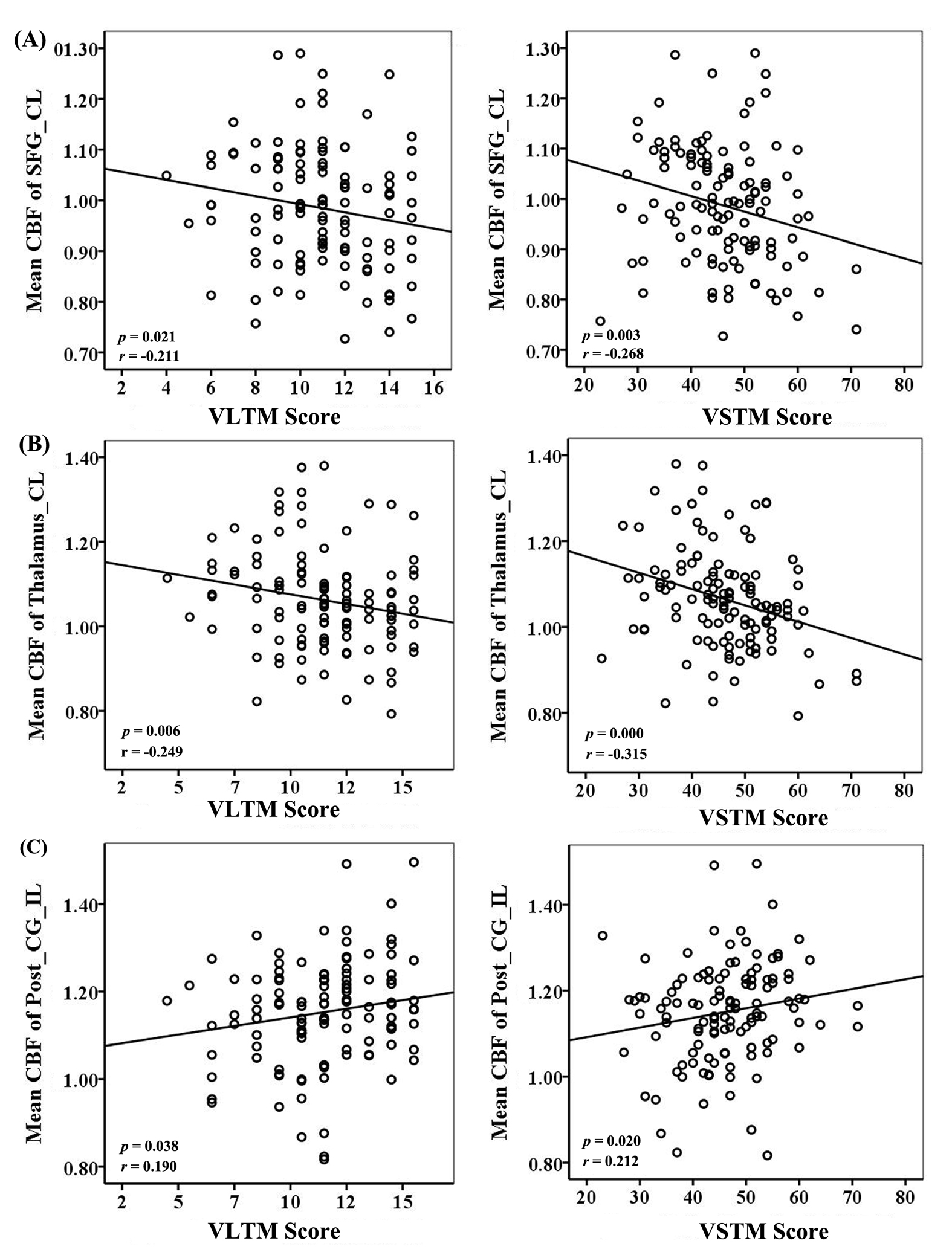
Compared with normal controls, the stroke patients showed worse performance in VSTM test (p = 0.003). The CBF differences between the stroke patients and normal controls were shown in Figure 1. The stroke patients exhibited increased CBF values in the contralesional superior frontal gyrus (SFG) and thalamus (Tha), and decreased CBF value in the ipsilesional postcentral gyrus (Post_CG) (p < 0.05, FWE corrected). The CBF-FCNs maps of each ROI were displayed in Figure 2 and Figure 3 (p < 0.05, FWE corrected). The SFG-associated FCN pattern in stroke patients (Figure 2A) was remarkably different from that in normal controls (Figure 2B). Notably, in the SFG-associated FCN, the stroke patients exhibited decreased CBF in the ipsilesional middle frontal gyrus (MFG), ipsilesional medial part of the superior frontal gyrus (mSFG) and ipsilesional precentral gyrus (Pre_CG) (p < 0.05, FWE corrected, Figure 2C). However, both in the patients (Figure 3 A) and in the normal controls (Figure 3 B), the thalamus-associated and Post_CG-associated FNs showed similar positive covariance patterns (Figure 3 C). Moreover, we could identify the stroke patients who had a remarkably altered CBF connection in the SFG-associated FCN (Figure 4). In general, the stroke patients had either activated or deactivated or absent CBF connections. In our study, the connection between the contralesional SFG and ipsilesional Pre_CG was deactivated, and the connections between the contralesional SFG and ipsilesional MFG, between the contralesional SFG and mSFG, and between the ipsilesional mSFG and ipsilesional Pre_CG were absent, different from the connection states in the normal control (NC) individuals (Figure 4). In addition, although the activated connection states between the ipsilesional MFG and ipsilesional mSFG, and between the ipsilesional MFG Pre_CG were the same with those in the NC group, the correlation were less strong than those in the NC group. Furthermore, in these stroke patients, the increased CBF in the contralesional SFG (pvstm = 0.003, pvltm = 0.021, Figure 5A ) and Tha (pvstm = 0.000, pvltm = 0.006, Figure 5B ) were both negatively correlated with VM scores. The decreased CBF in the ipsilesional Post_CG were positively correlated with VM scores (pvstm = 0.020, pvltm = 0.038, Figure 5C ). Additionally, we found that the chronic subcortical stroke patients who were well recovered in global motor function showed VSTM deficits compared with normal controls. In order to investigate the neurological mechanisms of the stroke-induced memory impairment in the subcortical stroke patients, we applied FCNs method to map brain connectivity patterns using resting-state CBF datasets. The results indicated that the connectivity was greatly impaired in the frontal-lobe network of the patients, implying interrupted connectivity and a chronic stage of cognitive functional impairment after a subcortical stroke.Conclusion
In this study, we identified the alterations of CBF brain connectivity in the frontal-lobe network. Quantitative results indicate that the subcortical stroke-induced functional deficits may involve the cognitive functional system beyond the motor system, suggesting that the disconnection of the frontal-lobe network may be the underlying mechanism of verbal memory impairment in subcortical stroke patients.Acknowledgements
We are indebted to our patients and their caregivers for generously supporting our study. This study was supported by the Natural Science Foundation of China (81601467, 81871327)References
1. Aslan S, Lu H. On the sensitivity of ASL MRI in detecting regional differences in cerebral blood flow. Magn Reson Imaging. 2010; 28(7): 928-935.
2. Brugnolo A, Morbelli S, Arnaldi D, et al. Metabolic correlates of Rey auditory verbal learning test in elderly subjects with memory complaints. J Alzheimers Dis. 2014; 39(1): 103-113.
3. Liu F, Zhuo C, Yu C. Altered cerebral blood fow covariance network in schizophrenia. Front Neurosci. 2016;10: 308.
Figures