3794
Working memory deficits in childhood cancer survivors: an fMRI studyPatricia Stefancin1, Christine Cahaney1, Robert Parker2, Thomas Preston1, Laura Hogan2, Rina Meyer2, Cara Giannillo1, Debra Giugliano1, and Tim Duong1
1Stony Brook Medicine, Stony Brook, NY, United States, 2Stony Brook Children's Hospital, Stony Brook, NY, United States
Synopsis
Little is known about the neural correlates of cognitive deficits in chemotherapy treated survivors of childhood cancer. We used event related fMRI to study working memory in childhood cancer survivors. Subjects underwent an n-back task to test working memory function. Patients showed reduced BOLD signal on correct responses compared to controls in the anterior cingulate, posterior cingulate, and sensory association cortices. Patients showed increased activation on error trials in the angular gyrus and superior parietal lobule. The affected brain regions are known to be involved in memory function, reward, motor planning and motivation.
Introduction
With the growing number of childhood cancer survivors 1-3, the negative effects of chemotherapy are becoming more important to understand. Many childhood cancer survivors have reported difficulties with memory, attention, visuospatial function, and executive function (chemo brain)1, 4. However, little is known about the neural correlates of these deficits in a pediatric population, in contrast to breast cancer survivors. To our knowledge there are only three published studies on the use of fMRI to understand chemobrain. We tested the hypothesis that childhood cancer survivors show working memory deficits by fMRI, and responses in affected regions are correlated with volumetric changes.Methods
Seventeen pediatric cancer survivors and seventeen healthy age matched controls were recruited for this study. Subjects underwent MRI scans with a 3.0T Siemens scanner. A T1-weighted 3D MPRAGE structural scan was obtained (TR= 2300ms, TE= 3.24ms, FOV= 223x223mm, voxel=0.9x0.9x0.9mm) followed by an echo planar BOLD fMRI sequence (TR= 2020ms, TE= 30ms, FOV= 195x195mm, slice thickness = 3.0mm, voxel=2.5x2.5x3.0mm, 32 slices) lasting five minutes. A working memory n-back task was performed during the fMRI sequence. The task consisted of both 0-back and 2-back blocks. Trials were recorded based on correct, incorrect, or no response. fMRI analysis employed event-related analysis using Statistical Parametric Mapping (SPM). Statistical analysis included t-test and correlation.Results
Patients showed lower numbers of correct answers compared to controls, as well as a higher number of incorrect and no response answers. Patients showed a longer average response time compared to controls, as well as longer response times on both incorrect and correct trials. Total intracranial volume was correlated with the number of correct and incorrect answers. White matter volume was also correlated with the number of correct and incorrect answers. Patients showed reduced BOLD signal on correct responses compared to controls in the anterior cingulate (T=-7.62, p=0.012), posterior cingulate (T=-6.26 p=0.023), and sensory association cortices. Compared to controls, patients showed higher activation on incorrect and no response trials in the angular gyrus (T=4.23, p=0.041) on incorrect trials and the superior parietal lobule on no response trials (T=3.98, p=0.033). For correct responses, fMRI responses in the anterior cingulate cortex negatively correlated with left amygdala volume and right thalamus volume. For error trials, fMRI responses in the angular gyrus negatively correlated with the right hippocampal volume and white-matter volume, whereas the fMRI responses in the superior parietal lobule were positively correlated with left putamen volume.Discussion
In working memory trials where participants answered correctly, patients showed lower activation within the anterior cingulate cortex, posterior cingulate cortex, and sensory association cortex compared to controls. The anterior cingulate cortex is associated with decision making, and error detection5. Reduced activation in this area could reflect difficulty in making decisions associated with the memory tasks. The posterior cingulate is associated with memory retrieval6. The reduction of activation within this area could reflect a struggle to recall previous experiences. The sensory association cortex, implicated in learning and memory, aides in shifting attention to important stimuli, as well as making complex association. Reduced activation in this area in patients could reflect difficulty in shifting and/or maintaining attention to image features patients were asked to remember. In working memory trials where participants answered incorrectly or not at all, patients showed higher activation within the angular gyrus and the superior parietal lobule. Our findings suggest patients required more effort on these trials, which could be a result of impaired memory recall or impaired sustaining attention. The angular gyrus is associated with memory retrieval, in particular, episodic memories7. The superior parietal lobule is activated when subjects perform mental rotations to solve puzzles or encounter visual pictures they are instructed to remember8, 9. In regards to correlation, it is interesting that different circuits were involved in the correct and error trials. Most correlations are negative and some negative correlations involved many circuits, suggesting a complex relationship of neural networks that contribute to working memory impairment in chemo brain. Further studies are needed to confirm these correlations.Conclusion
Event-related task fMRI identified the neural correlates of working memory impairment in childhood cancer survivors. The affected brain regions are known to be involved in memory function, reward, motor planning and motivation. fMRI responses in these brain regions are correlated with regional anatomical changes. The ability to quantitatively delineate the affected neural circuits associated with chemo brain could inform treatment strategies, identify patients at high risk of developing cognitive deficits, as well as pre-emptively tailor behavioral enrichment to overcome specific cognitive deficits to achieve normal functioning in schools and beyond.Acknowledgements
No acknowledgement found.References
1. Campbell LK, Scaduto M, Sharp W, et al. A meta-analysis of the neurocognitive sequelae of treatment for childhood acute lymphocytic leukemia. Pediatr Blood Cancer. 2007;49(1):65-73. 2. Peterson CC, Johnson CE, Ramirez LY, et al. A meta-analysis of the neuropsychological sequelae of chemotherapy-only treatment for pediatric acute lymphoblastic leukemia. Pediatr Blood Cancer. 2008;51(1):99-104. 3. Anderson FS, Kunin-Batson AS. Neurocognitive late effects of chemotherapy in children: the past 10 years of research on brain structure and function. Pediatr Blood Cancer. 2009;52(2):159-164. 4. Kaiser J, Bledowski C, Dietrich J. Neural correlates of chemotherapy-related cognitive impairment. Cortex. 2014;54:33-50. 5. Gasquoine PG. Localization of function in anterior cingulate cortex: from psychosurgery to functional neuroimaging. Neurosci Biobehav Rev. 2013;37(3):340-348. 6. Maddock RJ, Garrett AS, Buonocore MH. Remembering familiar people: the posterior cingulate cortex and autobiographical memory retrieval. Neuroscience. 2001;104(3):667-676. 7. Seghier ML. The angular gyrus: multiple functions and multiple subdivisions. Neuroscientist. 2013;19(1):43-61. 8. Koenigs M, Barbey AK, Postle BR, et al. Superior parietal cortex is critical for the manipulation of information in working memory. J Neurosci. 2009;29(47):14980-14986. 9. Lesourd M, Osiurak F, Navarro J, et al. Involvement of the Left Supramarginal Gyrus in Manipulation Judgment Tasks: Contributions to Theories of Tool Use. J Int Neuropsychol Soc. 2017;23(8):685-691.Figures
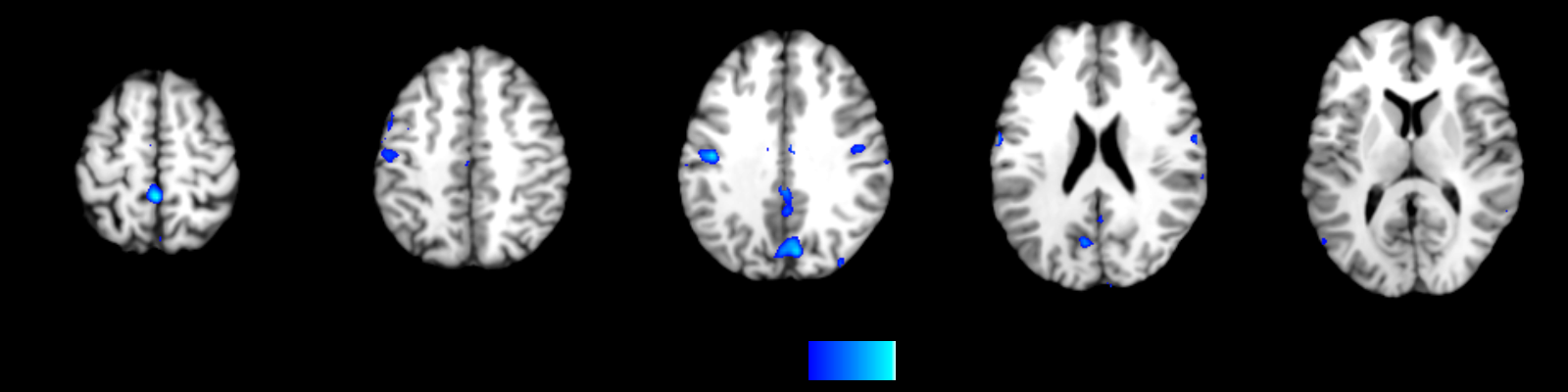
fMRI results for correct responses (Patients>Controls)
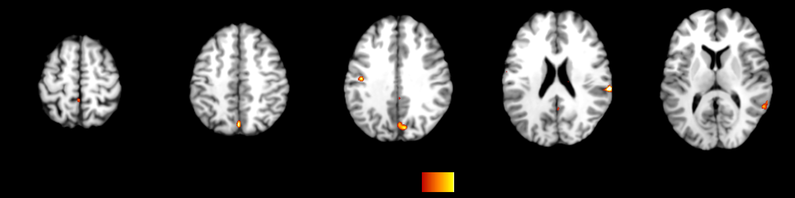
fMRI results for error responses (Patients>Controls)