3793
A combined task based fMRI connectivity and VBM study to lateralization of memory function in pre-and post-surgery mesial temporal lobe epilepsy patients.1Neurology, AIIMS, New Delhi, India, 2NMR, AIIMS, New Delhi, India, 3Neurosurgery, AIIMS, New Delhi, India
Synopsis
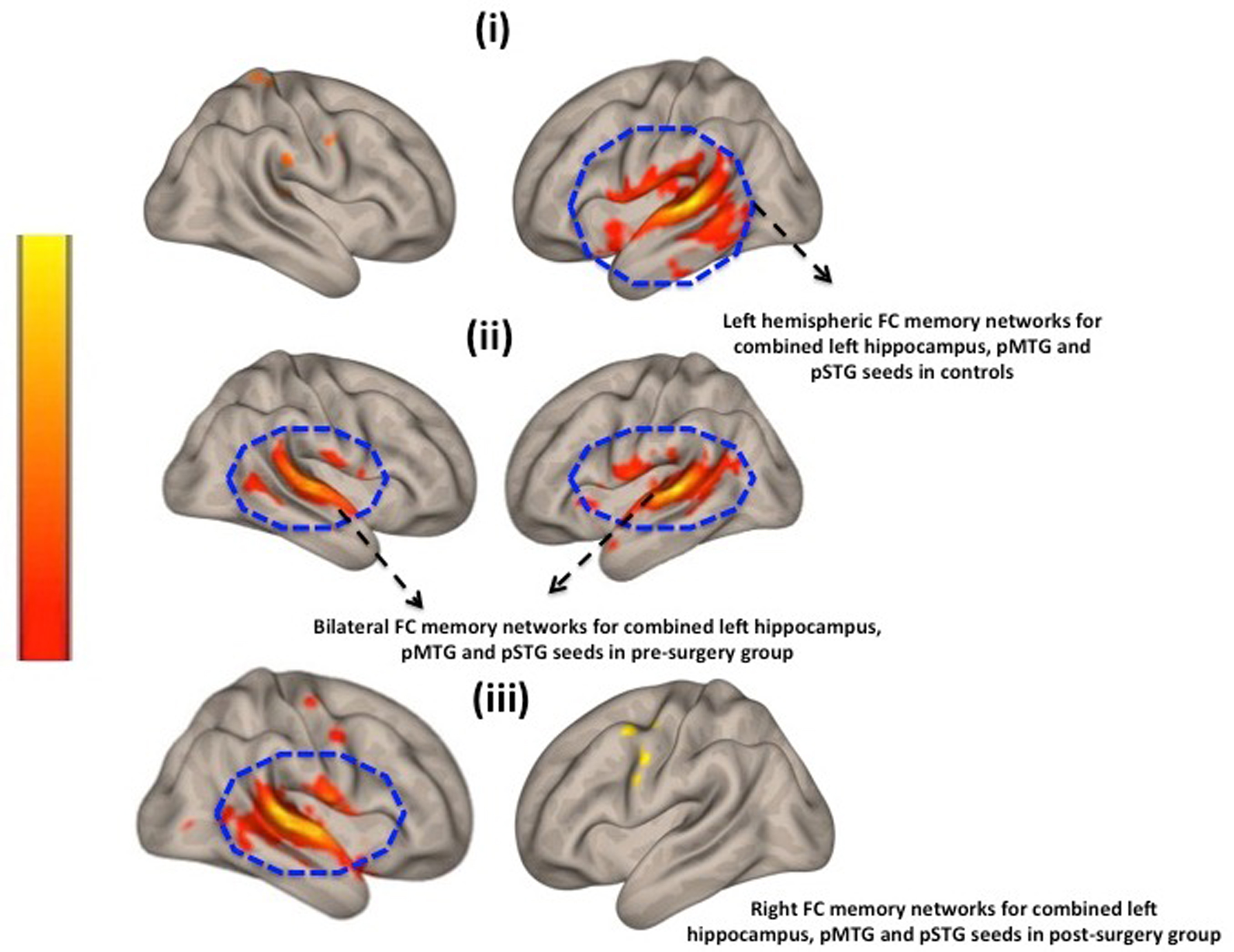
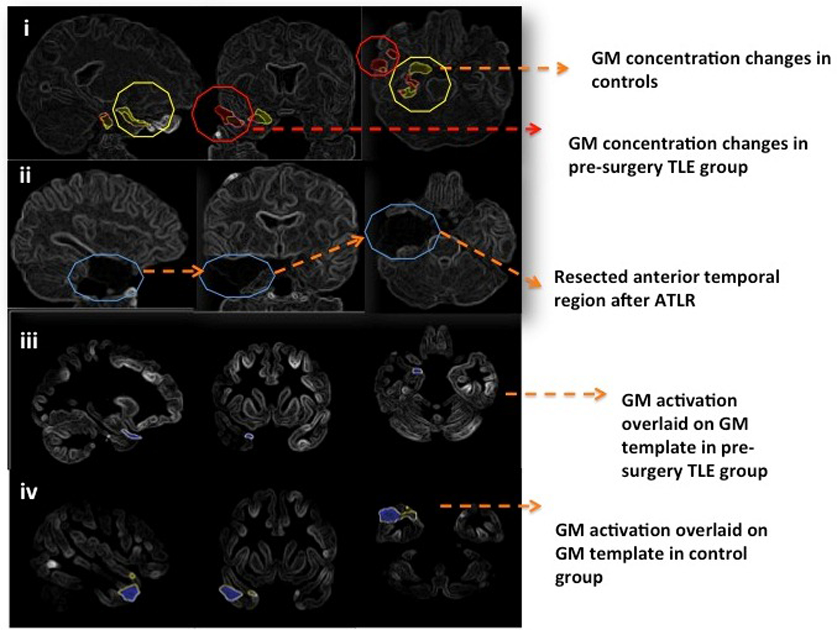
Patients with temporal lobe epilepsy (TLE) have associated cognitive dysfunction and memory impairment on anterior temporal lobe resection (ATLR). Semantic verbal memory reorganization was investigated using functional connectivity (FC) and VBM in pre-and post-operative TLE subjects and healthy controls. Reduction in GM concentration was observed in left temporal lobe post-operatively in comparison to pre-surgery session and healthy controls. Semantic verbal memory components revealed increased FC in right hemisphere with decreased memory scores at follow-up (6 months post-surgery) compared to that of pre-surgery, indicating damage in the medial temporal region due to surgical resection resulting in semantic verbal memory network dysfunction.
Introduction
Patients with drug resistant epilepsy (DRE) are frequently treated by surgical management, the most common substrate being mesial temporal sclerosis (MTS)1,2. Surgical treatment of DRE patients may cause memory impairment and naming difficulties 3,4. In temporal lobe epilepsy (TLE), memory is typically impaired when mesial-temporal and associated neocortical structures are affected by lesions, on-going epileptic activity, or undesired treatment effects 4,5. Patients with TLE have associated abnormalities in grey matter (GM), white matter, metabolic and functional blood oxygen level dependent (BOLD) networks. Relatively, structural and functional abnormalities can be studied by using voxel-based morphometry (VBM) and functional magnetic resonance imaging (fMRI). By using these methods, we can detect the changes in concentration of GM and functional connectivity in multiple brain regions such as temporal and extra-temporal brain areas in patients with DRE.Methods
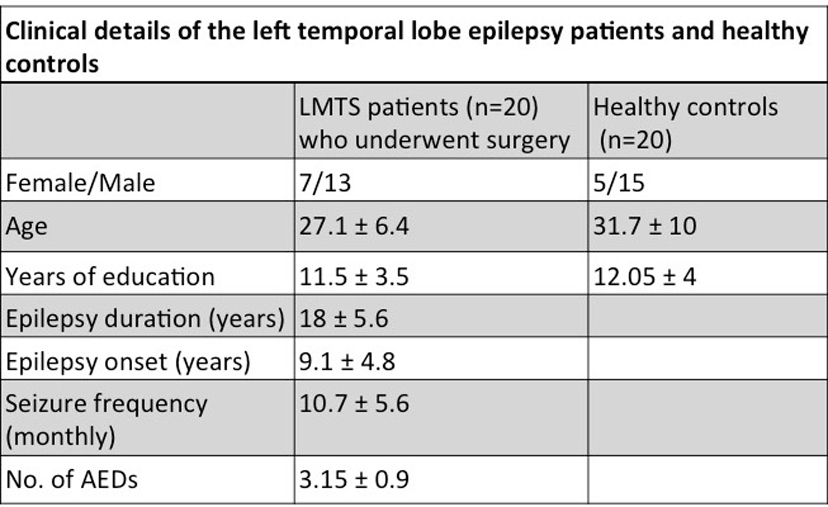
After obtaining the institute ethics approval, 20 consecutive patients with temporal (n=20, left mesial temporal sclerosis) and 20 healthy controls were recruited in this study (Table 1). Standard diagnostic and exclusion criteria were followed. Auditory cue of a standardized story in Hindi, using Super Lab presentation software was given to the subjects using MR compatible auditory interface system (Nordic Neuro Lab, Norway). After the story, patients were instructed to speak the answers in this story based questions during fMRI scan.The fMRI sessions were carried out using 1.5T MR scanner (Avanto, Siemens, Germany) using 8 channel head coil. The stimuli were presented using a MR compatible audio visual stimulus system with binocular goggles (Nordic Neuro Lab, Norway). Single-shot echo planar imaging (EPI) sequence was used for the BOLD studies (number of slices: 29, slice thickness 4.5 mm; TR: 2000 ms, TE: 24 ms, FOV: 230 mm, resolution: 64x64 and total number of measurements: 72). Data analysis and group comparisons were carried out using SPM12. We performed a seed-based correlation analysis for memory task, by defining a seed to voxel ROI on the left posterior and anterior MTG/STG, IFG/MFG, AG and hippocampus using conn functional connectivity toolbox (version17). Results were obtained in between three pre-surgery, post-surgery and control groups (height threshold p uncorrected < 0.001, cluster threshold P< 0.05 (cluster size, p-FDR corrected, T score >3). In order to preprocess each individual T1 sequence, we used the CAT12 toolbox for VBM analysis.Results and Discussion
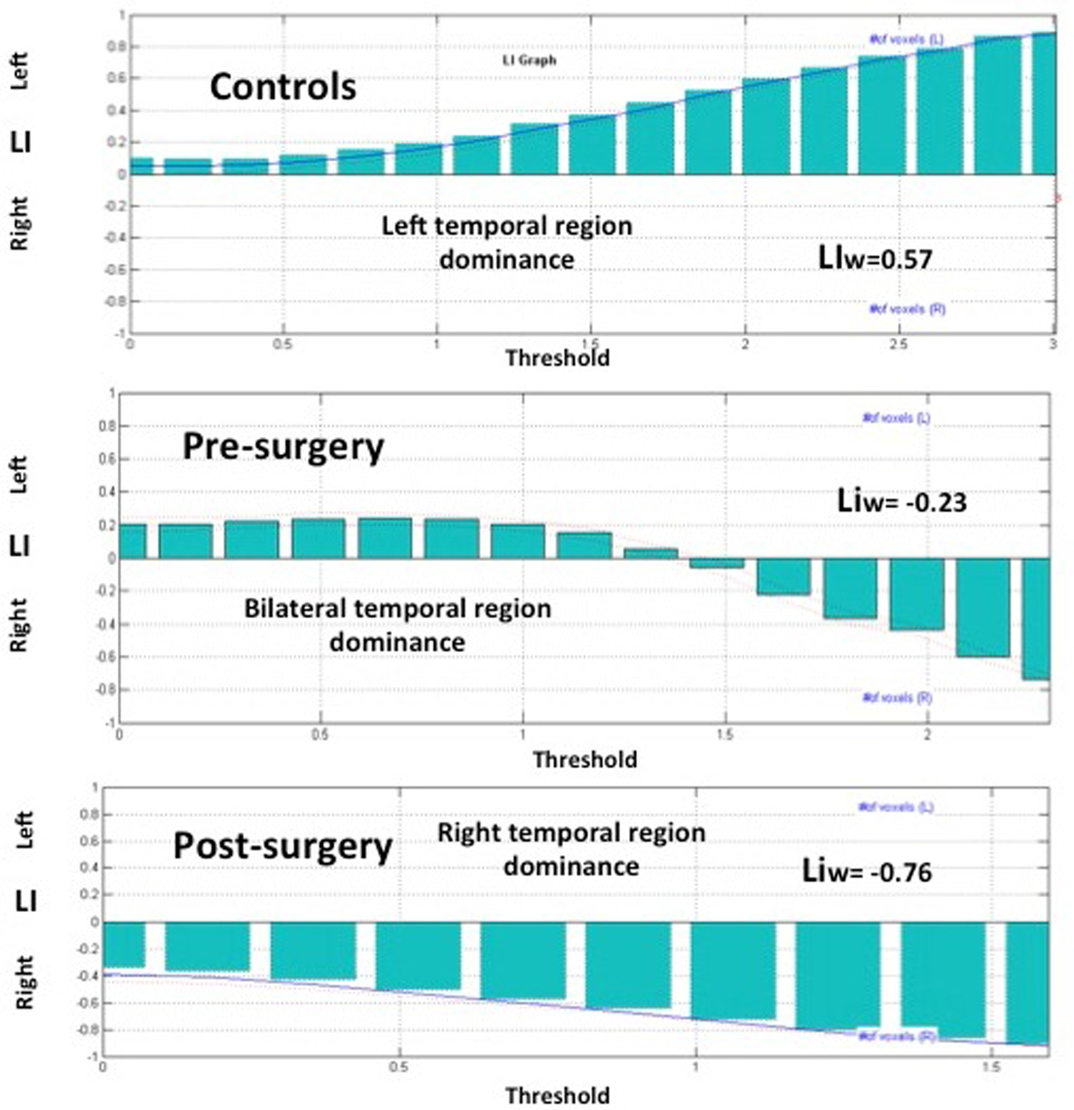
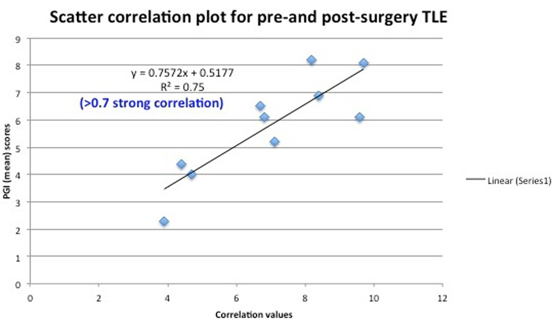
Significant reduction in GM concentration was observed in left temporal lobe post-operatively in comparison to pre-surgery and healthy control groups (Figure 2). Similarly, semantic verbal memory components showed increased FC in right hemisphere post-surgery in comparison to pre-surgery (Figure 1). Laterality index (LI) revealed left temporal (LIw=0.57) dominance for semantic verbal memory task in healthy controls. Bilateral temporal region (LIw = -0.23) was observed in pre-surgery group, whereas in post-surgery group, right hemispheric lateralization of right temporal region (LIw = -0.76) was observed (Figure 3). Our results suggest damage in the medial temporal region due to surgical resection that is responsible for semantic verbal memory network dysfunction. TLE patients had less GM volume compared to healthy controls similar to previously reports 6,7. This may guide neurosurgeons to understand brain recovery mechanisms after surgery 3,5. The patients with left TLE exhibited reduced memory (PGI) functioning after surgery as compared to pre-surgery session (Figure 4). They revealed significant decline scores in executive function, working memory, visual memory, attention and recognition, similar to earlier results in TLE patients post-ATLR 5,8. The results suggest that memory components are impaired in the follow-up session in our study at 6±2 months, which suggests that we need to carry out a longitudinal study beyond 12 months of post-surgery to observe any memory recovery/ reorganization. Conclusion: We investigated structural and functional networks for semantic verbal memory in pre and post-surgery TLE. The volume of GM was decreased in post-surgery patients compared to pre-surgery and healthy controls. Task based fMRI pre-operative data showed strong connections between dorsolateral medial frontal gyrus (MFG), hippocampus, MTG and STG brain regions in left hemisphere with increased memory scores. In post-surgery session, memory function was decreased.Conclusion
We investigated structural and functional networks for semantic verbal memory in pre and post-surgery TLE. The volume of GM was decreased in post-surgery patients compared to pre-surgery and healthy controls. Task based fMRI pre-operative data showed strong connections between dorsolateral medial frontal gyrus (MFG), hippocampus, MTG and STG brain regions in left hemisphere with increased memory scores. In post-surgery session, memory function was decreased.Acknowledgements
I thank to all our clinical research team and staff of Epilepsy clinic and NMR&MRI facility centre at the All India Institute of Medical Sciences, (New Delhi, India) for their invaluable help, and all our patients and controls for their participation in this study.References
1. Puka and Smith. Predictors of language skills in the long term after paediatric epilepsy surgery. Epilepsy Behav. 2016; 63:1-8.
2. Smith AD, Michelle A, Ensign E, et al. Epilepsy surgery in the underserved Hispanic population improves depression, anxiety, and quality of life. Epilepsy & Behavior. 2018; 83:1-6.
3. Helmstaedter C, Brosch T, Kurthen M, et al. The impact of sex and language dominance on material‐specific memory before and after left temporal lobe surgery. Brain. 2004; 127:1518-1525.
4. Sidhu MK, Stretton J, Winston GP, et al. A functional magnetic resonance imaging study mapping the episodic memory encoding network in temporal lobe epilepsy. Brain. 2013; 136: 1868-1888.
5. Bonelli S, Powell R, Yogarajah M, et al. Imaging memory in temporal lobe epilepsy: predicting the effects of temporal lobe resection. Brain. 2010; 133:1186-1199.
6. Gaelle E, Xiaosong He, Sperling M, et al. Gray Matter Abnormalities in Temporal Lobe Epilepsy: Relationships with Resting-State Functional Connectivity and Episodic Memory Performance. PloS One. 2016; 11: e0154660.
7. Mueller SG, Laxer KD, Cashdollar N, et al. Voxel-based optimized morphometry (VBM) of gray and white matter in temporal lobe epilepsy (TLE) with and without mesial temporal sclerosis. Epilepsia.2006; 47,:900–7.
8. Wong SW, Jong L, Bandur D, et al. Cortical reorganization following anterior temporal lobectomy in patients with temporal lobe epilepsy. Neurology. 2009; 73:518-25.
Figures