3789
The faster the better: onsets of pre-SMA and right inferior frontal activations predict successful stopping in a stop-signal task1National Taiwan University, Taipei, Taiwan, 2National Yang-Ming University, Taipei, Taiwan, 3University of Toronto, Toronto, ON, Canada, 4Department of Neuroscience and Biomedical Engineering, Aalto University, Espoo, Finland
Synopsis
Using fast fMRI (whole-brain 10 Hz sampling), we revealed BOLD activities in the pre-SMA and right inferior frontal gyrus are activated faster for successful than for unsuccessful stopping.
Introduction
Functional MRI (fMRI) has previously been demonstrated for detecting hundred-millisecond differences of evoked response onsets, even though the underlying blood oxygenation level-dependent (BOLD) response is delayed and dispersed on the order of seconds. In this study, we exploited a well-established paradigm for motor control, i.e., the stop-signal task, together with the fast functional imaging to explore how our brain implements a precise, dynamic control on action inhibition. By taking advantage of a cutting-edge fMRI protocol, i.e., the simultaneous multi-slice inverse imaging (SMS-InI)1,2, we are able to describe temporal characteristics of the related brain areas and, therefore, are allowed to pinpoint possible causes for stopping efficiency.
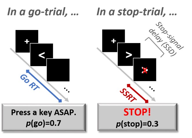
In a stop-signal task, participants are asked to make a motor response as fast as possible (Go), and occasionally, before action execution, a stop-signal appears to call for stopping the intended action (Stop). For stopping, the time interval between the response cue and the stop-signal is critical. The shorter the interval, the easier the stopping. According to a computational model3, we can calculate the processing time of a stop-signal (i.e., the stop-signal reaction time; SSRT) to denote stopping efficiency. By combining a stop-signal task and SMS-InI, we were allowed to characterize the temporal characteristics of these brain areas important for stop-signal processing. Especially, the onsets of BOLD activity in the pre-supplementary area (pre-SMA) and right posterior inferior frontal gyrus (rpIFG) were estimated to examine if these onsets correlate with successful (SST) and unsuccessful stopping (USST).
Methods
14 participants (age, 23.3±2.7; 10 males) took part in the experiment. Figure 1 illustrates the trial procedure of the experiment.
Data were collected on a 3T MRI scanner (Skyra, Siemens) using a 32-channel head coil array and the SMS-InI (TR=0.1 s; TE= 27.9 ms; flip angle=30°). Five runs of data were collected, including about 80 trials in each run. Each run lasted 270 s.fMRI data were reconstructed with the minimum-norm estimates. They were further analyzed by the General Linear Model, where hemodynamic responses were modeled by finite impulse response functions. We chose the time-to-half (TTH), the time reaching 50% of the maximal response to index the timing of BOLD activity onset. TTHs of SST and USST BOLD responses in ROIs were analyzed.
Results
At the behavioral level, RT of go trials is 484±12 ms. RT of stop trials (i.e., signal-respond RT) was faster than RT of go trials (t(14) = 5.5, p < .001). The SSRT is 293±9 ms.
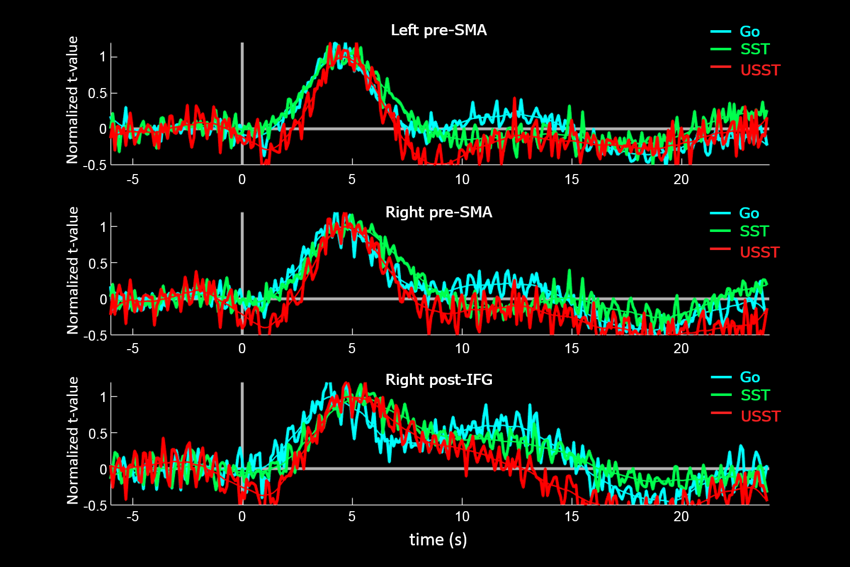
While the stop-trials caused cortical activities in the precentral gyrus, medial frontal gyrus, anterior cingulate cortex, middle prefrontal gyrus, inferior frontal gyrus, inferior parietal cortex, and middle occipital gyrus, the go trials caused activities in the precentral gyrus, premotor cortex, SMA, superior temporal cortex, and right superior parietal cortex. Hemodynamic response function (HRF) of the three ROIs was estimated (figure 2). TTHs of the three ROIs in the pre-SMA and rpIFG were consistently shorter for SST than for USST (figure 3). TTHs of the go trials were not consistent among the ROIs.
Discussion
HRF onsets of SST in the pre-SMA and right inferior frontal gyrus were consistently shorter than those of USST. Activations of these two areas were key to successfully stopping an action, the quicker the better. In the stop-signal task with a tracking procedure to adjust the stop signal delay, the participants were not able to predict when the stop trials occurred. Therefore, our findings suggested that when activities of these areas in response to a stop-signal had a shorter rising time, they could work to inhibit an action more accurately.Acknowledgements
This work was partially supported by Ministry of Science and Technology, Taiwan (103-2628-B-002-002-MY3, 105-2221-E-002- 104), the National Health Research Institutes, Taiwan (NHRI-EX107-10727EI), and the Academy of Finland (No. 298131).References
1. Hsu Y. C., Chu Y. H., Tsai S. Y., et al. Simultaneous multi-slice inverse imaging of the human brain. Scientific reports. 2017;7(1): 17019.
2. Lin F. H., Tsai K. W. K., Chu Y. H., et al. Ultrafast inverse imaging techniques for fMRI. NeuroImage. 2012;62(2):699-705.
3. Verbruggen F., Chambers C. D. & Logan, G. D. Fictitious inhibitory differences how skewness and slowing distort the estimation of stopping latencies. Psychol Sci. 2013; 24:352-362.
Figures