3788
Effect of cue induced craving on brain morphometry and function in adolescent inhalant users1Department of Psychiatry, All India Institute of Medical Sciences, New Delhi, India, 2Department of NMR & MRI Facility, All India Institute of Medical Sciences, New Delhi, India, 3National Drug Dependence Treatment Centre, All India Institute of Medical Sciences, New Delhi, India
Synopsis
Inhalants are legal, inexpensive substances that are often abused by adolescents. This study compared the BOLD changes in brain associated with cue-induced craving in adolescent inhalant users (n=13) and healthy controls (n=12) to elucidate the neural mechanisms associated with cue-induced craving. The cases exhibited an increased activation of superior occipital gyrus, inferior parietal lobule, cingulate gyrus, thalamus, and culmen and a decreased activation of insula as compared to control group for craving cues. Visual cue reactivity was associated with activation of the areas responsible for visual perception, visuo-spatial attention and working memory, control and motivation.
INTRODUCTION
Prevalence of inhalant abuse in the age group 12-29 years has been reported to be around 10–25%[1,2] Certain objects, environments, emotions or pictures which are closely linked with the naturalistic setting of use which is regularly associated with drug use may act as drug cues, which may induce craving even in the absence of drug and may be influenced by the severity of use rather than last dose or recent use[3]. The study estimated BOLD and morphometric changes in the brain on cue-induced craving among adolescent inhalant users and healthy controls.METHODS
Adolescents (aged 12-18 years) with inhalants as their primary substance use, with current use (defined as at least one use within past one month) were recruited from clinics. Controls were healthy adolescents matched for gender and age (±2years; age range 12-18 years), who had not used inhalants during their lifetime. Participants (both cases and controls) with any substance use in preceding 48 hours (either by self report or by urine drug screening), lifetime dependence on any other drug (except tobacco), psychiatric disorder (using MINI-KID) except for conduct disorder, low intellectual ability (using cut-off <5th percentile Raven’s Coloured Progressive Matrices), neurological or medical disorders, and psychotropic medications were excluded. Imaging was carried out on a 3T MR scanner (Achieva 3.0T TX, M/s. Philips Medical Systems) and a 32-channel head coil. 3D TFE multi-shot T1W spin echo sequence was acquired. Visual Cue Paradigm consisted of eight cycles of active (pictures of adolescents using inhalants, either with plastic bag or cloth, both alone and as part of group, in diverse settings resembling real-life settings) and baseline (pictures of adolescents in their naturalistic setting involved in day to day activities as studying, playing, praying, eating, etc.). Data were processed using SPM12 for BOLD and CAT12 toolbox of SPM12 for T1.RESULTS
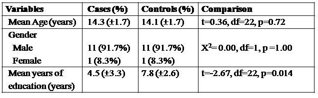
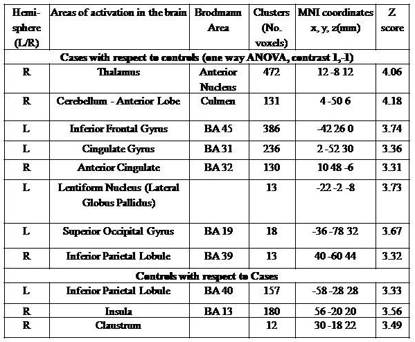
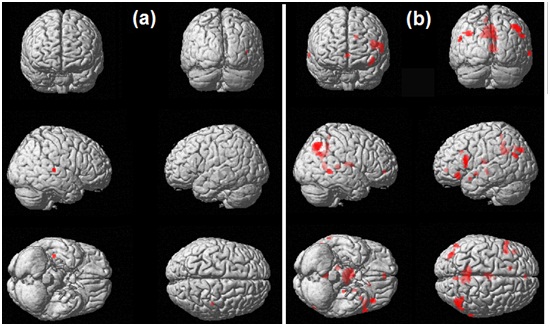
A total of 19 cases and 18 controls were initially recruited, but after exclusion, a final sample of 13 cases and 12 controls were taken up for study. Both the groups were comparable with respect to place of residence (urban/ rural) and religion, and controls had a significantly higher mean number of years of education compared to cases (Table 1). There were no significant difference in degree of handedness score (EHI score) and physical examination or laboratory tests between cases and controls. All the study participants scored ≥5th percentile in Raven's colored progressive matrices, however the percentile score for cases was suggestive of grade IV ‘below average’ intellectual ability compared to controls which was suggestive of grade III ‘average’ intellectual ability (t=-3.32, df=22, p<0.01). All (100%) selected cases had inhalant (volatile solvents) as primary substance of use. As per DSM-IV-TR, 10 (83.3%) subjects met inhalant dependence and 1 (8.3%) met abuse criteria. The mean amount of correction fluid and glue used per day was approximately 29 ml (S.D.=12.5) and 60 ml (S.D.=40) respectively. Due to lack of availability, only 33.3% used correction fluid in previous month. Apart from fluid and glue, cases have never used any other types of inhalants. Mean period of abstinence on inhalants before fMRI session was 6.33 days (S.D.=7.83). On being exposed to cue pictures (with respect to neutral pictures and baseline), cases exhibited differences in BOLD activation in left hemispheric middle temporal gyrus, inferior frontal gyrus and thalamus, right cerebral parahippocampal gyrus and inferior temporal gyrus and healthy controls showed enhanced activation in left inferior occipital gyrus and inferior parietal lobule, right hemispheric precuneus, superior parietal lobule, middle frontal gyrus, fusiform gyrus, and postcentral gyrus (Figure 1, Table 2). Total intracranial volume (p=0.042), gray matter (p=0.035) and cortical thickness (in middle, superior frontal gyrus) were significantly different between cases and controls.DISCUSSION and CONCLUSION
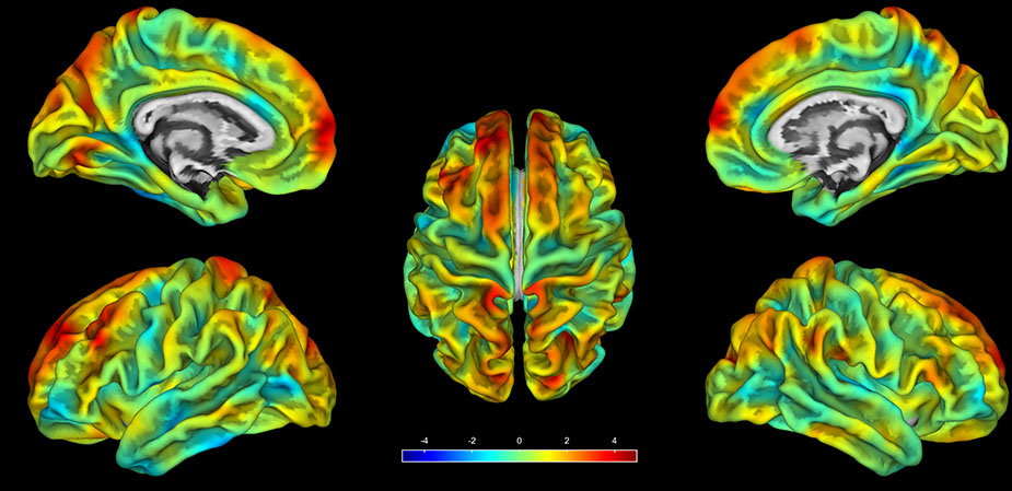
Exposure to cue pictures in inhalant users was associated with activation of the areas involved in visual perception (superior occipital gyrus), followed by visuospatial attention (superior occipital gyrus, inferior parietal lobule, posterior cingulate, cerebellum) and working memory (anterior nucleus of thalamus, posterior cingulate) followed by stimulation of circuits related to control and motivational area. Cerebellum activation may be attributed to its role as coordinator between these circuit and also play a role in selective attention[4], as it presumably reflects lower stimulus salience[5]. Activation of posterior cingulate is known to be associated with visuospatial attention and information processing[7,8] and correlated with the magnitude of arousal on exposure to stimuli[8]. Reduced cortical thickness (figure 2) suggests altered attention network in inhalant users.
The study presented visual cue reactivity associated with activation of the areas responsible for visual perception, visuospatial attention, working memory, motivational area and cerebellum.
Acknowledgements
No acknowledgement found.References
1. Tikoo, V.K., Dhawan, A., Pattanayak, R.D., Chopra, A., 2013. Assessment and pattern, profiles and correlates of substance use among children in India. Natl. Comm. Prot. Child Rights NCPCR New Delhi.
2. White, V.M., Bariola, E., 2012. Australian Secondary School Students’ Use of Tobacco, Alcohol, and Over-the-counter and Illicit Substances in 2011: Report. National Drug Strategy, Department of Health and Ageing.
3. Khazaal, Y., Zullino, D., Billieux, J., 2012. The Geneva Smoking Pictures: development and preliminary validation. Eur. Addict. Res. 18, 103–109.
4. Anderson, C.M., Maas, L.C., deB Frederick, B., Bendor, J.T., Spencer, T.J., Livni, E., Lukas, S.E., Fischman, A.J., Madras, B.K., Renshaw, P.F., 2006. Cerebellar vermis involvement in cocaine-related behaviors. Neuropsychopharmacology 31, 1318–1326.
5. Lou, M., Wang, E., Shen, Y., Wang, J., 2012. Cue-elicited craving in heroin addicts at different abstinent time: an fMRI pilot study. Subst. Use Misuse 47, 631–639.
6. Grön, G., Wunderlich, A.P., Spitzer, M., Tomczak, R., Riepe, M.W., 2000. Brain activation during human navigation: gender-different neural networks as substrate of performance. Nat. Neurosci. 3, 404–408.
7. Vogt, B.A., Finch, D.M., Olson, C.R., 1992. Functional heterogeneity in cingulate cortex: the anterior executive and posterior evaluative regions. Cereb. Cortex 2, 435–443.
8. Maddock, R.J., 1999. The retrosplenial cortex and emotion: new insights from functional neuroimaging of the human brain. Trends Neurosci. 22, 310–316.
Figures