3787
7T functioanl MRI reveals frequency-dependent responses during vibrotactile stimulation at somatosensory cortex1Interdisciplinary Institute of Neuroscience and Technology, Qiushi Academy for Advanced Studies, Zhejiang University, Hangzhou, China, 2College of Biomedical Engineering & Instrument Science,Zhejiang University, Hangzhou, China, 3Department of Neurobiology, Zhejiang University School of Medicine, Hangzhou, China, 4The Second Affiliated Hospital, Zhejiang University School of Medicine, Hangzhou, China
Synopsis
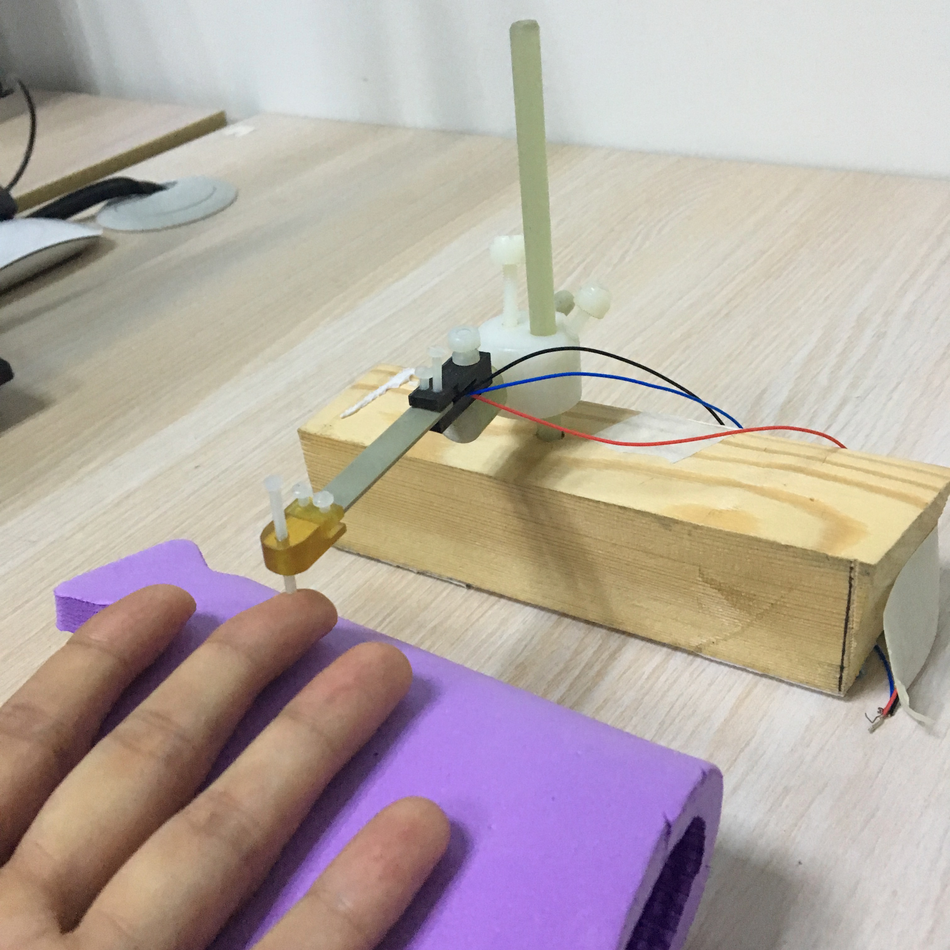
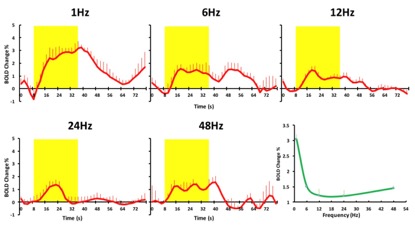
To investigate whether the brain can directly encode different vibrotactile frequencies, we made a MR-compatible lab-designed vibrotactilestimulator to presented pulse stimuli on middle finger (with a frequency difference in the same duration) andusing BOLD fMRI to explore the effectsofvibrotactilefrequencyin somatosensory cortex with 7T MR research system. The results showedthat the lab-designed MR compatible vibrotactile stimulator could elicit BOLD responsesin the somatosensory cortex. Also, the BOLD response was frequency-dependent with the peak at 6Hz.
Introduction
Primary somatosensory cortex(S1)is sensitive to the tactile frequency1.Mapping functional location of the S1 is necessary for understanding of the properties of tactile perception in human2-4. Generally, the ultra-high field functional magnetic resonance imaging (fMRI) could provide the reliable cortical responses of digits in the S1 because of the higher signal-to-noise ratioand spatial resolution in blood-oxygen-level dependent (BOLD) signals. The previous study demonstrated BOLD fMRIisa valid tool for mapping the somatosensory systemand the study mentioned thatit is unlear aboutwhat the effect of vibrotactile frequency responded to the cortical function in S1 and how did the BOLD signals present the effects of vary frequency in vibrotactile.The goal of this study is to make MR-compatible lab-designed vibrotactilestimulator and combined with ultra-high field7T research MRI for investigating the vibrotactile frequencyin human S1.Methods
Results and Discussion
Conclusion
This study demonstratedBOLD response was frequency-dependent and the low-frequency stimuli may affect the recover time of BOLD curve.This MR-compatible lab-designed vibrotactilestimulatorcould be a useful tool to evaluate brain functional recoverin patients withstrokein the future.Acknowledgements
No acknowledgement found.References
1. YG Chung, J Kim, SW Han, HS Kim SP Kim et al. Frequency-dependent patterns of somatosensory cortical responses to vibrotactile stimulation in humans: A fMRI study[J]// Brain research, 2013:47-57.
2.YH Li, Ying Lee, Wolfgang Grodd, Christoph Brarun. Comparing tactile pattern and vibrotactile frequency discrimination: a human fMRI study[J]//Journal of neurophysiology, 2010.
3.Renate Schweizer, Dirk Voit, and Jens Frahm. Finger representations in human primary somatosensory cortex as revealed by high-resolution functional MRI of tactile stimulation[J]// Neruoimage, 2008:28-35
4.Christian Kalberlah, ArnoVillringer, Burkhard Pleger. Dynamic causal modeling suggests serial processing of tactile vibratory stimuli in the human somatosensory cortex—An fMRI study[J]// Neruoimage, 2013:164-171
Figures