3785
Dual-route mechanism for pictorial visual perception in developmental dyslexia1Department of NMR and MRI Facility, All India Institute of Medical Sciences, New Delhi, India, 2Department of Psychiatry, All India Institute of Medical Sciences, New Delhi, India, 3Department of Neurology, Fortis Hospital, New Delhi, India, 4Department of Psychiatry (Psychology Unit), All India Institute of Medical Sciences, New Delhi, India, 5Department of Linguistics, School of Language, Jawahar Lal Nehru University, New Delhi, India, 6Department of Biostatistics, All India Institute of Medical Sciences, New Delhi, India
Synopsis
Reading a window to world’s information, is a coordinated skill of grapheme-phoneme interface (letter-sound) at symbolic level. Inefficient mapping of this alphabets-horizontal string is labeled as dyslexia (reading problem). It can be developmental or acquired (post-stroke) disorder. Developmental dyslexia (DD) is heterogeneous state with disputed underlying mechanism(s) constraining optimal remediation. Compared to typical readers, DD literature is bifurcated into children with strength and impairment for visual perception. To unfold it neurobiologically, nonsymbolic visual processing role was studied. Observed bilateral brain activation (functional MRI) of ventral stream (inferior occipital and fusiform) and modified dorsal route-gating for figure-ground filtering in DD.
Purpose
Dyslexia is a neurodevelopmental disorder affecting reading, writing and calculating performance (Vidyasagar and Pammer, 2010). Despite its description since century ago (Pringle-Morgan, 1896) it’s mechanism is yet unclear (Johnston et al, 2017; Zoccolotti, et al, 2016), with broad range of performance deficits and tasks inconsistencies (Ramus and Ahisaar, 2012; Roach and Hogben, 2007). These persistent deficits lead to emotional, academic, social consequences (Vidyasagar and Pammer, 2010). In children with developmental dyslexia (DD) symbolic visual performance has variability (Zoccolotti, et al, 2016), whether non-symbolic level plays any role is crucial to untangle the phenomena (Johnston et al, 2017) especially at neurobiological level. Dual-route model at lexicosemantic level is involved in reading deficits (Perrone-Bertolotti et al, 2017) but whether has any participation for pictorial visual perception is yet not revealed. So this study was planned to understand picture processing with functional magnetic resonance imaging (fMRI) comparing reading impaired (DD) and typical readers (age-matched healthy controls, HC). Literature also report visual processing strength in DD (Wang et al, 2016) so to observe the cognitive grown-up perception (adult cognitive trajectory) healthy adults were compared.Introduction
DD is primarily being associated with auditory or phonological processing and has been studied as the strongest predictor of reading skill (Jaffe-Dax et al, 2018; Ziegler, et al, 2009; Ziegler and Goswami, 2005). In DD visual processing with non-verbal stimuli need to be assessed independently of phonological ability (Zoccolotti, et al, 2016; Lobier et al., 2011) for better understanding the heterogeneity (Peterson and Pennington, 2012). In reading, ventral pathway participate in lexicosemantic and dorsal-route in grapho-phonological processing (letter-sound conversion) (Perrone-Bertolotti et al, 2017) but how these play role in dyslexic prelexical visual (pictorial) perception is not being explored. Performance on the picture-completion task (visual perception at behavioral level), measures deliberate focusing of attention on small-miniature details and recognizing specific feature that is ‘missing’ for completing whole picture (Wechsler, 2004). FMRI visual search paradigms are useful tool for investigating brain areas for identification of target, distractors, spatial localization, development and allocation of attention (Ellison et al, 2014). So, the present study included fMRI picture-completion paradigm for understanding dual-route mechanism at prelexical level.Methods
Sample consisted of healthy adults (n = 16), dyslexics (n = 20) age-matched typical readers (healthy controls, HC; n = 20). After ethics approval from IEC and informed written consent subjects were recruited. fMRI for visual processing task was acquired in 3T MR scanner (Achieva, M/s. Philips HealthCare, The Netherlands) 32 channel head coil using single shot EPI sequence with parameters of FOV 230; 31 slices, slice thickness of 5 mm without any slice gap; TR of 2 s; echo train length of 33; and 163 dynamic. Paradigm was standardized (outside and inside the MR scanner) for presentation time. Visual images were black-white line drawings (taken from picture-completion subtest of local standardized version of WISC-R) presented on the MR compatible LCD monitor with E-Prime & ESys System (version 1.1, Psychology Software Tools Inc, USA). The task paradigm was block design (4 events x 4 blocks) of total acquisition time of 326 s. fMRI data was processed in SPM12 and clinical data using SPSS.Results & Discussion
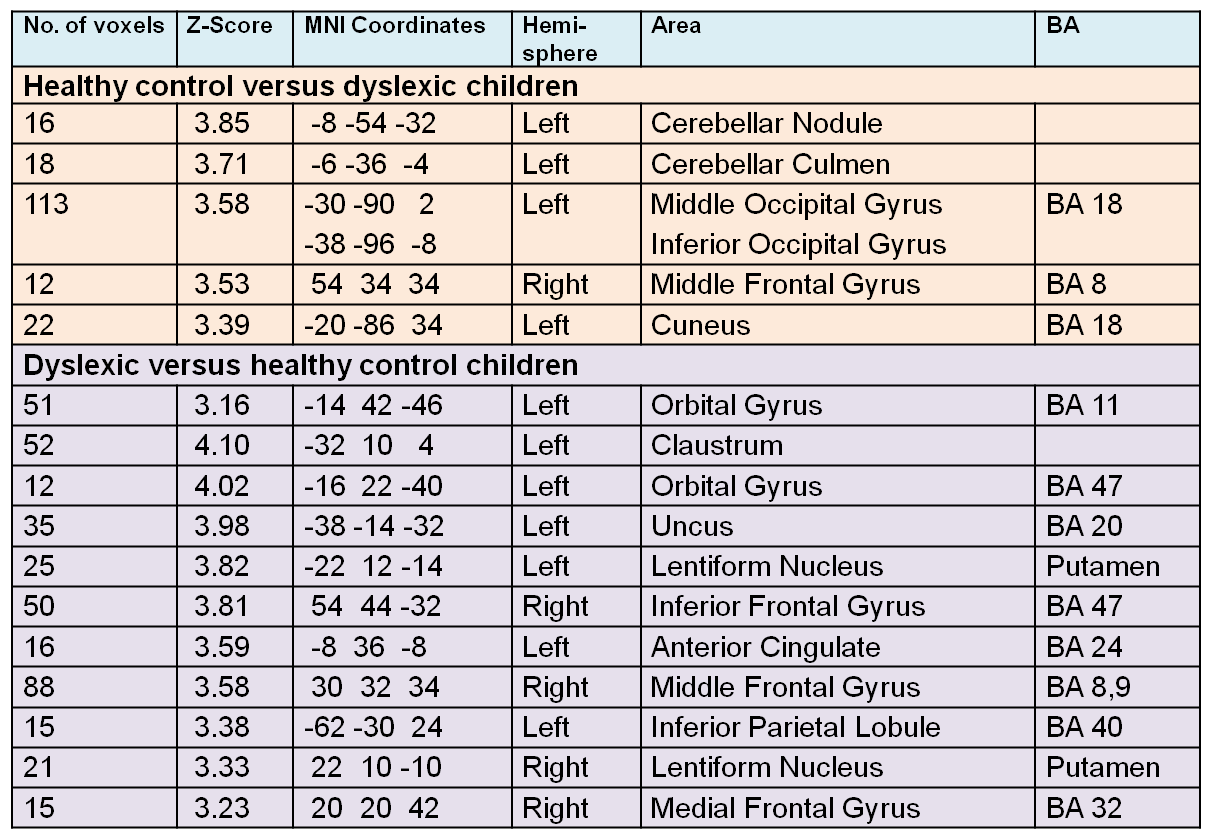
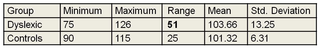
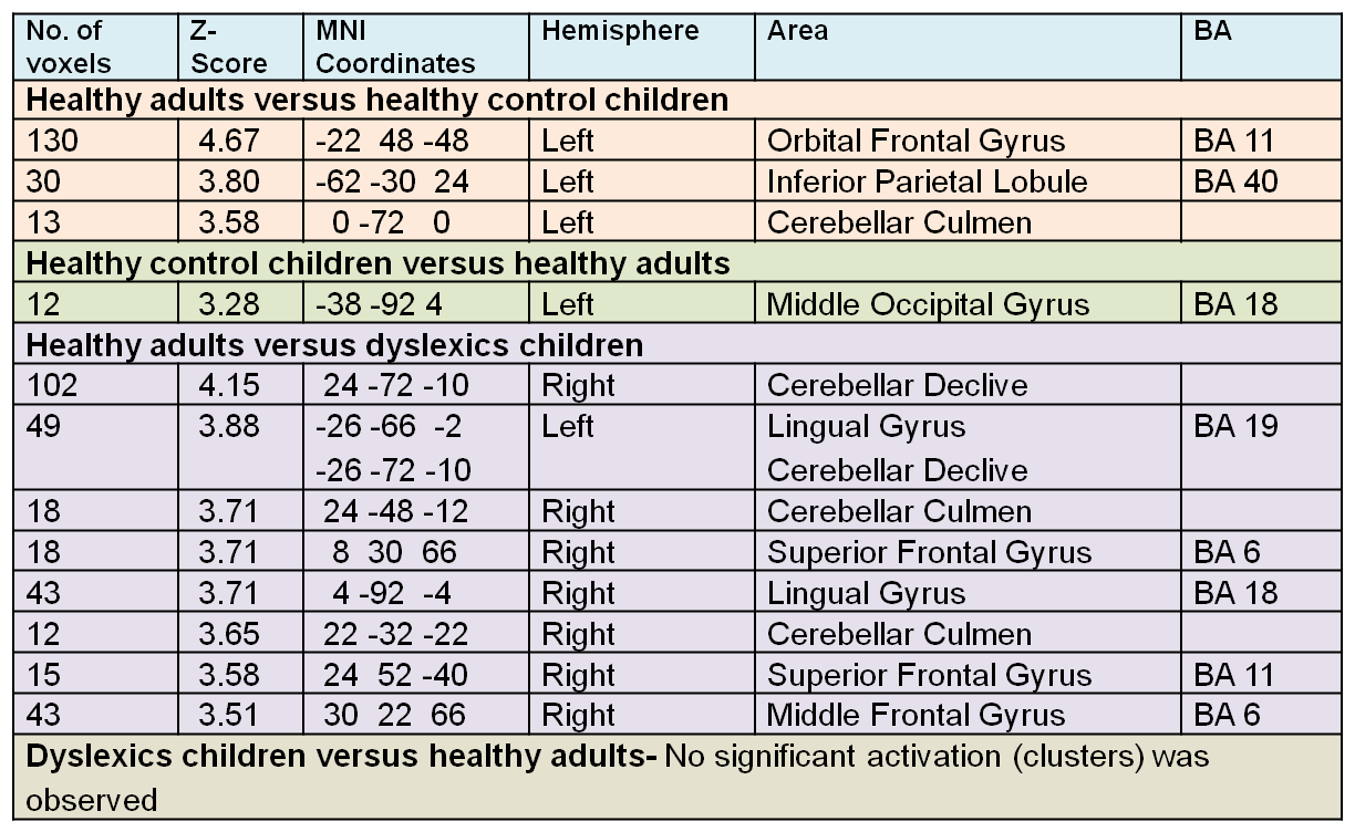
In children (DD and HC) similar BOLD activation observed in right inferior occipital (BA 18) and fusiform gyri (BA 37) precuneus (BA 7), but in DD it was being complemented by left fusiform (BA 18, 19) and left precuneus. Activation volume (based on sum-cluster-count) in frontal areas and parietal areas was greater in dyslexic group as compared to healthy children. Group comparison DD > HC revealed significant activation in left BA 47, 11, 20, 40 with uncus (BA 20), left anterior cingulate and in right BA 8, 9, 32 (Table-1). Performance mean scores of dyslexic and HC were statistically not significant (t = -0.790; p = 0.432, two-tailed) but variability was observed in DD group on performance distribution (Table-2). Comparing healthy adult with healthy children activation observed in left BA 11, BA 40 and culmen (anterior cerebellum) suggesting some similarity with DD group (Table-3) but the greater volume of BOLD activation in DD (Figure-1) indicate higher effort involved in attention allocation, distracter inhibition and/or figure-ground filtering (Roe et al, 2012) though with equated performance mean scores as in HC. Indicating modified interface of top-down gating of dorsal-ventral stream in DD (Ding et al, 2016; Nash et al, 2017; Xia et al, 2016).Conclusion
In dyslexia performance variability and visual perception at nonsymbolic level (pictorial) was associated with bilateral activation in ventral route and hyperactivation of dorsal-stream, indicating modified top-down gating that may attribute to the reading skill.Acknowledgements
No acknowledgement found.References
Ding, Y., Zhao, J., He, T., Tan, Y., Zheng, L., Wang, Z. (2016). Selective impairments in covert shifts of attention in Chinese dyslexic children. Dyslexia, 22, 362-378 doi:10.1002/dys.1541
Ellison, A., Ball, K.L., Moseley, P., Dowsett, J., Smith, D.T., Weis, S., Lane, A.R. (2014). Functional interaction between right parietal and bilateral frontal cortices during visual search tasks revealed using functional magnetic imaging and transcranial direct current stimulation. PLoS ONE, 9(4), e93767. doi:10.1371/journal.pone.0093767
Jaffe-Dax S, Kimel E, Ahissar M, (2018).Shorter cortical adaptation in dyslexia is broadly distributed in the superior temporal lobe and includes the primary auditory cortex. eLife 2018;7:e30018 DOI: 10.7554/eLife.30018
Johnston, R., Pitchford, N.J., Roach, N.W., Ledgeway, T. (2017) Visual perception in dyslexia is limited by sub-optimal scale selection. Sci Rep.,7(1):6593. doi: 10.1038/s41598-017-06967-6.
Lobier, M., Zoubrinetzky, R., Valdois, S. (2011). The visual attention span deficit in dyslexia is visual and not verbal. Cortex, 1-6. doi:10.1016/j.cortex.2011.09.003
Nash, H.M., Gooch, D., Hulme, C., Mahajan, Y., McArthur, G., Steinmetzger, K., Snowling, M.J. (2017). Are the literacy difficulties that characterize developmental dyslexia associated with a failure to integrate letters and speech sounds? Developmental Science, 20, e12423. doi: 10.1111/desc.12423
Perrone-Bertolotti, M., Kauffmann, L., Pichat, C., Vidal, J.R., Baciu, M. (2017). Effective Connectivity between Ventral Occipito-Temporal and Ventral Inferior Frontal Cortex during Lexico-Semantic Processing. A Dynamic Causal Modeling Study. Front Hum Neurosci., 11:325. doi:10.3389/fnhum.2017.00325.
Peterson, R.L., Pennington, B.F. (2012). Developmental dyslexia. Lancet, 379, 1997–2007. Pringle-Morgan, W. (1896). A case of congenital word blindness. BMJ., ii,178.
Ramus, F., Ahissar, M. (2012). Developmental dyslexia: The difficulties of interpreting poor performance, and the importance of normal performance. Cognitive Neuropsychology, 29 (1–2), 104-122.
Roach, N.W., Hogben, J.H. (2007). Impaired filtering of behaviourally irrelevant visual information in dyslexia. Brain, 130, 771-785. doi:10.1093/brain/awl353
Roe A.W., Chelazzi, L., Connor, C.E., Conway, B.R., Fujita, I., Gallant, J.L., Lu, H., Vanduffel, W. (2012). Toward a unified theory of visual area V4. Neuron, 74, 1-18. doi:10.1016/j.neuron.2012.03.011
Vidyasagar, T.R., Pammer, K. (2010). Dyslexia: a deficit in visuo-spatial attention, not in phonological processing. Trends in Cognitive Sciences, 14 (2), 57-63
Wang, J., Schneps, M. H., Antonenko, P. D., Chen, C., Pomplun, M. (2016). Is reading impairment associated with enhanced holistic processing in comparative visual search? Dyslexia, 22, 345-361. doi:10.1002/dys.1540
Wechsler, D. (2004). Wechsler Intelligence Scale for Children-IV Conceptual and Interpretive Guide. http://www.iupui.edu/~flip/wiscdescription.pdf
Xia, Z., Hoeft, F., Zhang, L., Shu, H. (2016). Neuroanatomical anomalies of dyslexia: disambiguating the effects of disorder, performance, and maturation. Neuropsychologia, 81, 68-78. doi:10.1016/j.neuropsychologia.2015.12.003
Ziegler, J.C., Goswami, U. (2005). Reading acquisition, developmental dyslexia, and skilled reading across languages: A psycholinguistic grain size theory. Psychol. Bull., 131, 3-29.
Ziegler, J.C., Pech-Georgel, C., George, F., Lorenzi, C. (2009). Speech-perception-in-noise deficits in dyslexia. Developmental Science, 12(5), 732-745. doi:10.1111/j.1467-7687.2009.00817.x
Zoccolotti, P., de Jong, P.F. and Spinelli, D. (2016). Editorial: understanding developmental dyslexia: linking perceptual and cognitive deficits to reading processes. Front Hum Neurosci., 10, 140. doi:10.3389/fnhum.2016.00140
Figures
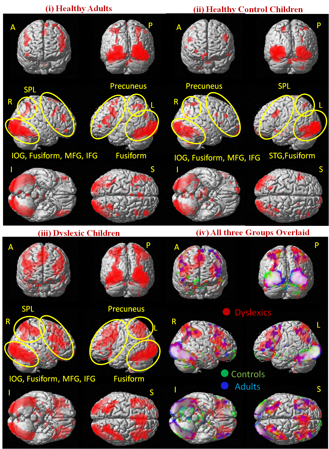
Figure-1: BOLD (fMRI) activation during visual processing (searching missing part in the picture) in (i) healthy adults (n=16), (ii) age-matched typical reading children (healthy control,n = 20) and (iii) dyslexia children (n = 20) [analysed using one-way one-way ANOVA (p < 0.001, voxel threshold: 10, 1 voxel: 2x2x2 mm3); A- anterior view; P- posterior; R-right; L- left; I- inferior; S- superior view; MFG-middle frontal gyrus, IFG-inferior frontal gyrus, STG-superior temporal gyrus, IOG-inferior occipital gyrus, SPL-superior parietal lobule]