3783
Engagement of multiple networks during meditation: an fMRI study1Indraprastha Institute of Information Technology, New Delhi, India, 2Indian Institute of Technology, New Delhi, India, 3Centre for Advanced Research in Imaging, Neuroscience and Genomics, Mahajan Imaging, New Delhi, India, 4All India Institute of Medical Sciences, New Delhi, India
Synopsis
Long-term effects of meditation and the differences between meditators and non-meditators have been studied extensively. In order to understand the corresponding mechanisms of action, brain areas activated while meditating need to be studied. Such studies are limited due to lack of a suitable experimental paradigm and analysis technique. We investigated experienced meditators who underwent a 20 minutes audio-guided meditation in the fMRI scanner. Using the Intersubject correlation (ISC) analysis, correlated activity across the subjects was found in frontal pole, middle frontal gyrus, precuneus, primary somatosensory cortex, visual cortex, lingual gyrus, and cerebellum.
Introduction
In recent years, interest in meditation has increased dramatically. Many benefits such as increase in cortical thickness1, reduction in stress2, reduction in fatigue, anxiety and increased mindfulness3, increased positive mood states4,5 etc have been reported. Using fMRI, differences between long-term meditators and novices in terms of emotional regulation6 and emotional reaction7 to different stimuli have been established. Meditators have higher activation in prefrontal cortex (mPFC), ventromedial prefrontal cortex (vmPFC), hippocampus which signify a state of meta-awareness.
All these studies have observed the effect of long-term meditation on physiological and behavioral characteristics. But there have been a dearth of studies which observe the immediate effects of meditation on the brain. The dynamics of meditation are still unknown. An MRI scanner environment is not inherently conducive to a meditative practice. Also, supine position is not a default for meditation. We chose Yoga Nidra meditation, a unique practice meant to be done in the supine posture, to study the brain dynamics.
Methods
24 experienced yogic practitioners (average yogic experience of 2,989 hours, mean age 27.53 years, 20 males) volunteered from local meditation centers. The total yogic experience included time spent in yogic asanas (postures), pranayama (breathing exercises), meditation and chanting. Participants were subject to Mini International Neuropsychiatric Interview (MINI) screen and only those with no history of mental illness were recruited for the study. Ethical clearance was taken from the IIT Delhi Institute Ethical committee.
We acquired the MRI data on a Philips Ingenia 3.0T whole-body scanner at Mahajan Imaging, Gurugram. The anatomical data were acquired using a T1-weighted 3D MP-RAGE protocol (TR = 2250 ms, TE = 4.18 ms, FOV = 256 mm, flip angle = 9°, voxel size = 1×1×1 mm). Functional data were acquired using a multi-band echo-planar imaging pulse sequence (TR = 1000 ms, TE=25 ms, 65° flip angle) with a multi band factor of 8. Forty-eight 4 mm thick oblique-axial slices were acquired in head first order with no gap. The acquisition matrix was 73x73 with 3x3x4 mm voxels.
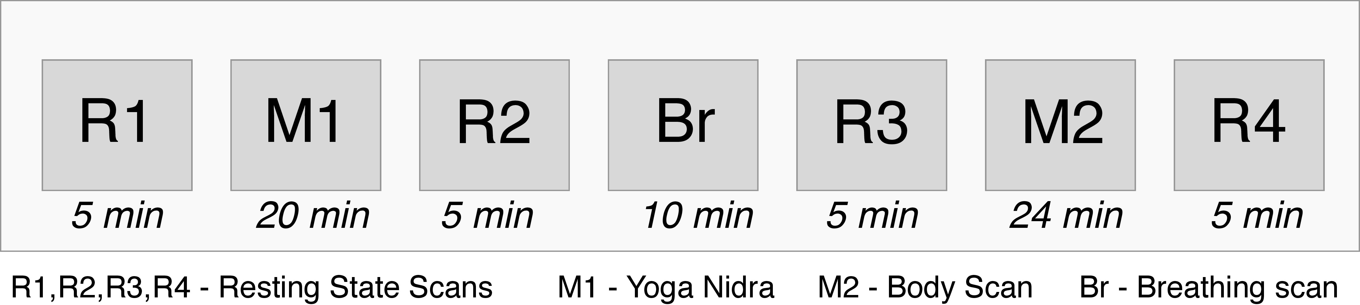
The protocol employed is given in Figure 1. We used FSL for preprocessing8. The brain was extracted from the high resolution structural file using the repeated version of the FSL-BET. Custom Python code was used for slice-time correction for multi band acquisition. This was followed by motion correction, temporal filtering (100s), spatial smoothing with 6mm and co-registration across the dataset. Normalization was done with an MNI 2mm template.
Results
Auditory signal was downsampled, convolved with HRF, and then correlated with BOLD signal voxelwise across the whole brain to determine a subject’s auditory response to the guided instructions. Using a bootstrapping method to create a null distribution, correlation values were converted to p-values. A correlation greater than 0.15 corresponded to uncorrected p<0.05, 2 subjects having insignificant auditory correlation were excluded from further analysis Fig 2. shows the auditory correlations.
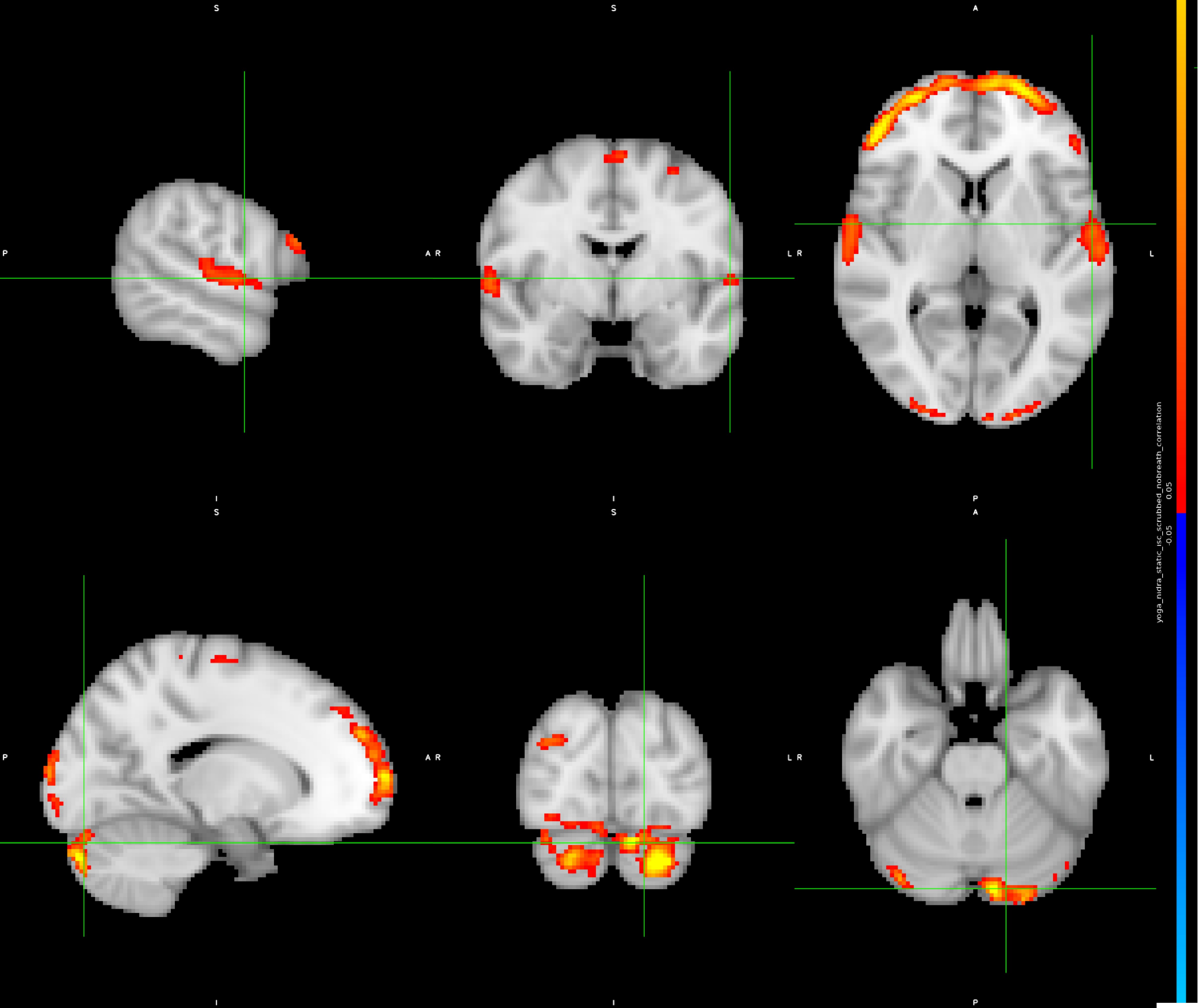
The Inter Subject Correlation9 analysis was performed on the selected subjects. A bootstrapping method was used to create a null distribution, the voxel wise correlation value was converted to a p-value which was corrected(p<0.05) for multiple comparisons. Figure 3 shows the corrected p-values for the correlated activity in the auditory cortex, primary somatosensory cortex, visual cortex, middle frontal gyrus, precuneus cortex, frontal pole, lingual gyrus, and cerebellum.
Discussion
As the meditation involved taking attention to different parts of the body, we see activations in the auditory cortex, the superior temporal sulcus, inferior temporal gyrus, lingual gyrus, somatosensory cortex and postcentral gyrus. This is related to the auditory processing and visualization. The effects of meditation can be observed in the middle frontal gyrus associated with attention network, precuneus and posterior cingulate which are part of the default mode network associated with self referential thoughts, insula which is related to emotional processing, frontal pole involved in working memory and the cerebellum which has been determined in motor movements as well as dorsal attention network. Meditation is a dynamic process that involves seemingly opposing regions like the attention network and the default mode network.Conclusions
The study of mindfulness and meditation need new fMRI paradigms and analysis methods. We show that for audio guided meditation, ISC analysis can detect the regions involved in meditation. The spatiotemporal dynamics of such activations needs to be explored in detail to determine the ways in which meditation affects the brain.Acknowledgements
We wish to thank Ms. Niharika Singh and her team at Mahajan Imaging, Gurugram for helping coordinate MRI scanning of our subjects. We would also like to thank Ms. Rupsa Bhattacharjee and Dr. Indrajit Saha from Philips India Ltd. for providing multi-band EPI capability and for helping configure the MR scanner.References
- Lazar, S. W., Kerr, C. E., Wasserman, R. H., Gray, J. R., Douglas, N., Treadway, M. T., Mcgarvey, M., Quinn, B. T., and Dusek, J. A. (2006). Meditation experience is associated with increased cortical thickness. Neuroreport, 16(17):1893–1897.
- Seppala, E. M., Nitschke, J. B., L., T. D., Hayes, A., Goldstein, M., Nguyen, D. T., Perlman, D., and Davidson, R. J. (2014). Breathing-Based Meditation Decreases Posttraumatic Stress Disorder Symptoms in U.S. Military Veterans: A Randomized Controlled Longitudinal Study. Journal of traumatic stress, 27(1):397–405.
- Zeidan, F., Johnson, S. K., Diamond, B. J., David, Z., and Goolkasian, P. (2010). Mindfulness meditation improves cognition: Evidence of brief mental training. Consciousness and Cognition, 19(2):597–605.
- Ding, X., Tang, Y. Y., Tang, R., and Posner, M. I. (2014). Improving creativity performance by short-term meditation. Behavioral and Brain Functions, 10(1):1–8.
- Jain,S.,Shapiro,S.L.,Swanick,S.,Roesch,S.C.,Mills,P.J.,Bell,I.,and Schwartz,G. E. R. (2007). A randomized controlled trial of mindfulness meditation versus relaxation training: Effects on distress, positive states of mind, rumination, and distraction. Annals of Behavioral Medicine, 33(1):11–21.
- Lutz, J., Herwig, U., Opialla, S., Hittmeyer, A., Jäncke, L., Rufer, M., Holtforth, M. G., and Brühl, A. B. (2013). Mindfulness and emotion regulation-an fMRI study. Social Cognitive and Affective Neuroscience, 9(6):776–785
- Taylor, V. A., Grant, J., Daneault, V., Scavone, G., Breton, E., Roffe-Vidal, S., Courtemanche, J., Lavarenne, A. S., and Beauregard, M. (2011). Impact of mindfulness on the neural responses to emotional pictures in experienced and beginner meditators. NeuroImage, 57(4):1524–1533.
- Jenkinson, M., Beckmann, C. F., Behrens, T. E., Woolrich, M. W., and Smith, S. M. (2012). Fsl. NeuroImage, 62(2):782 – 790. 20 YEARS OF fMRI.
- Hasson, U. (2004). Intersubject Synchronization of Cortical Activity During Natural Vision. Science, 303(5664):1634–1640.
Figures