3782
Cerebral blood flow changes associated with meditation practice time: a longitudinal study1State University of New York at Binghamton, Binghamton, NY, United States, 2Cornell University, Ithaca, NY, United States
Synopsis
Longitudinal cerebral blood flow (CBF) images were acquired in ten college students after 2-month meditation practice. The CBF difference before and after meditation practice and the association between CBF change and meditation practice time were analyzed on a voxel-by-voxel basis using SPM8. We found significant perfusion increase and decrease in the occipital and thalamus regions respectively. However, the CBF increase and decrease in the corresponding regions exhibited negative and positive association with practice time respectively. Regional analysis confirmed that the direction of CBF change may switch after some amount of meditation practice time, indicating a complex neural pathway of meditation.
Introduction
Meditation has been established as effective practice in reducing symptoms of anxiety [1, 2], depression [3, 4], panic disorder [5], emotional distress [5], and in enhancing cognitive abilities [6]. Neuroimaging studies [7-10] have revealed brain functional changes when subjects were in active meditation. However, the literature for studying the longitudinal effects of meditation practice is rather scarce. This preliminary study is to determine the change of resting cerebral blood flow (CBF) after 2-month meditation practice and whether the changes of CBF are associated with practice time.Method
All studies were conducted at the Cornell University MR Facility using a GE 3T MR 750. Volunteers received 3D T1-weighted Magnetization Prepared RApid Gradient Echo (MPRAGE) images for image registration and resting-state 3D pseudo-continuous arterial spin labeling (PCASL) sequence [11] for CBF and functional connectivity measurements before and after approximately 2-month meditation practice (practice duration: 66.50 ± 4.14days, practice time: 574.00 ± 312.30 minutes). Ten college students(19.20 ± 0.28 years old, age range: 19 to 20 years old, 4 females) were recruited from a university meditation class. Subjects were instructed to practice focused meditation for a minimum of 10 minutes each time and at least 5 times per week. They were allowed to choose their own focus: their breath, a point on the wall, a phrase, or anything else as they saw fit. Volunteers reported their total practice time at the end of the follow-up scan.
For each subject, the ASL difference signal time series was used to derive the absolute CBF map using the flow kinetic model [12], and then normalized by the whole-brain CBF to produce the normalized CBF map to adjust for the variation of the global CBF between baseline and follow-up. The CBF maps were normalized to a standard MNI template using the T1-weighted images as an intermediate, and smoothed with a 6×6×6 mm Gaussian kernel. The CBF maps before and after practicing meditation were compared using SPM8 via a paired t test with gender as a covariate. Age was not considered as a covariate because the maximum difference among ages of our volunteers is 1. The difference of the CBF maps before and after meditation practice was modeled using SPM8 via multiple linear regression with gender and practice time as independent variables. The statistical maps were thresholded using a voxel-level p value of 0.01. A cluster-level p value of 0.05 was used to correct for multiple comparisons. Regional analysis has been used to verify the correlation of the CBF changes with practice time. Regional CBF values at the clusters which showed significance in the voxel-based analysis were calculated both at the baseline and follow-up. The change of regional CBF values were modeled via a linear regression model with gender and practice time as variables.
Results
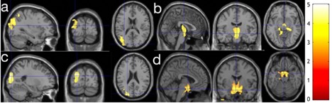
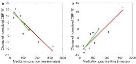
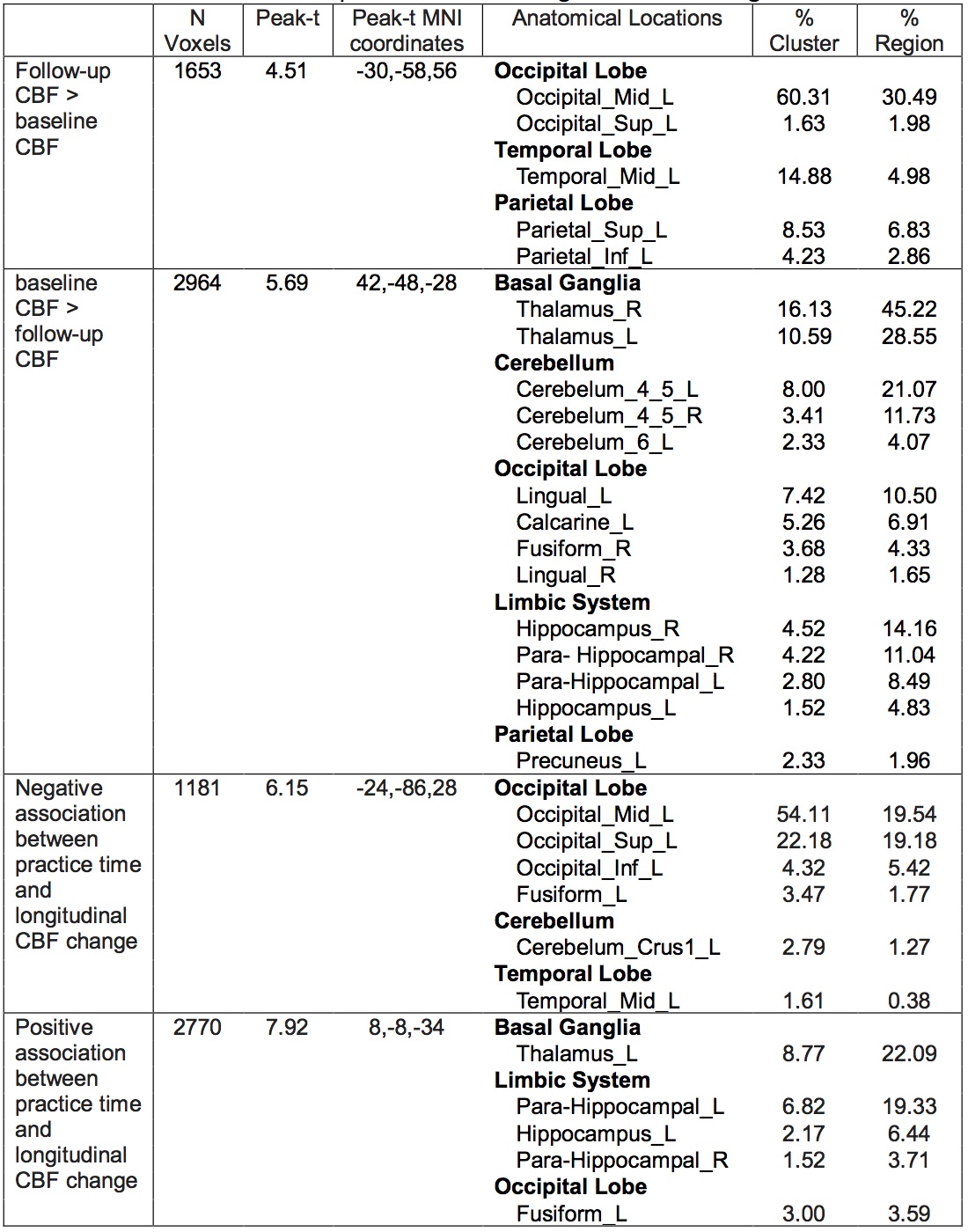
Following 2-month meditation practice, a significant CBF increase was observed mainly in the left occipital region (corrected P = 0.010) (Fig. 1a and Table 1) and a significant CBF decrease was observed primarily in the thalamus region (corrected P < 0.001) (Fig. 1b and Table 1). However, we also found a significant negative association between CBF change and practice time in the left occipital region (corrected P = 0.013) (Fig. 1c and Table 1), and a positive association between CBF change and practice time mainly in the left thalamus region (corrected P = 0.020) (Fig. 1d and Table 1). Regional analysis confirmed the association of the CBF changes in the left occipital and left thalamus region with practice time (Fig. 2). Two subjects have spent a much longer time in meditation than the rest of the subjects. Even after we removed the two subjects from the dataset, the observed association was still significant (Fig. 2). Interestingly, the two subjects who practiced for a much longer time had almost an opposite CBF change from that of the rest of the subjects, which indicates the direction of CBF change may switch after some amount of meditation practice time (in our cohort, approximately 800 to 1000 minutes). The comparisons of CBF functional connectivity and the association with practice time are in progress.Discussion
The baseline CBF increase in the occipital/parietal region and decrease in the thalamus region after 2-month meditation practice agrees with the baseline CBF change in a SPECT study after 8-week meditation practice of patients with memory issues [13]. The baseline CBF increase in the occipital/parietal region has been related to tension [14], supporting the neural change of meditation in handling emotional stress. The association of CBF change in the occipital/parietal and thalamus regions indicates a complex neural pathway of meditation.Acknowledgements
This research was supported by the State University of New York at Binghamton.References
1. Delmonte, M.M., Biochemical indices associated with meditation practice: a literature review. Neurosci Biobehav Rev, 1985. 9(4): p. 557-61.
2. Roemer, L., S.M. Orsillo, and K. Salters-Pedneault, Efficacy of an acceptance-based behavior therapy for generalized anxiety disorder: evaluation in a randomized controlled trial. J Consult Clin Psychol, 2008. 76(6): p. 1083-9.
3. Grossman, P., et al., Mindfulness-based stress reduction and health benefits. A meta-analysis. J Psychosom Res, 2004. 57(1): p. 35-43.
4. Teasdale, J.D., et al., Prevention of relapse/recurrence in major depression by mindfulness-based cognitive therapy. J Consult Clin Psychol, 2000. 68(4): p. 615-23.
5. Bishop, S.R., What do we really know about mindfulness-based stress reduction? Psychosom Med, 2002. 64(1): p. 71-83.
6. Canter, P.H. and E. Ernst, The cumulative effects of Transcendental Meditation on cognitive function--a systematic review of randomised controlled trials. Wien Klin Wochenschr, 2003. 115(21-22): p. 758-66.
7. Lazar, S.W., et al., Functional brain mapping of the relaxation response and meditation. Neuroreport, 2000. 11(7): p. 1581-5.
8. Lou, H.C., et al., A 15O-H2O PET study of meditation and the resting state of normal consciousness. Hum Brain Mapp, 1999. 7(2): p. 98-105.
9. Newberg, A., et al., The measurement of regional cerebral blood flow during the complex cognitive task of meditation: a preliminary SPECT study. Psychiatry Res, 2001. 106(2): p. 113-22.
10. Herzog, H., et al., Changed pattern of regional glucose metabolism during yoga meditative relaxation. Neuropsychobiology, 1990. 23(4): p. 182-7.
11. Dai, W., et al., Quantifying fluctuations of resting state networks using arterial spin labeling perfusion MRI. J Cereb Blood Flow Metab, 2016. 36(3): p. 463-73.
12. Buxton, R.B., et al., A general kinetic model for quantitative perfusion imaging with arterial spin labeling. Magn Reson Med, 1998. 40(3): p. 383-96.
13. Newberg, A.B., et al., Meditation effects on cognitive function and cerebral blood flow in subjects with memory loss: a preliminary study. J Alzheimers Dis, 2010. 20(2): p. 517-26.
14. Moss, A.S., et al., Effects of an 8-week meditation program on mood and anxiety in patients with memory loss. J Altern Complement Med, 2012. 18(1): p. 48-53.
Figures