3780
fMRI of Radiologists with a Diagnosis Task1Biomedical Engineering, Stony Brook University, Stony Brook, NY, United States, 2Radiology, Stony Brook University, Stony Brook, NY, United States
Synopsis
Radiologists display superior perception in the analysis of medical images, but there are currently no fMRI studies of radiologists during a complex diagnosis task. In this study, radiologists read a variety of medical images and chose a diagnosis during fMRI. Compared to age- and education-matched controls, radiologists had lower functional activation in several visuospatial areas, including the lateral ocipital cortex and lingual gyrus.
Introduction
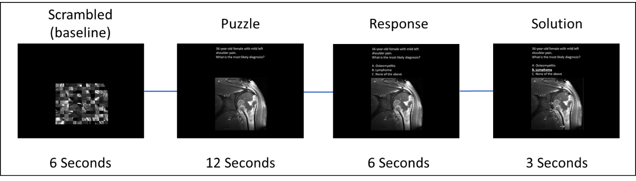
Radiologists train for thousands of hours to be able to recognize subtle abnormalities in medical images. Intense practice in a specific task can lead to structural and functional changes in the brain regions related to that task. While functional changes in the brain associated with sport and music expertise have been well-studied, there have only been a handful of task-based fMRI studies associated with radiology expertise [1, 2]. These two particular functional studies use simple recognition task such as identifying an image as an x-ray or locating lesions on a chest x-ray displayed for 2 seconds. In this novel study we use a problem solving task, where a series of medical images were presented during fMRI acquisition for 12s and participants were asked to quickly choose a diagnosis. This task was designed to engage all of the neural networks that are commonly activated by radiologists in their normal practice by mimicking the day-to-day application of radiology expertise.Methods
Board-certified radiologists (N=12) and age- and education-matched controls (N=16) were recruited for this study. Subjects were shown a series of medical images and then asked to choose a diagnosis. Subjects completed four runs of twenty trials each of the radiology task paradigm summarized in Figure 1. fMRI was acquired on a Siemens clinical 3T scanner with 2.5x2.5x3 mm voxels and TR= 2000 ms. Each run lasted approximately 9 minutes. Image realignment and normalization was performed with the FSL toolbox, and General Linear Model regression analysis was performed with FEAT. Significant activation was defined as a z-score greater than 3.1 and a cluster probability p-value of less than 0.05. Task-related activation was compared between radiologists and non-radiologists during the puzzle phase.Results
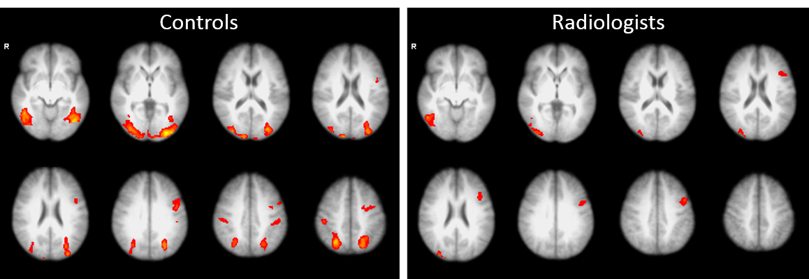
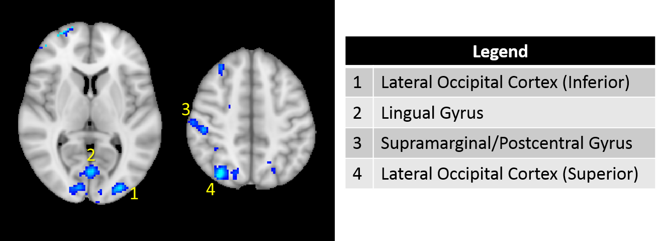
Non-radiologists had higher mean activation than radiologists during the puzzle phase of the task, including in some areas that were not active on average in radiologists (Figure 2). When compared in higher-level analysis, radiologists had significantly lower activation in several areas, including the lateral occipital cortex (inferior and superior divisions), lingual gyrus, and postcentral/supramarginal gyrus (Figure 3).Discussion
Radiologists showed significantly lower activation of visual areas, including the lingual gyrus and lateral occipital cortex. It has been shown that radiologists can locate lung nodules within a fraction of a second with reasonable accuracy [3] , so we may interpret this lowered visual activation as the experts needing less time and/or effort to fully analyze each image. In particular, radiologists displayed less activation in the left inferior lateral occipital cortex, which is associated with object and shape recognition [4]. This disengagement of the lateral occipital cortex also agrees with the findings of Harley et al. in a lung-nodule recognition task [2]. There was also lower activation in the lingual gyrus, which is associated with visual encoding and recall of complex images [5] Lower activation in the lingual gyrus indicates that radiology training affects such encoding and recall with regards to viewing medical images.
Conclusion
Radiology training results in functional changes in primarily visual/spatial networks. The efficiency of these regions while performing a radiology task is higher in trained radiologists than in non-radiologists.Acknowledgements
No acknowledgement found.References
1. Bilalic, M., et al., The Faces in Radiological Images: Fusiform Face Area Supports Radiological Expertise. Cereb Cortex, 2016. 26(3): p. 1004-1014.
2. Harley, E.M., et al., Engagement of fusiform cortex and disengagement of lateral occipital cortex in the acquisition of radiological expertise. Cereb Cortex, 2009. 19(11): p. 2746-54.
3. Kundel, H.L. and C.F. Nodine, Interpreting chest radiographs without visual search. Radiology, 1975. 116(3): p. 527-32.
4. Grill-Spector, K., Z. Kourtzi, and N. Kanwisher, The lateral occipital complex and its role in object recognition. Vision Research, 2001. 41(10): p. 1409-1422.
5. C.M., M.W., et al., fMRI of visual encoding: Reproducibility of activation. Human Brain Mapping, 2000. 9(3): p. 156-164.
Figures