3774
Influence of multiband factor on temporal SNR limits using a clinical BOLD-EPI sequence1Department of Radiology, Ghent University Hospital, Gent, Belgium, 2Department of Diagnostic Sciences, Ghent University, Ghent, Belgium, 3Department of Data Analysis, Ghent University, Ghent, Belgium, 4Department of Experimental Psychology, Ghent University, Ghent, Belgium
Synopsis
Detectability of BOLD signal change is determined by temporal SNR (tSNR, mean divided by standard deviation of the signal over time), and ultimately determines how long needs to be scanned to obtain significant results in a fMRI experiment. With the clinical availability of multiband BOLD-EPI, TR can be shortened significantly, but it is unclear how MB factor influences tSNR. We show in a human subject that tSNR limits up to 192 can be reached and that multiple combinations of MB factor and voxel size can be used to reach similar tSNR values.
Introduction
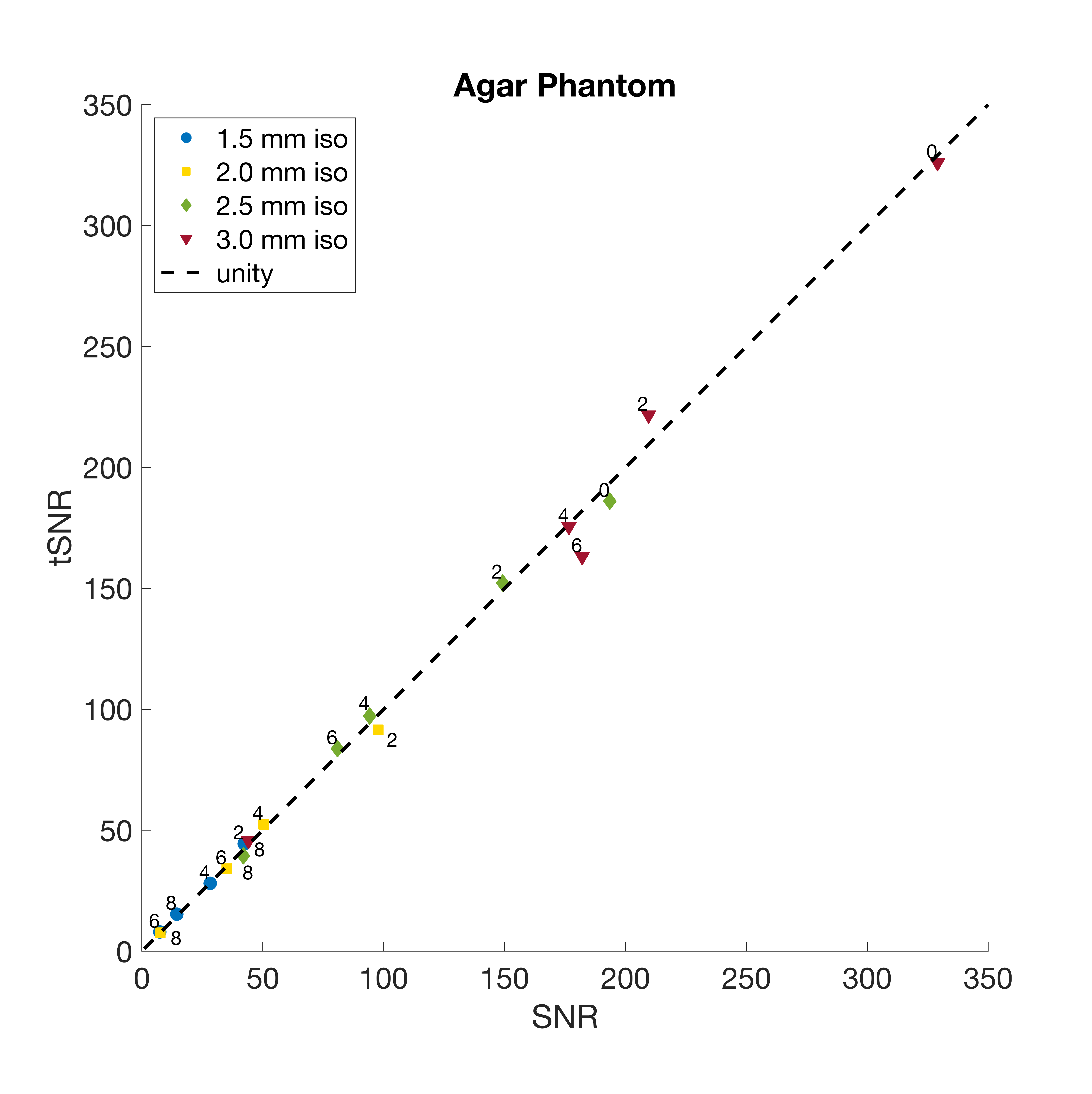
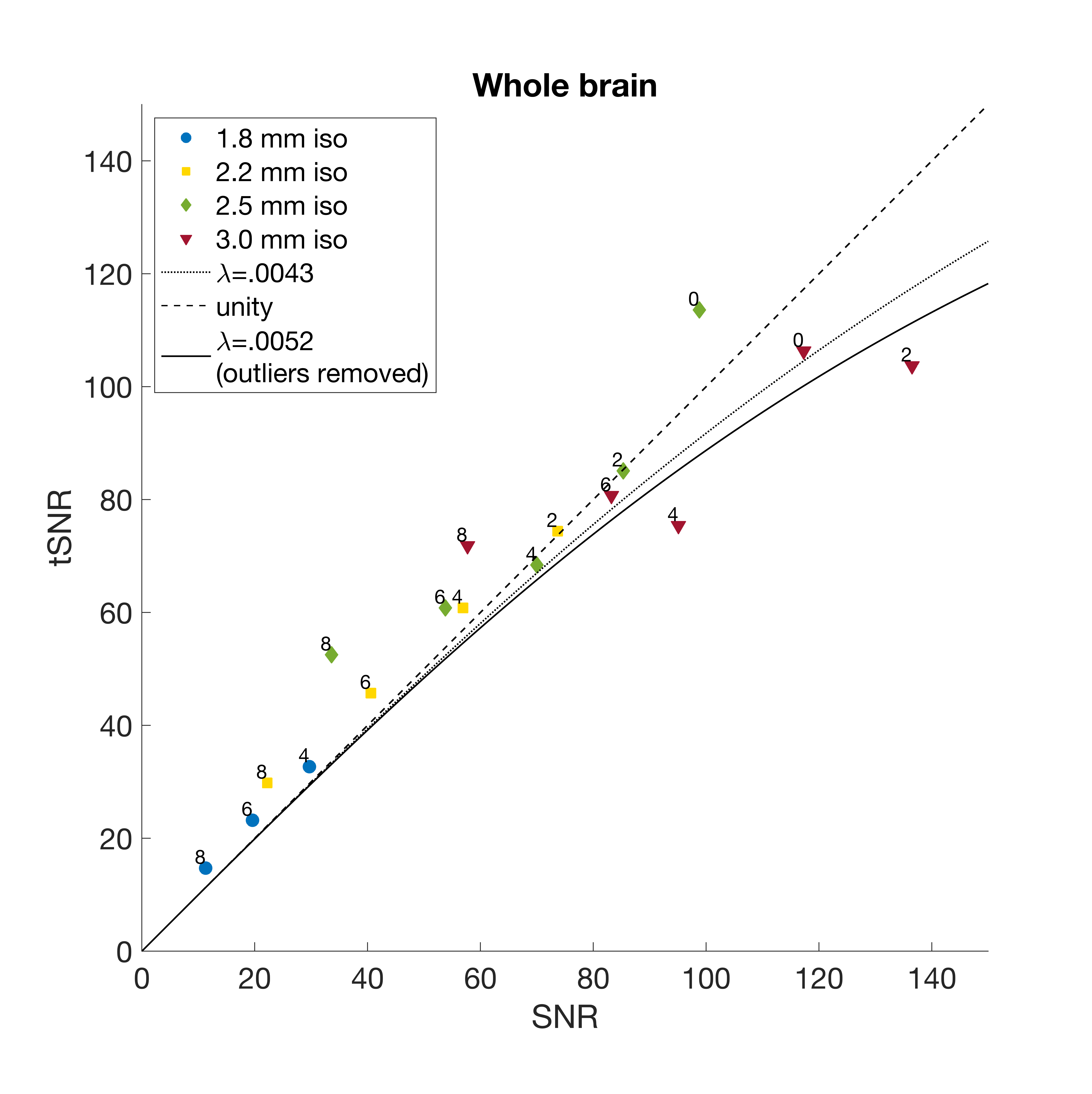
Detectability of BOLD signal change is determined by field strength, echo time and temporal SNR (tSNR, mean divided by standard deviation of the signal over time)[1]. Acquisition parameters such as field strength, voxel size, TE/TR, and multiband factor, influence image SNR (SNR0) and tSNR[2], [3]. Ultimately, tSNR determines the amount of volumes that needs to be scanned in order to obtain significant results from a fMRI experiment [4]. Total noise in a fMRI time series consists of physiological noise (generated by the subject) and thermal noise (generated by the system): $$$\sigma=\sqrt{\sigma_0^2+\sigma_P^2}$$$ [5]. In absence of physiological noise, tSNR and SNR are equal. tSNR can be defined mathematically as $$$tSNR=\frac{SNR_0}{\sqrt{1+\lambda^2\cdot SNR_0}}$$$. It was shown previously in [5] that, when physiological noise is present, tSNR has an asymptotic behaviour as SNR increases, with tSNR upper limit $$$tSNR=1/\lambda$$$ as $$$SNR\rightarrow\infty$$$. This behaviour was studied earlier by changing the resolution, different field strengths [5] or different coils [1] at fixed TRs. With the availability of simultaneous multislice imaging or multiband (MB), however, TR can be shortened significantly. It is not exactly known what the effect of SMS is on tSNR, and what the upper limit of tSNR is at different MB factors. We have investigated the effect of changing MB factor and resolution on SNR0, tSNR and $$$\lambda$$$ in an agar phantom and in a healthy volunteer.Methods
Both the phantom and healthy volunteer (M, 27y) were scanned on a 3T Siemens PrismaFit VE11C AP1, with a 64ch headcoil. Using the Siemens MB-EPI implementation, MB factor was varied from 0, 2, 4, 6 and 8. TR was adjusted to the MB factor, and flip angle was the Ernst angle at each TR. Parallel Fourier of 7/8 or 6/8 was used in MB 4,6,8 to keep TE constant. The experiment was repeated for isotropic resolutions of 1.5, 1.8, 2.0, 2.2., 2.5 and 3.0mm. Not all resolutions could be scanned in the phantom and human subject because of SAR limitations. Per MB factor and resolution, 160 volumes per run were acquired and a T1-weighted MP-RAGE structural brain scan was made for white and gray matter segmentation. Phantom SNR and tSNR were calculated using the bxh/xcede tools 1.11.14 [6] for FBIRN stability analysis using default settings. Healthy subject EPI images were co-registered to the T1w image with FSL’s [7] epi_reg script , which also produces a white and gray matter segmentation. The white and gray matter mask were co-registered back to the EPI images using the inverse transformation matrix using FSL-FLIRT. SNR and tSNR for each EPI timeseries were calculated on the full brain mask and using binarized white and gray matter masks as input to bxh/xcede tools for FBIRN stability analysis. $$$\lambda$$$ was calculated by non-linear least-squares fitting of the function $$$tSNR=\frac{SNR_0}{\sqrt{1+\lambda^2\cdot SNR_0}}$$$ in Matlab R2018a.Results
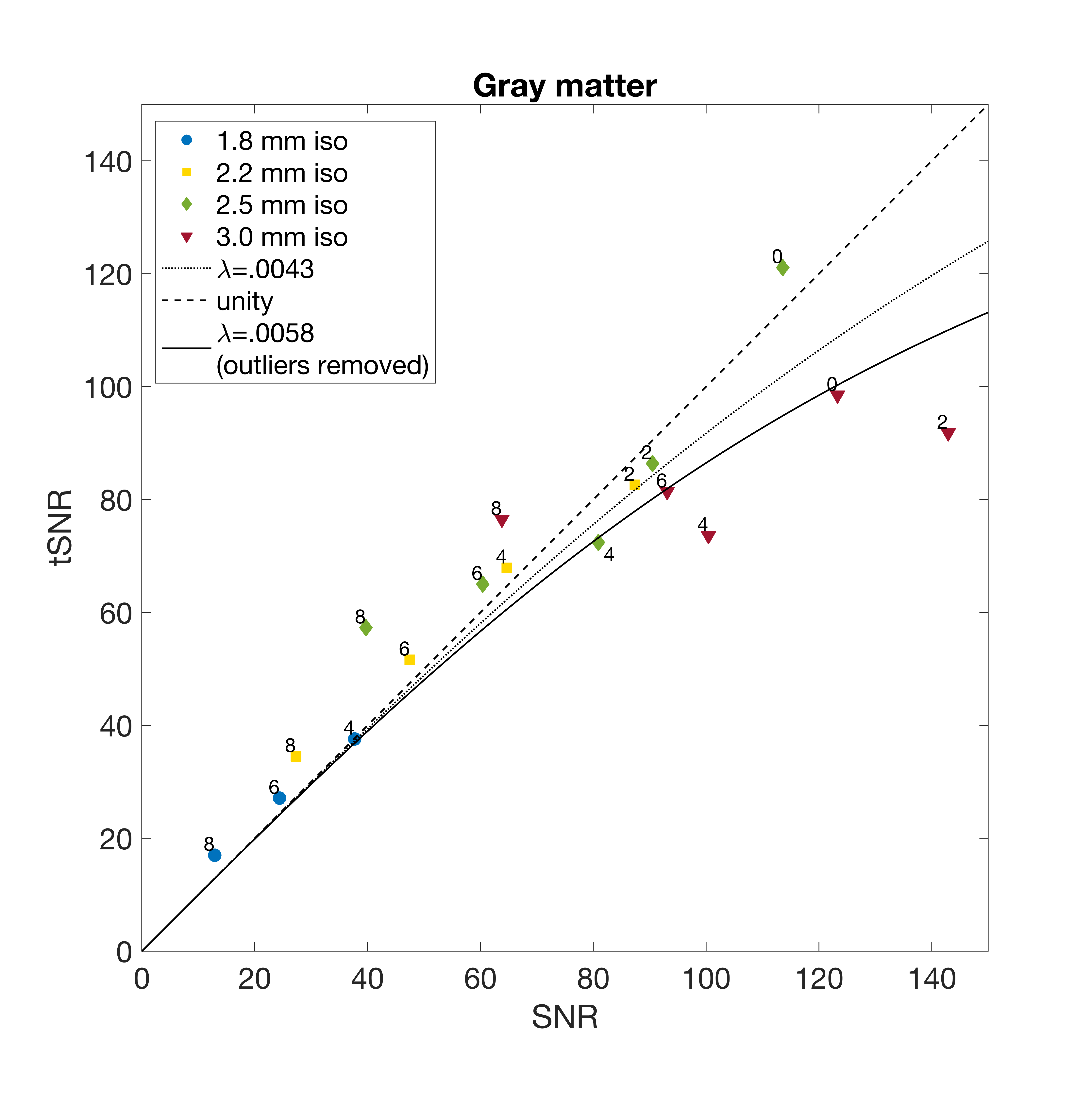
Fig 1 shows SNR vs tSNR in the agar phantom. As expected by theory, in the absence of physiological noise tSNR is equal to SNR for all multiband factors and resolutions. At 3 mm resolution and MB 0, a very high tSNR (325) and SNR of 329 can be achieved. Fig 2 shows whole brain SNR vs tSNR for MB=0,2,4,6,8 and various voxel sizes. Part of the points are located left of the unity line, which is theoretically implausible and likely caused by ghosting artifacts, resulting in an overestimation of image SNR. Highest tSNR of 106 and SNR of 117 is achieved at MB 0 and 3 mm resolution. At increasing multiband factors, but fixed resolution, both SNR and tSNR drop. The upper tSNR limit is 233. Fig 3 shows SNR vs tSNR in gray matter, where again at increasing MB factor, both SNR and tSNR drop. The upper tSNR limit is identical to the whole brain upper tSNR limit of 233. If the most implausible points (MB0, 2.5mm iso and MB8, 2.5 and 3mm iso) are not included in the estimation of $$$\lambda$$$, the whole brain tSNR limit is 192 and thegray matter upper limit is 172.Discussion
With today’s 3T hardware very high tSNR can be achieved. Depending on the application (high image resolution or high temporal resolution) multiple combinations of MB factor and resolution are possible to reach a desired tSNR. The observed upper tSNR limit of 172 to 192 is considerably higher than the upper tSNR limit of 124 that was reported earlier in [1], which potentially can lead to shorter fMRI trials. However, improvements need to be made in the SNR calculations and confidence intervals need to be calculated for SNR, tSNR and the $$$\lambda$$$ estimates, which is not possible with the currently used tools. Secondly, our measurements need to be confirmed in multiple subjects.Acknowledgements
References
[1] C. Triantafyllou, J. R. Polimeni, and L. L. Wald, “Physiological noise and signal-to-noise ratio in fMRI with multi-channel array coils,” Neuroimage, vol. 55, no. 2, pp. 597–606, Mar. 2011.
[2] M. Barth, F. Breuer, P. J. Koopmans, D. G. Norris, and B. A. Poser, “Simultaneous multislice (SMS) imaging techniques,” Magn. Reson. Med., vol. 75, no. 1, pp. 63–81, 2016.
[3] L. Chen et al., “Evaluation of highly accelerated simultaneous multi-slice EPI for fMRI,” Neuroimage, vol. 104, pp. 452–459, Jan. 2015.
[4] K. Murphy, J. Bodurka, and P. A. Bandettini, “How long to scan? The relationship between fMRI temporal signal to noise ratio and necessary scan duration,” Neuroimage, vol. 34, no. 2, pp. 565–574, Jan. 2007.
[5] C. Triantafyllou et al., “Comparison of physiological noise at 1.5 T, 3 T and 7 T and optimization of fMRI acquisition parameters,” Neuroimage, vol. 26, no. 1, pp. 243–250, May 2005.
[6] “BXH/XCEDE tools.” NITRC. https://www.nitrc.org/projects/bxh_xcede_tools/
[7] S. M. S. Smithet al., “Advances in functional and structural MR image analysis and implementation as FSL.,” Neuroimage, vol. 23 Suppl 1, pp. S208-19, Jan. 2004.
Figures