3771
Dynamic Model-based Reconstruction for Oscillating Steady State fMRI1Biomedical Engineering, University of Michigan, Ann Arbor, MI, United States
Synopsis
We introduce Oscillating Steady State (OSS) fMRI, a method that uses quadratic phase cycling and balanced gradients to create an oscillating T2* sensitive signal. We also present a model-based reconstruction that converts these signal oscillations into B0 estimates, enabling correction for respiratory or scanner induced signal changes. OSS fMRI enables the SNR benefits of bSSFP-based methods, while adding T2* sensitivity and B0 encoding into a unique oscillating signal.
Introduction
Balanced steady state free precession (bSSFP) has seen widespread clinical adoption, having the highest SNR efficiency of all pulse sequences1. However it has yet to catch on within the fMRI community because of limited BOLD contrast due to T2 rather than T2*-weighting, strong sensitivity to scanner or respiratory induced B0 shifts, and non-uniform spectral sensitivity leading to the need for linear phase cycling2. To address both T2* sensitivity and the frequency sensitivity, we propose Oscillating Steady State (OSS) fMRI: a novel SSFP technique which uses quadratic phase cycling to create frequency-invariant T2* sensitivity, even with long TRs and an unstable B0 field. While quadratic phase cycling with gradient dephasing ordinarily produces RF spoiling3, quadratic phase in conjunction with balanced gradients creates high SNR images with spatially varying oscillations. Herein, we present a method which simultaneously converts these oscillations into a stable signal and frame-by-frame B0 maps using a dynamic model-based dictionary matching reconstruction, resulting in a T2* weighted signal which is robust against temporal and spatial B0 variation.Methods
An oscillating steady-state of period $$$n_c$$$ was created by linearly increasing the phase increment between RF pulses by $$$\phi(n) - \phi(n-1)=\frac{2\pi}{n_c}n$$$, a pattern which is periodic for $$$n_c$$$ TRs. For large phase steps ($$$n_c < 15$$$), the resulting oscillating signal becomes T2*-weighted across its entire frequency response. As shown by Bloch simulation, these oscillations vary non-linearly in shape and magnitude as a function of B0, and are thus non-trivial to remove using traditional filtering methods. To address this, we implemented a reconstruction that uses a pre-computed dictionary of complex signals to match each period of unique oscillations to their latent B0 values, enabling reconstruction of a stable signal magnitude despite time varying B0. First, our method was used to correct worst-case respiration drift in an in vivo time course, where the human subject was instructed to inhale and exhale to maximum at 5 second intervals, indicated by visual cue. Next, our method was used to correct a functional time series of a motor task, consisting of 20 second blocks of finger tapping and rest. All experiments were performed on a 3T GE MR 750 scanner using 2-shot balanced EPI encoding, with matrix = 64x64 and a TR of 22.5 ms.Results
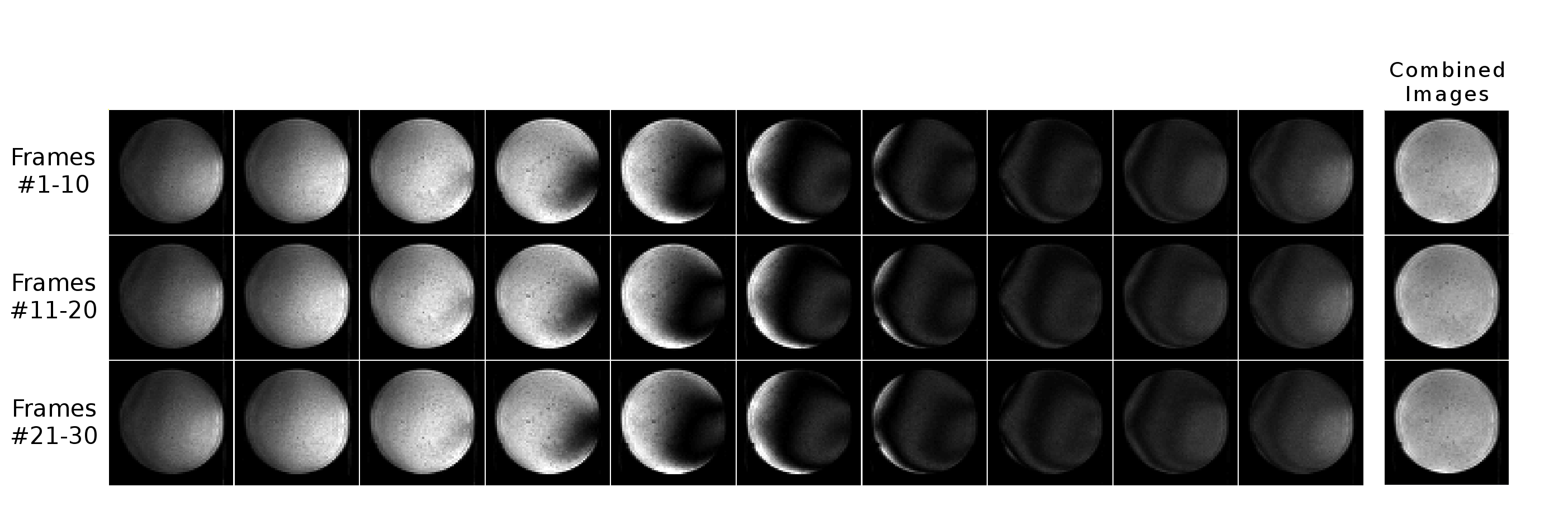
Fig. 1 shows the first 30 TRs of a phantom time course, demonstrating spatial variation of oscillations due to imperfect B0 shim.
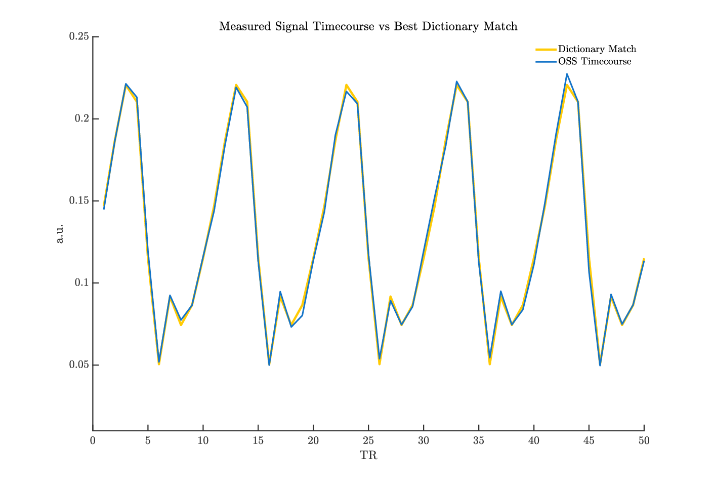
Fig. 2 shows a 50 TR time course in a phantom compared to best dictionary match, with very good agreement of the model.
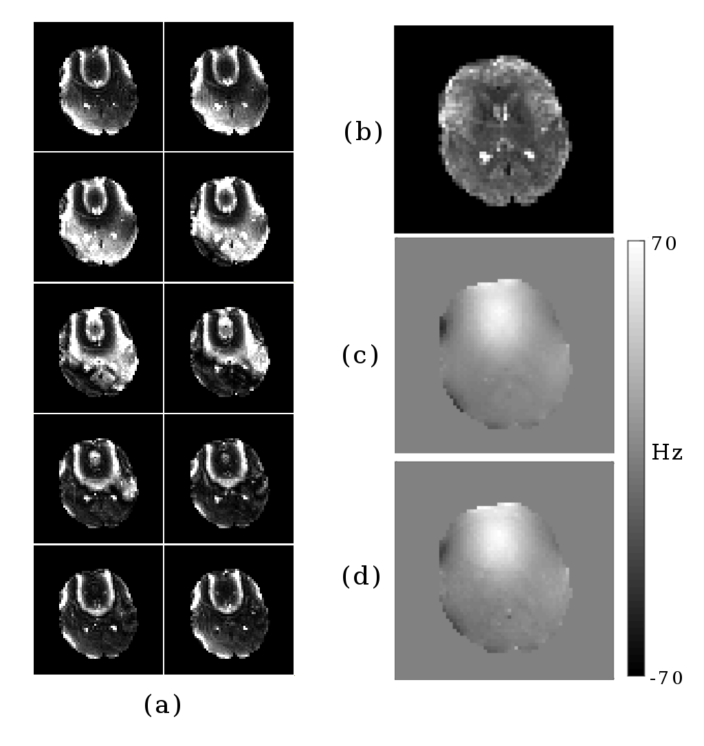
Fig. 3 shows a series of individual in vivo images from one oscillation period, with the recovered magnitude and B0 images.
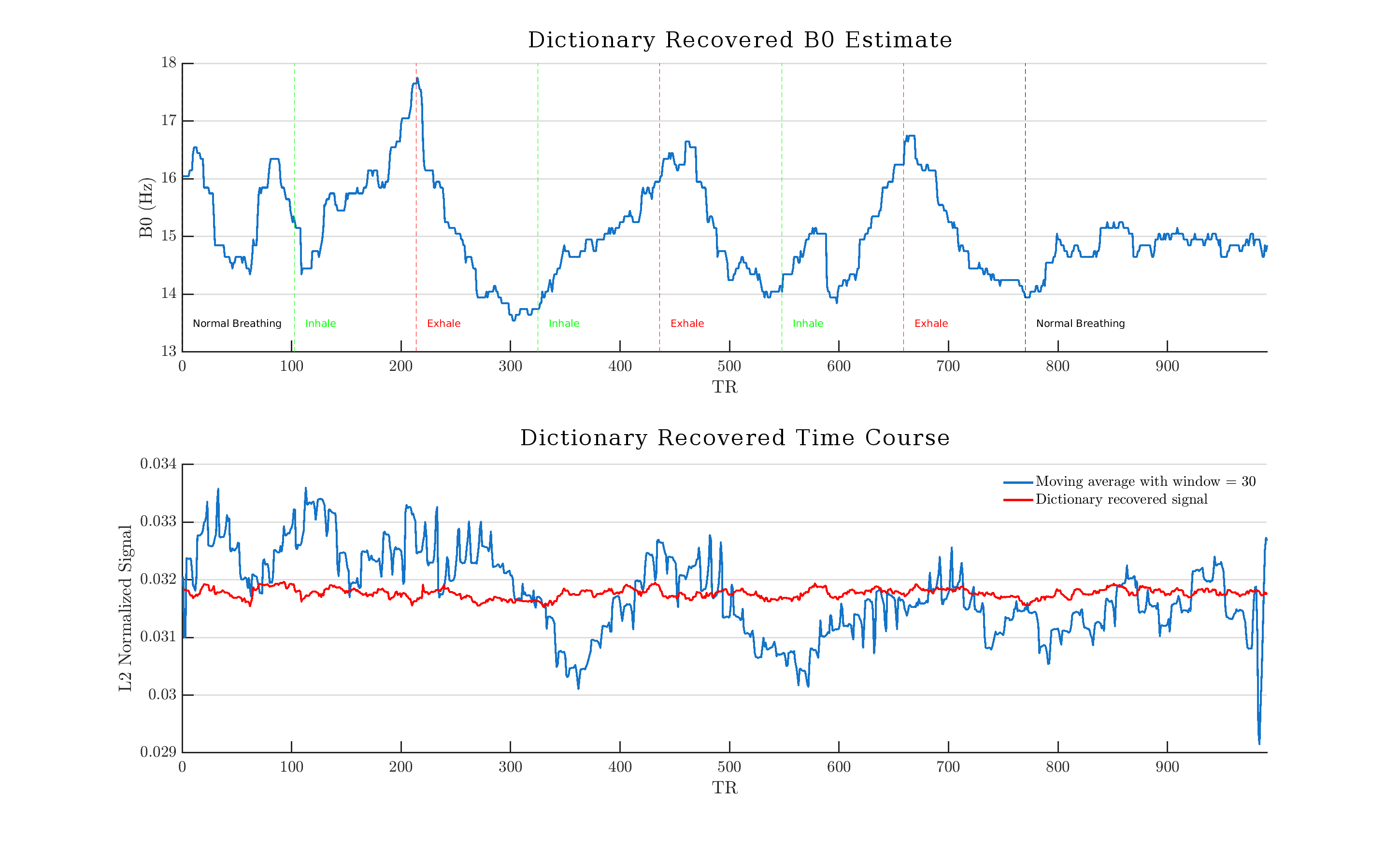
Fig. 4 shows a reconstructed magnitude and B0 time course during severe respiratory induced B0 distortion.
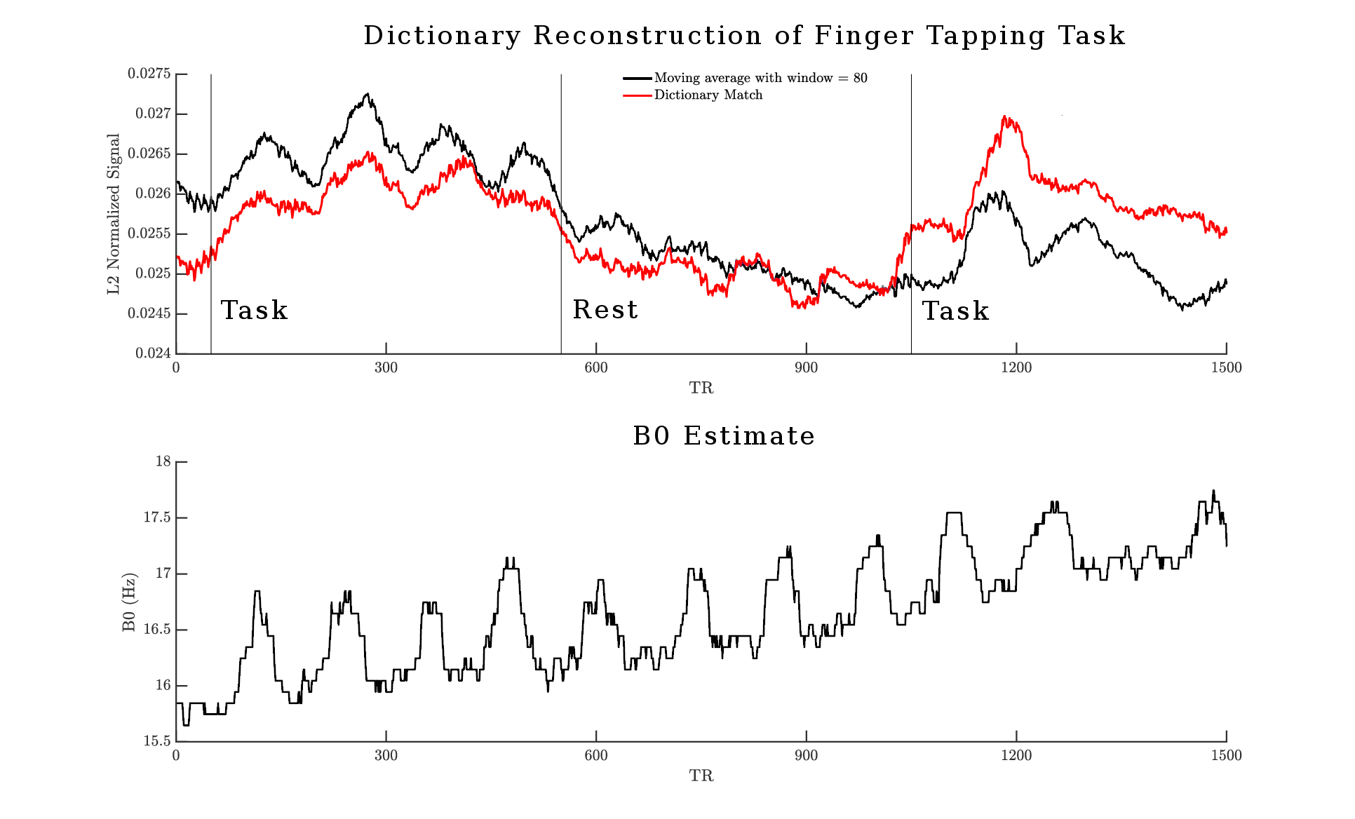
Fig. 5 shows a reconstructed magnitude and B0 time course during activation from a functional motor task.
Discussion
A proof-of-concept dictionary-based reconstruction is able to dynamically retrieve latent signal parameters from a oscillating steady state signal. Though scanner / respiration B0 drift affect the oscillations, computational modeling and data demonstrate that spins rapidly adjust to modest field changes with transient response less than one full oscillation cycle. This enables a quasi-static modeling approach to accurately capture and compensate for temporally varying field strength, and overcome a primary limitation of bSSFP for fMRI applications.
Furthermore, this method is able to accurately estimate voxel-level B0 values over time, despite modeling the signal behavior as a single isochromat. The underlying assumption is that shifts in intervoxel inhomogeneity result in signal magnitude changes, but will still retain the same relative shape. The dictionary therefor only models and corrects for macroscopic field variation, while maintaining T2* sensitivity.
The current implementation reconstructs all oscillation periods independently, and does not utilize parameter regularization. Further development in the reconstruction of OSS signals will be able to harness the smoothness of the B0 field in up to 3 spatial / 1 temporal dimension.
Conclusion
This work demonstrates that OSS fMRI retains the desirable SNR efficiency of bSSFP, while adding T2* weighting and the ability to encodes B0 information into a unique an oscillating magnetization. These oscillations can then be dynamically converted into B0 estimates and then used to compensate for the deleterious effects of respiration or scanner drift.Acknowledgements
We wish to acknowledge the support of NIH Grants R01EB023618 and U01EB026977.References
1. Klaus Scheffler and Stefan Lehnhardt. Principles and applications of balanced SSFP techniques. Eur Radiol, 13:2409–2418, 2003. doi: 10.1007/s00330-003-1957-x.
2. Karla L Miller. FMRI using balanced steady-state free precession (SSFP). NeuroImage, 62(2):713–9, 8 2012. ISSN 1095-9572. doi: 10.1016/j.neuroimage.2011.10.040.
3. Y. Zur, M. L. Wood, and L. J. Neuringer. Spoiling of transverse magnetization in steady-state sequences. Magnetic Resonance in Medicine, 21(2):251–263, 10 1991. ISSN 07403194. doi: 10.1002/mrm.1910210210.
Figures