3770
MP-PCA denoising to improve detection of task-based fMRI activation in brain tumor patients1Radiology, Center for Biomedical Imaging, New York University School of Medicine, New York, NY, United States
Synopsis
Functional MRI improves preoperative planning in brain tumor patients, however BOLD signal changes for task-based 3T fMRI are only 2-4% and the tumor often compromises patient performance. This retrospective study evaluated Marchenko-Pastur Principle Component Analysis (MP-PCA) denoising to increase sensitivity for cortical regions recruited during language task-based fMRI paradigms in brain tumor patients. MP-PCA denoising is shown to improve the clinical value and practical utility of performing preoperative fMRI in brain tumor patients.
Introduction
In fMRI, the BOLD effect typically accounts for only 2-4% of signal amplitude at 3T, and the remaining 95-98% of fluctuations are contributions from outside influences1. Besides neurovascular coupling related to cognition performing the task, the BOLD signal may be altered by head motion, cardiac pulsation noise, respiratory noise, low frequency changes in oxygenation, venous flow signal contamination and B0 field drift1. Furthermore, task-based fMRI also can suffer from low SNR when patient responses are compromised by sedation, altered cognition or neurovascular uncoupling from the underlying tumor2,3.
Marchenko-Pastur-Principal-Component-Analysis (MP-PCA) is a model-free noise removal method4 that exploits redundancy in MRI series, thereby improving the precision of parameter estimators5. This study tested the hypothesis that task-based fMRI SNR and Z-score sensitivity can be improved using MP-PCA in patients undergoing imaging for neurosurgical planning for glial neoplasms. We compared fMRI histograms and residuals in four common fMRI paradigms commonly used for language and motor mapping from a tertiary care clinical pre-operative brain mapping service to measure the magnitude and distribution of activated signal gained from denoising2.
Methods
This retrospective analysis was approved by institutional review board and included 23 brain tumor patients (13 female, mean age = 42.5 ± 18.1) that underwent verb generation, sentence completion and listening comprehension task-based fMRI for language assessment3, along with a sequential finger movement motor tasks using the hand contralateral to the tumor location. All tasks consisted of a 20-second on/off block design. Imaging was performed on a Siemens Magnetom Skyra 3-T scanner (Siemens Healthineers, Erlangen, Germany). All task-based fMRI data were acquired prior to contrast administration (Gradient echo EPI acquisitions with 160 time points, TR/TE = 1000/300ms, flip angle 62°, resolution of 3×3×3mm, matrix size = 66×66×34 voxels).
Raw fMRI data was processed using MP-PCA in MATLAB. Both raw and denoised volumes underwent motion correction, slice timing correction, brain extraction, spatial smoothing, high pass temporal filtering and BOLD signal modeling using FSL-FEAT6. A board-certified neuroradiologist with 5 year’s fMRI experience placed a single voxel seed on the surface of the precentral gyrus hand knob, pars triangularis and planum temporale (near Broca’s and Wernicke’s areas respectively), and medial frontal gyrus (in the coronal plan of the anterior commissure). The seeds were inflated to 20mm radius spherical ROIs to ensure adequate anatomical coverage.
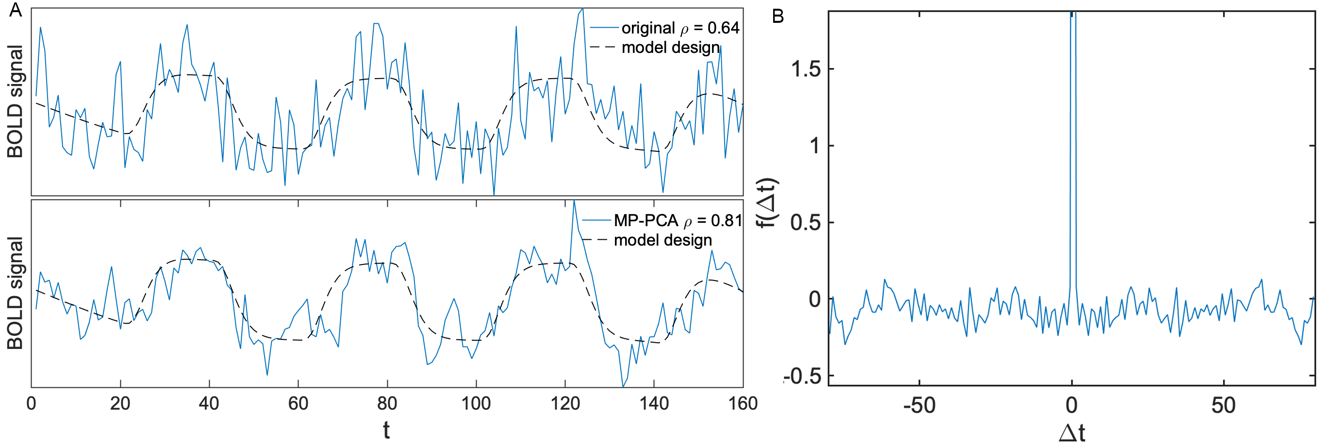
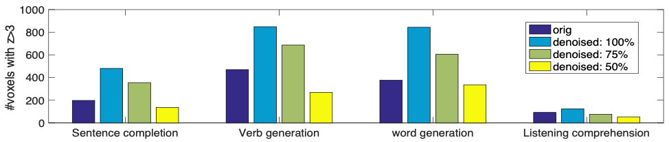
Z-score is used as a measure of task-related cortical activation and histograms of Z-scores within each region were compared to the contra-lateral homolog. Paired 2-sided t-tests determined whether mean activation in each region was different between groups. We tested the capacity of MP-PCA to reduce the scan duration required to reach the same level of sensitivity as non-denoised data by truncating a representative case 74 y/o female brain tumor patient to 50%, 75%, and 100% of the length of the raw dataset. We compared the sensitivity of resulting maps using the number of voxels with Z>3. We also test the accuracy of fMRI denoising using the temporal correlation function given by $$$f(\Delta t) = \int dt \left\langle\epsilon(t) (\epsilon(t+\Delta t))\right\rangle$$$ where $$$\epsilon(t)$$$ = denoised – original.
Results
Figure 1A shows the fMRI signal before and after denoising, demonstrating a 60% increase in SNR, and 1B shows the temporal correlation function of residual signal as a function of lag time. The memoryless Poissonian distribution of the residuals shows that MP-PCA is removing only thermal noise and no task related temporal fluctations.
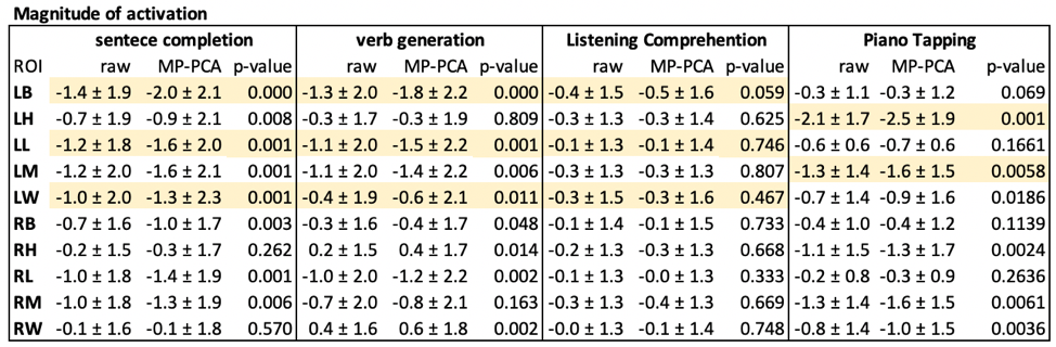
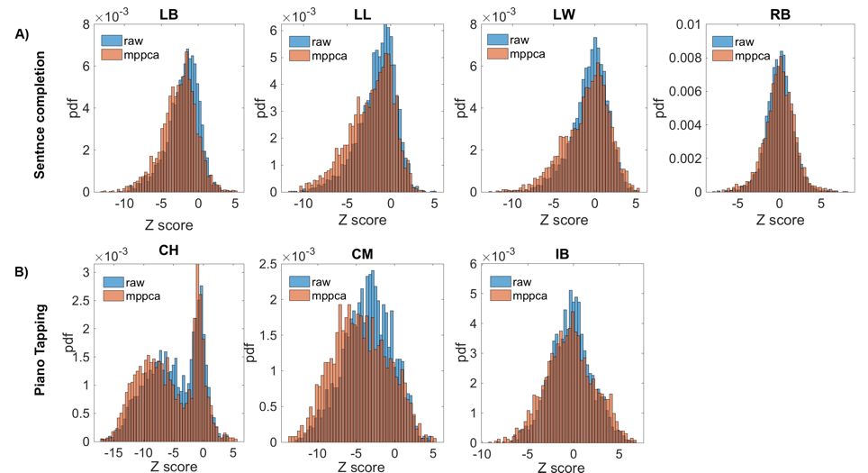
Denoising resulted in increased volume of task-related activation (volume of voxels with |Z|>3) and increased Z-score magnitude for region-specific correlations with all language tasks. Figure 2 shows histograms over all subjects for sentence completion (2A) and finger tapping (2B) for regions where we expect to see task-related activation along with control regions. Table 1 shows mean and standard deviation z-scores within each region analyzed, as well as p-values showing paired t-test results. For sentence completion, denoised data showed 42% greater activation (p=0.001) and 30% greater activation (p=0.001) in Broca’s and Wernicke’s regions respectively.
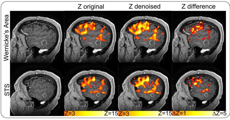
Figure 3 shows Z-statistic maps overlaid on an MP-RAGE for a representative subject with a brain tumor in the posterior-left-middle-temporal-gyrus near receptive language regions while performing the sentence completion task. There is increased activation in the cortex adjacent to the tumor after the images have undergone processing with MP-PCA. Figure 4 shows the sensitivity of activation maps when varying the amount of time series data used for denoising. We show here that language tasks can be reduced by about 40% without loss of sensitivity compared to the non-denoised fMRI data.
Discussion
We$$$\,$$$demonstrate$$$\,$$$here$$$\,$$$that$$$\,$$$MP-PCA$$$\,$$$is$$$\,$$$capable$$$\,$$$of$$$\,$$$increasing$$$\,$$$the$$$\,$$$SNR$$$\,$$$of$$$\,$$$raw$$$\,$$$fMRI$$$\,$$$data$$$\,$$$by$$$\,$$$a$$$\,$$$large$$$\,$$$degree$$$\,$$$and$$$\,$$$does$$$\,$$$so$$$\,$$$with$$$\,$$$high$$$\,$$$accuracy.$$$\,$$$This$$$\,$$$increased$$$\,$$$SNR$$$\,$$$leads$$$\,$$$to$$$\,$$$activation$$$\,$$$maps$$$\,$$$that$$$\,$$$are$$$\,$$$more$$$\,$$$sensitive$$$\,$$$to$$$\,$$$tasks$$$\,$$$(particularly$$$\,$$$in$$$\,$$$regions$$$\,$$$with$$$\,$$$pathology)$$$\,$$$and$$$\,$$$can$$$\,$$$detect$$$\,$$$activation$$$\,$$$that$$$\,$$$may$$$\,$$$have$$$\,$$$previously$$$\,$$$been$$$\,$$$invisible$$$\,$$$due$$$\,$$$to$$$\,$$$noise.$$$\,$$$Finally$$$\,$$$we$$$\,$$$show$$$\,$$$that$$$\,$$$MP-PCA$$$\,$$$can$$$\,$$$reduce$$$\,$$$the$$$\,$$$duration$$$\,$$$required$$$\,$$$for$$$\,$$$adequate$$$\,$$$intraoperative$$$\,$$$mapping$$$\,$$$in$$$\,$$$patients$$$\,$$$with$$$\,$$$tumors$$$\,$$$near$$$\,$$$eloquent$$$\,$$$language$$$\,$$$cortical$$$\,$$$regions$$$\,$$$by$$$\,$$$about$$$\,$$$40%.
Conclusion
Comparison of raw and denoised activation maps demonstrates increased sensitivity for MP-PCA processed fMRI to detect cortical regions involved in language production and motor function.Acknowledgements
Research was supported by The National Institute of Neurological Disorders and Stroke of the NIH under award number R01 NS088040, and was performed at the Center of Advanced Imaging Innovation and Research (CAI2R, www.cai2r.net), and NIBIB Biomedical Technology Resource Center P41 EB017183.References
1. Caballero-Gaudes, C., & Reynolds, R. C. (2017). Methods for cleaning the BOLD fMRI signal. Neuroimage, 154, 128-149.
2. Zacà, D., Nickerson, J. P., Deib, G., & Pillai, J. J. (2012). Effectiveness of four different clinical fMRI paradigms for preoperative regional determination of language lateralization in patients with brain tumors. Neuroradiology, 54(9), 1015-1025.
3. Stippich, C., Rapps, N., Dreyhaupt, J., Durst, A., Kress, B., Nennig, E., ... & Sartor, K. (2007). Localizing and lateralizing language in patients with brain tumors: feasibility of routine preoperative functional MR imaging in 81 consecutive patients. Radiology, 243(3), 828-836.
4. Veraart, J., Fieremans, E., & Novikov, D. S. (2016). Diffusion MRI noise mapping using random matrix theory. Magnetic resonance in medicine, 76(5), 1582-1593.
5. Ades-Aron, B., Veraart, J., Kochunov, P., McGuire, S., Sherman, P., Kellner, E., ... & Fieremans, E. (2018). Evaluation of the accuracy and precision of the diffusion parameter EStImation with Gibbs and NoisE removal pipeline. NeuroImage, 183, 532-543.
6. Woolrich, M. W., Ripley, B. D., Brady, M., & Smith, S. M. (2001). Temporal autocorrelation in univariate linear modeling of FMRI data. NeuroImage, 14(6), 1370–1386.
Figures