3768
Voxel-Specific Modeling of Physiological Fluctuations in BOLD fMRI Signal1University of California, Irvine, CA, United States
Synopsis
Cardiac pulsation and respiration have significant contributions to the BOLD signal. This is particularly challenging given the long TRs typically used in BOLD experiments since these fluctuations are at higher frequency than the sampling rate and therefore aliased to lower frequency components. This study presents a new voxel-specific method accounting for the physiological effects in BOLD time series signal. We show that this approach is able to improve the estimation of physiological effects compared to the widely used RETROICOR method.
Introduction
Cardiac pulsation and respiration have been shown to produce significant contributions to the BOLD signal(1,2). Particularly for BOLD experiment with long TR (~2s), the challenges are to identify and to remove those effects since generally they are fluctuating at a higher frequency than the BOLD signal. This study aims to propose a new voxel-based method using a quasi-sinusoidal function to assess and reduce those effects in the BOLD signal. To assess the performance of the proposed method, we also implemented the widely used RETROICOR method for comparison (3). Our new method results in better estimation of the physiological effects.Methods
MRI scans were performed on 5 healthy subjects (all males, 37±11 years) on a Siemens Prisma 3T scanner. Anatomical MPRAGE, resting-state, and task-based BOLD (TR=2s) MRI data were collected using parameters as previously described (4). During the task-based BOLD MRI, a flashing black-and-white checkerboard was presented 8 times with 30 s on and 30 s off periods. In addition, we also collected resting-state fMRI with short TR (287ms) on one of the subjects. Cardiac and respiratory pulsations were monitored using the scanner’s built-in photoplethysmograph and respiratory belt.
To assess the voxel-specific aliasing effects of lower frequency components of the BOLD signal due to the cardiac and respiratory pulsations, the BOLD time series data was reordered according to the phase lag relative to the cardiac and respiratory cycle. It was assumed that the fluctuations of BOLD signal follow those physiological pulsations. The phase distribution of BOLD samples was than fitted with a cosine function to assess the amplitude and phase of the cardiac and respiratory contributions to the BOLD signal. The BOLD images with and without physiological correction were then co-registered and processed using AFNI software (5). In addition, we implemented the RETROICOR method (order=1) to assess physiological contributions for comparison.
Results & Discussion
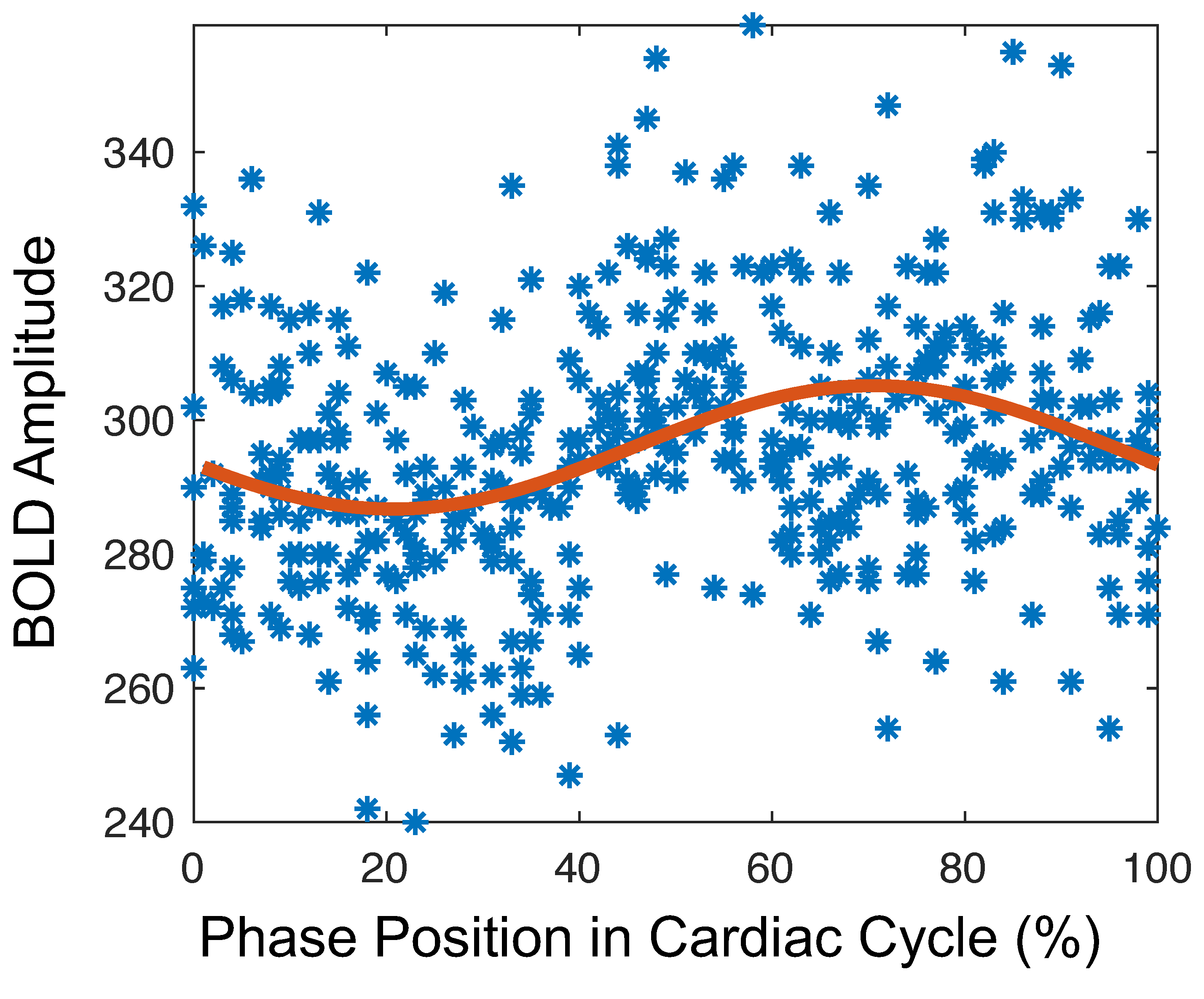
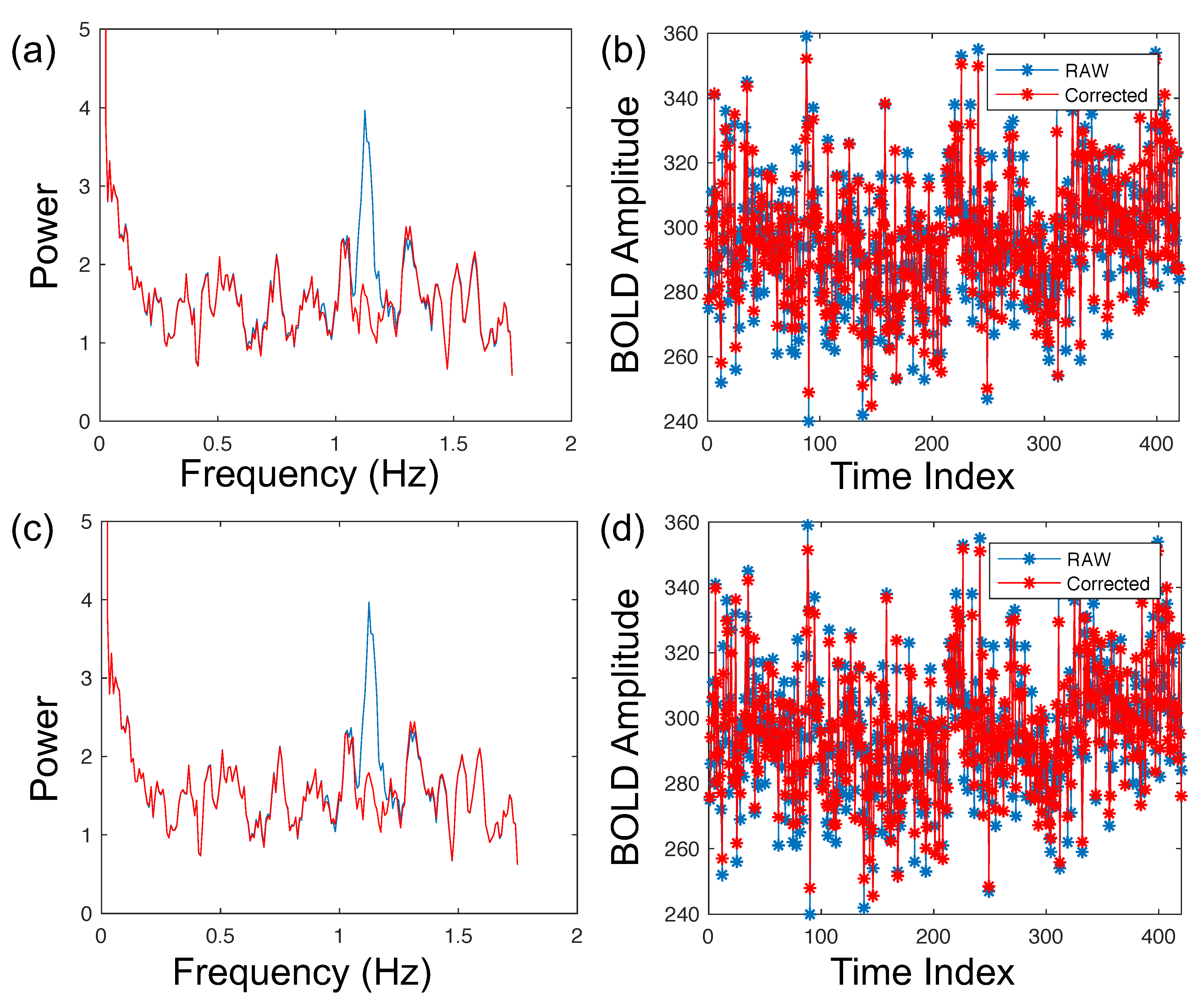
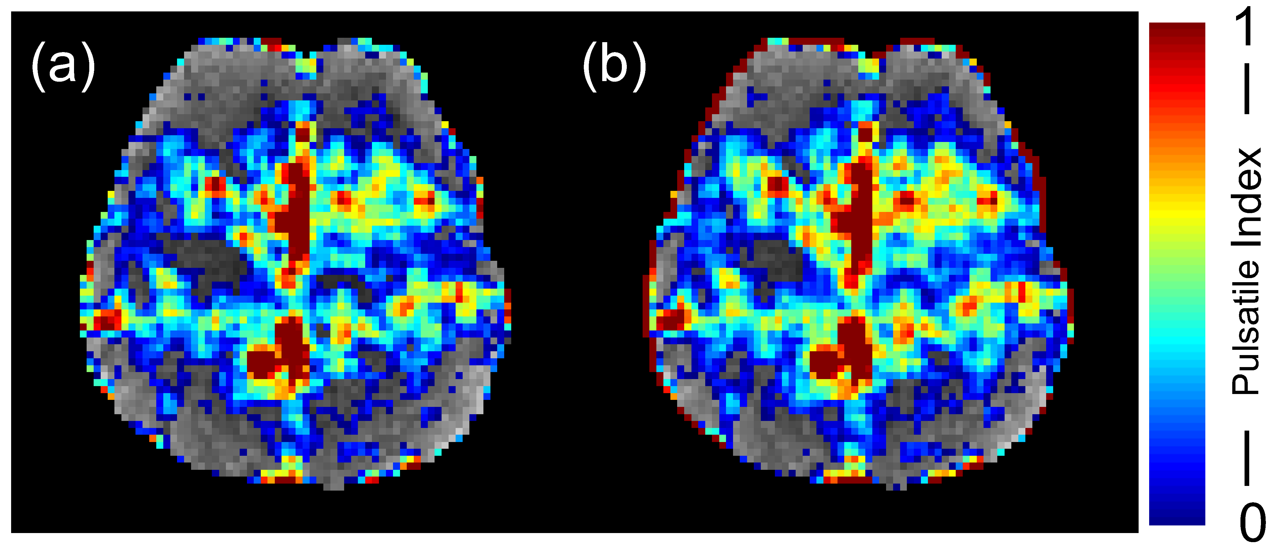
In brain regions that were susceptible to physiological pulsations, the signal fluctuations were either in-phase or lagged relative to the physiological pulsation cycles. Fig. 1 shows a representative BOLD signal in a voxel that fluctuated significantly due to cardiac activities, collected using the short TR data that allows us to view the modulation without aliasing. By averaging the data time-locked to the cardiac phase, we can see that the amplitude and phase of the cardiac effect in BOLD data can be estimated by a quasi-cosine function (Fig. 1). Fig. 2 shows the power spectrum of BOLD signal from the same voxel shown in Fig. 1 before and after the cardiac effect correction. These results demonstrate that the cardiac pulsation effect was reduced effectively by both our approach and the RETROICOR method (Fig. 2). The number of cardiac effected voxels across the entire BOLD data set shows no significant difference from the RETROICOR method (p>0.05); however, the amplitude of the estimated pulsation effect using our method is smaller than when using RETROICOR. Representative maps of the cardiac effected voxels are shown in Fig. 3. These results indicate that our approach is capable of modeling the pulsatile cardiac activity in the BOLD time series data.
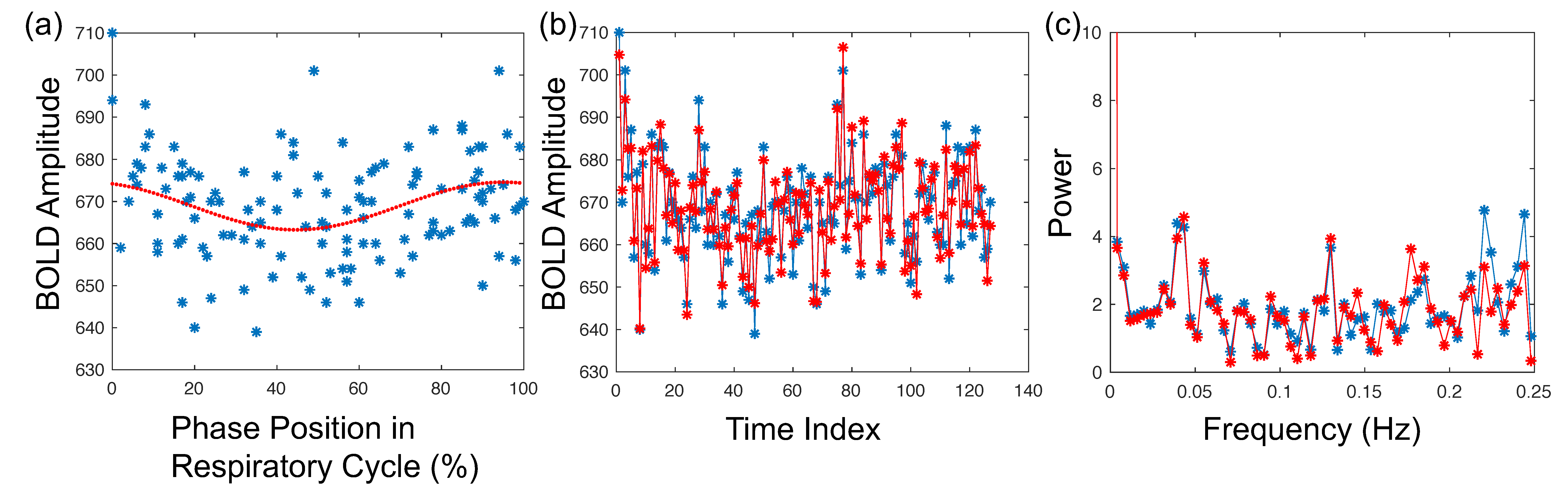
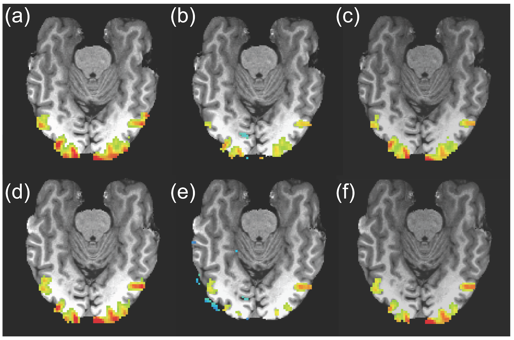
We also found that our approach can effectively remove both the cardiac and respiratory contamination at a lower frequency of BOLD signal acquired using a long TR (~2s). The assessment of the respiratory effect of a representative voxel in the temporal and frequency domains is shown in Fig. 4. For the task-based runs, the application of both our approach and the RETROICOR resulted in significantly fewer activated voxels compared to no corrections for physiological effects (p<0.05) (Fig. 5). Note that there is no significant difference in activated voxels between our approach and the RETROICOR. However, some of image slices after applying the RETROICOR method show fewer activated voxels compared to our approach (Fig. 5). This may imply that our voxel-based approach can provide better estimation of the physiological effect.
Conclusion
In summary, the proposed voxel-based method can provide substantial reduction of additive effect accounting for the physiological components in BOLD signal as confirmed by the results obtained from the long- and short-TR scans as well as from the resting and event-related fMRI experiments. We also suggest that this method may provide better assessment of physiological effects that contaminate the BOLD signal.
Acknowledgements
No acknowledgement found.References
1. Birn RM. The role of physiological noise in resting-state functional connectivity. Neuroimage 2012;62(2):864-870.
2. Caballero-Gaudes C, Reynolds RC. Methods for cleaning the BOLD fMRI signal. Neuroimage 2017;154:128-149.
3. Glover GH, Li TQ, Ress D. Image-based method for retrospective correction of physiological motion effects in fMRI: RETROICOR. Magn Reson Med 2000;44(1):162-167.
4. Stark SM, Frithsen A, Mattfeld AT, Stark CEL. Modulation of associative learning in the hippocampal-striatal circuit based on item-set similarity. Cortex 2018;109:60-73.
5. Cox RW. AFNI: software for analysis and visualization of functional magnetic resonance neuroimages. Comput Biomed Res 1996;29(3):162-173.
Figures