3765
Longitudinal study of BOLD HRF in human cortex1Neuroscience, Baylor College of Medicine, Houston, TX, United States, 2Baylor College of Medicine, Houston, TX, United States
Synopsis
A simple audiovisual stimulus combined with a fast-paced task evoked a strong HRF across a majority of cerebral cortex during four scanning sessions to measure the repeatability of BOLD over time. HRFs were characterized by spatial and temporal parameters and showed remarkable correlation across the time points, which spanned three hours, three days, and three months. Results showed that HRFs are repeatable for consistent quantification of neurovascular coupling.
Introduction
In previous studies, BOLD fMRI characterization revealed hemodynamic response functions (HRFs) with significant variation in magnitude and profile across subjects and sessions1,2. However, these experiments were performed in small regions of cortex and in large voxels. In our previous work, we found consistent spatial patterns across many healthy subjects by using a brief stimulus and high functional resolution across whole cortex3. Sensory regions, driven heavily by our multimodal stimulus, showed strong activation amplitudes and temporal dynamics. To quantify neurovascular function across the brain, HRF measurements should be consistent within a healthy subject over time. In this study, we measured the BOLD HRF in four sessions with time points that span intervals of three hours, three days, and three months. High resolution HRF data from each session were compared directly on a voxel-wise basis within the subject to quantify variation across time.Methods
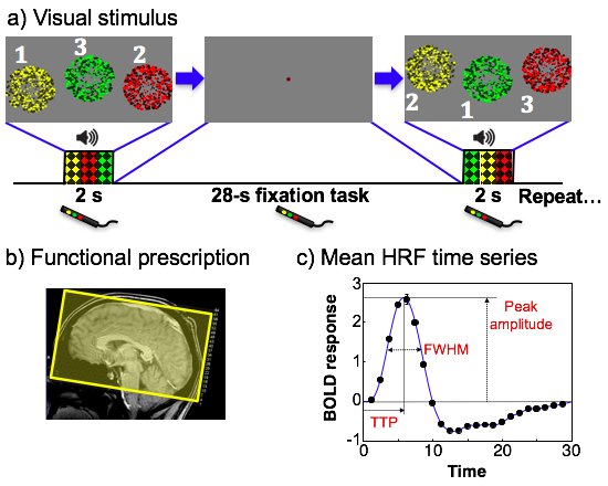
Subjects (N = 8) fixated upon a central colored dot on a display. Stimulus onset was cued by a change of dot color for 0.5 seconds before a 2-second stimulation period. Visual stimulation consisted of three circular regions of randomly flickering colored dots appearing sequentially for 667 ms (Figure 1a). Each region was colored red, yellow, or green, and had a corresponding audio stimulus of filtered white noise, medium pitch for yellow; low pitch for red; and high pitch for green. Subjects were tasked with responding to each dot display by pressing the button that matched the color and sound presented. 16 HRF measurements were collected in each run, and 5 runs were collected per session to yield 80 measurements for each subject. Imaging was performed on a 3T Siemens Trio scanner equipped with a 32-channel head coil with prescriptions covering all of cortex (Figure 1b). FMRI data were collected using an SMS-accelerated EPI sequence (G = 2, SMS = 3) to obtain 2-mm isotropic voxels across the entire brain with TR = 1.25 s. Functional data were registered to individual volume anatomies, collected with an MP-RAGE sequence (0.7-mm isotropic voxels). Each anatomy was segmented using FreeSurfer4 to extract gray and white matter. After image processing, data were analyzed to obtain HRF time series and parameters including peak amplitude, peak onset time, time-to-peak, and full-width-at-half-maximum (FWHM) in gray-matter voxels (Figure 1c).Results
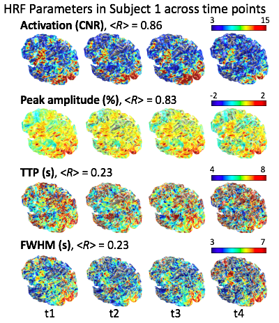
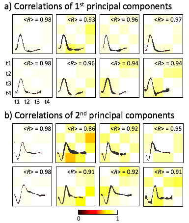
HRF parameter maps show highly correlated spatial patterns of activation and peak amplitude across the four time points, shown for one subject in Figure 2. Temporal parameters, TTP and FWHM, are much less correlated. Analysis of the HRF time series with principal component analysis was performed for eight subjects that reveal highly correlative temporal dynamics across time points. The first component (Figure 3a) represents the dominant temporal HRF features; and the second component (Figure 3b) projects the majority of residual HRF variation.Discussion and Conclusion
Our results demonstrate that our stimulus and task protocol evoked a strong and repeatable HRF across the majority of cortex. High spatial correlations of HRF parameters is consistent with our previous findings in which amplitude and activation was very similar across subjects, but temporal parameters were less correlative. However, principal component analysis revealed that underlying temporal variance is consistent across time points within a subject. This longitudinal HRF study can give insight into the physiology of any observed changes in the HRF, such as distinguishing CBF from CMRO2, and provide a further exploration in neurovascular coupling.Acknowledgements
Work supported by NIH R21HL108143, NSF BCS1063774, NIH R01NS095933References
1. Aguirre, G.K., Zarahn, E., D'esposito, M., 1998. The variability of human, BOLD hemodynamic responses. Neuroimage, 8, 360-9.
2. Handwerker, D.A., Ollinger, J.M., D'Esposito, M., 2004. Variation of BOLD hemodynamic responses across subjects and brain regions and their effects on statistical analyses. Neuroimage 21, 1639-51.
3. Taylor, A.J., Kim, J.H., Ress, D., 2018. Characterization of the hemodynamic response function across the majority of human cerebral cortex. Neuroimage 173, 322-331.
4. Fischl, B., Sereno, M.I., Dale, A.M., 1999. Cortical surface-based analysis. II: Inflation, flattening, and a surface-based coordinate system. NeuroImage 9, 195-207.
Figures