3759
Electrical Properties Tomography based Functional Magnetic Resonance Imaging (EPT-fMRI)1Philips Research, Hamburg, Germany
Synopsis
This study presents a new approach for the assessment of functional brain areas by using Electrical Properties Tomography (EPT). The measurements are performed similarly to conventional fMRI experiments to investigate brain activation for primary motor cortex areas, whereas EPT maps are generated for activation and resting periods and compared to BOLD fMRI images.
Introduction
The hemodynamic response of brain activation causes magnetic and electric changes in the activated brain area. MRI allows visualizing magnetic changes, e.g. based on the cerebral blood flow or blood-oxygen-level dependent (BOLD) effect. The latter is usually referred to as functional MRI (fMRI)1. In this study, for the first time, Electric Properties Tomography (EPT)2 is used to measure electric conductivity during brain activation. The measurements are performed similar to conventional fMRI experiments, whereas EPT maps are generated for activation and resting periods. Electrical properties were quantified during activation and resting states and the data was also analyzed by using statistical methods such as t-test.Methods
Measurements were performed in five healthy volunteers on a 3T Achieva Scanner (Philips, Best, The Netherlands) using an 8-element head-coil. The study investigated brain activation for three primary motor cortex areas according to the following tasks: fist clenching for right hand, left hand, and toe dorsiflexion with both feet. Each task was performed twice and imaged with a conventional BOLD fMRI sequence based on echo planar imaging (EPI) as well as with a balanced steady-state free precision (bSSFP) sequence for subsequent electrical properties analysis. Scan parameters for EPI acquisition were: FOV 230x230x96mm3, voxel size 2.4x2.4x4mm3 (reconstructed 1.8x1.8x4mm3), single-shot fast-field echo acquisition, FA 90°,TR/TE 3000/35ms, SENSE factor 1.8, 24 slices, 60 dynamics, total scan time 3:09min. Scan parameters for bSSFP acquisition were: FOV 230x201x28mm3, voxel size 1.8x1.8x2mm3 (reconstructed 1.8x1.8x2mm3), 3D fast-field echo bSSFP acquisition, FA 30°,TR/TE 3.4/1.7ms, 14 slices, 60 dynamics, total scan time 4:42min. All scans were performed using a block design with 6 alternating periods of rest and activation. Each block was acquired over a length of 10 dynamics (60 dynamics in total). The EPI images were analyzed for brain activation right after image acquisition on the scanner console with the Philips commercial software and statistical t-test model (IViewBOLD)3. Subsequently, the image volumes of the EPT scans were planned according to the activated regions in the EPI images as the coverage of the image volume of the EPT scans was smaller compared to the EPI sequences. In addition to the reconstructed magnitude images also the phase images of the scans are stored for subsequent reconstruction of electrical conductivity maps σ(r) via
σ(r) = Δφ(r)/(2μω2), (1)
where ω = Larmor frequency, Δ = Laplacian operator (second spatial derivative in 3D), φ(r) = bSSFP phase map, and μ = magnetic permeability of the body2. Conductivity is reconstructed for all 60 dynamics separately using Eq. (1) and results are investigated using a statistical t-test model1.
Results
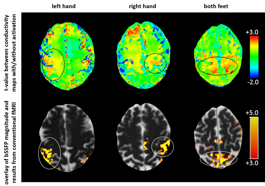
Figure 1 presents results for three different experiments of the left hand, the right hand, and the feet of one volunteer. The difference in conductivity in the respective motor cortex area with and without activation is of the order of 0.1 S/m for all experiments. The area of brain activation is almost the same for fMRI using conventional EPI and using EPT as proposed. However, using EPT, the activated region appears larger and blurred towards the inner part of the brain. Similar results have been obtained for the other volunteers.Discussion & Conclusion
In all experiments, the difference of about 0.1 S/m in conductivity between activation and resting periods in the activated brain areas corresponds roughly to a blood volume change of 10%, which is in line with expectations1. The blurring of the activated areas towards the inner part of the brain can be explained by the following issue: Solving the Laplacian of Eq. (1) numerically requires an ensemble of voxels (“kernel”) around the target voxel. This kernel has to be based on voxels with the same conductivity as the target voxel to avoid reconstruction errors2. Usually, this is realized by taking tissue boundaries from the bSSFP magnitude image into account, to match local geometric shape of kernel and tissue. In the case of fMRI, no tissue boundary between activated and non-activated areas was available, which is the reason for the blurred appearance of the activation area. The blurring appears only towards the inner part of the brain (since no clear boundary is given on this side of the activation area), but blurring does not appear towards the outer part of the brain (since a clear boundary is given here). More accurate quantification and delineation of brain conductivity during activation and rest may be accomplished by taking only voxels from brain structures into account which can be attributed to the underlying task, e.g. segmentation of motor cortex.
In conclusion, EPT-fMRI allows to measure and quantify electrical conductivity during brain activation and resting periods similar to BOLD fMRI.
Acknowledgements
No acknowledgement found.References
1. Chow MSM, Wu SL, Webb SE, Gluskin K, Yew DT. Functional magnetic resonance imaging and the brain: A brief review. World J Radiol. 2017;28: 5-9.
2. Katscher U, van den Berg CAT. Electric properties tomography: Biochemical, physical and technical background, evaluation and clinical applications. NMR Biomed. 2017;30: 3729.
3. Ulmer S, Jansen O. fMRI: Basics and Clinical Applications. Berlin Heidelberg, Germany: Springer Verlag; 2010.
Figures