3750
Line Scanning fMRI combined with optical calcium recordings to resolve the direct BOLD response to underlying slow oscillation-associated traveling waves1AG Molecular Imaging and Optogenetics of the Institute of Pathophysiology, University Medical School of Johannes Gutenberg-University Mainz, Mainz, Germany
Synopsis
In this multimodal approach consisting of line scanning (ls)-fMRI combined with optical calcium recordings, we investigated the direct BOLD response on a sensory stimulus in the slow wave brain state in anesthetized rats. We were able to observe the fMRI correlate of a slow wave recruiting the field of view with a significant latency between the recruitment of posterior and anterior voxels. This latency suggest the identification of the propagation of slow waves, as described with optical methods.
Introduction
While the relation of
neuronal activity to the BOLD response remains elusive, we have provided
conceptual evidence, that by simultaneous optic-fiber based calcium recordings
and fMRI, individual calcium events can be used as a regressor for the
subsequent fMRI analysis. However, brain-wide fMRI methods still lack temporal
resolution, in contrast to optical or electrophysiological methods. This limits
the detection of propagating neurophysiological events such as
slow-oscillation-associated calcium waves. Fast line scanning (ls-) fMRI (1)
with high temporal resolution and a reasonable spatial coverage provides the
opportunity to close the gap between fMRI and optical or electrophysiological
methods. In this study, we conducted a multimodal approach combining ls-fMRI and
simultaneously acquired optical calcium recordings in slow wave brain state,
characterized by a typical cortex-wide activation pattern (2,3), in contrast to
the awake-like persistent state.Methods
One female Lewis rat (220g) was anesthetized by 2.4% isoflurane to maintain a stable slow wave brain state during ls-fMRI. For simultaneous optical calcium recordings during the ls-fMRI experiment OGB-1 was bolus-loaded in both visual cortices (AP:-5.5; ML: +/-3.8; DV:0.9/0.7/0.5). Subsequently, a fiber was implanted into the left visual cortex (V1) for the recording of the calcium signal (2), and the animal was transferred into a 9.4T MR system (Bruker BioSpin, Germany). To evoke slow waves during the ls-fMRI experiments a light flash with a duration of 10 ms, every 10s was delivered to both eyes. For ls-fMRI we used a 2D-FLASH (fast low angle shot) sequence without phase encoding (4). Field of view (FOV) was restricted by saturation slabs, TR/TE= 50/18ms; the line was placed in anterior-posterior axis along the cortex (first in left, then in the right cortex) as shown in Fig. (1). For analysis the onsets of slow wave events were extracted from the recorded calcium signal using the algorithm described in (2). Only those stimulation intervals were chosen for further analysis, where the slow wave events were locked to the stimulus within 300ms, and without spontaneously evoked slow wave event in the previous stimulation interval to avoid a BOLD signal overlap from previous slow waves (Fig. 2a-c). For line scanning analysis the time courses per voxel were filtered (low pass filter, median filter with a 5-time frame-kernel, smoothing over 5 time frames) and separated into time courses of 10s corresponding to the stimulation interval. Only voxels with sufficient signal intensity were chosen. Then, the mean ls-fMRI signal per voxel was calculated from selected stimulation intervals (as described above).Results
The BOLD responses of evoked slow waves were investigated in both cortical hemispheres with ls-fMRI. Voxels containing artefacts of the fiber or injection were dismissed from further analysis (Fig. 3a). In slow wave state only, we observe the fMRI correlate of a slow wave recruiting the field of view (Fig 3b). Notably, we could identify a significant latency between the recruitment of posterior and anterior voxels.Discussion
In this multimodal approach consisting of ls-fMRI combined with optical calcium recordings we investigated the cortex-wide BOLD responses on a sensory stimulus in the slow wave brain state in anesthetized rats. For both hemispheres, we show a direct dependency of BOLD response to the neurophysiologically defined induced slow wave events. The latencies between posterior and anterior voxels suggest the identification of the propagation of slow waves, as described with optical methods (3).Conclusion
Therefore, this multimodal approach is able to bridge the gap between local methods with high temporal resolution like optical calcium recording and brain-wide fMRI methods with low temporal resolution for a given distinct brain state and a known neurophysiological event. As slow wave propagation is significantly disturbed in early Alzheimer Disease (5), these brain-state-informed BOLD response patterns have the potential to serve as an early biomarker.Acknowledgements
No acknowledgement found.References
(1) Yu X, Qian C, Chen DY, Dodd SJ, Koretsky AP. Deciphering lamina-specific neural inputs with line-scanning fMRI. Nat Methods. 2014 Jan;11(1):55-8.
(2) Schwalm M, Schmid F, Wachsmuth L, Backhaus H, Kronfeld A, Aedo Jury F, Prouvot PH, Fois C, Albers F, van Alst T, Faber C, Stroh A. Cortex-wide BOLD fMRI activity reflects locally-recorded slow oscillation-associated calcium waves. Elife. 2017 Sep 15; 6. pii: e27602.
(3) Stroh A, Adelsberger H, Groh A, Rühlmann C, Fischer S, Schierloh A, Deisseroth K, Konnerth A. Making waves: initiation and propagation of corticothalamic CA2+ waves in vivo. Neuron. 2013 Mar 20; 77(6): 1136-50.
(4) Kronfeld A, Aedo-Jury F, Hamzehpour L, Stroh A. Cortical propagation of slow ascillation-associated travelling waves resolved by fast line scanning in brain-state-informed BOLD fMRI. ISMRM 2018
(5) Busche MA, Kekus M, Adelsberger H, Noda T, Förstl H, Nelken I, Konnerth A. Rescue of long-range circuit dysfunction in Alzheimer's disease models. Nat Neurosci. 2015 Nov; 18(11):1623-30.
Figures
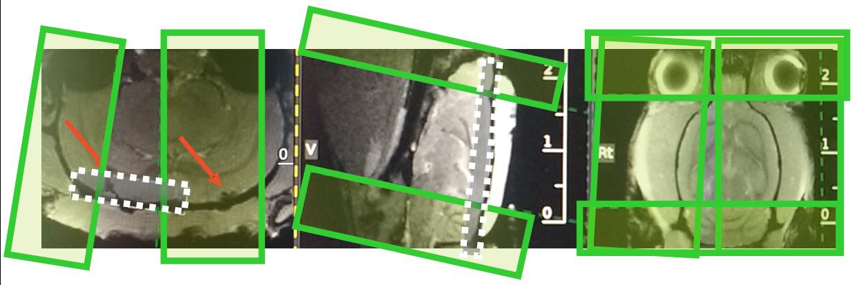
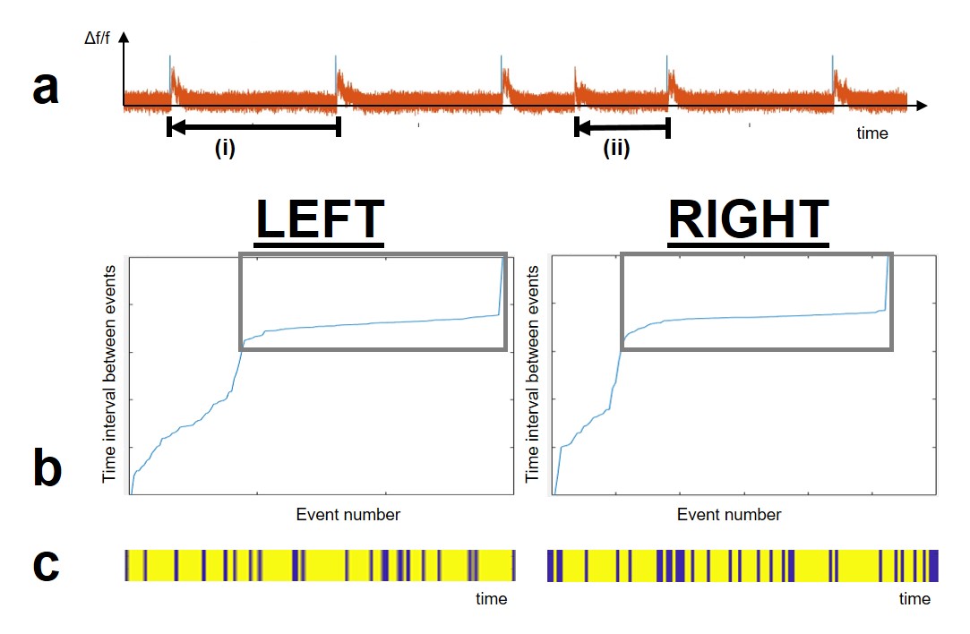
Figure 2: a) Depiction of stimulated and spontaneously evoked slow wave events (orange) after visual stimulation (blue). Stimulation intervals with a locked slow wave to the stimulus and no spontaneously evoked slow waves in the previous interval were used in further analysis (i), other stimulation intervals were dismissed (ii).
b) Events sorted by the time between to consecutive slow wave events for both hemispheres. Events within long time intervals were analyzed (gray rectangular).
c) Depiction of chosen stimulation intervals (yellow) for further analysis contra dismissed intervals (blue) over the complete measurement for both hemispheres.
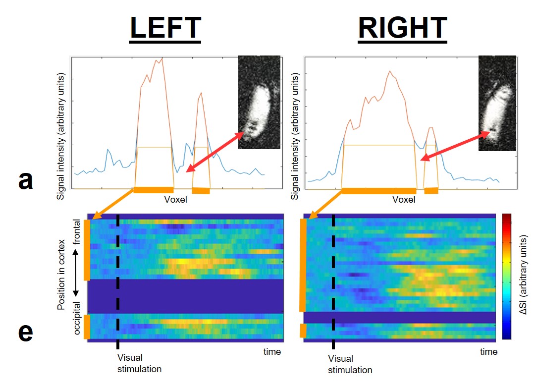
Figure 3: a) Signal intensities over the FOV of the line scanning experiments for both hemispheres. For further analysis, only voxels with high signal were chosen (orange). Small 2D images show each FOV with signal drops in the injection area.
b) Mean signal intensity over stimulation interval for each hemisphere. The fMRI correlate of a slow wave recruit the FOV in both measurements. Notably, we could identify a significant latency between the recruitment of posterior and anterior voxels.