3747
Adipose Tissue Evaluation in Economic Fish with MRI Imaging Translation via Conditional Generative Adversarial Networks1Ocean University of China, qingdao, China, 2Radiology Department,Bosto Children's Hospital; and Harvard Medical School, Boston, MA, United States
Synopsis
High presence of excess fat makes strong implications in obesity and its complications. High-fat diet could trigger fat accumulation. In this paper, we investigated and developed one quantitative online assessment strategy for adipose tissues in economic fish with Conditional Generative Adversarial Networks (CGAN) by means of magnetic resonance imaging (MRI), which could automatically help insights into the morphology of the adipose tissues in fish.
Introduction
Excess fat accumulation, mostly derived from unbalanced diets, can be seen as one of human health indicators. In most economic fish species, adipose tissues constitute the leading types of fat deposit, and the knowledge of its anatomic distribution and total mass is the basic premise to explore the mechanism of fat accumulation. Here we propose one online adipose tissue evaluation framework for major economic fish, with the help of T2-weighted MRI imaging translation in three-dimensional (3D) CGAN, which could provide a potential possibility to automatically access and visualize the combination and distribution of physiological healthy cues in fat deposit.Methods
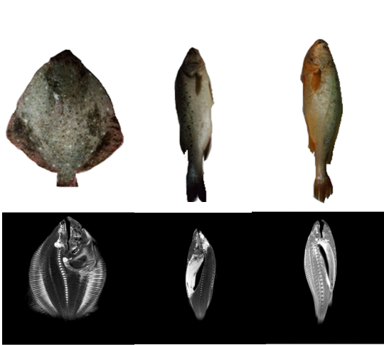
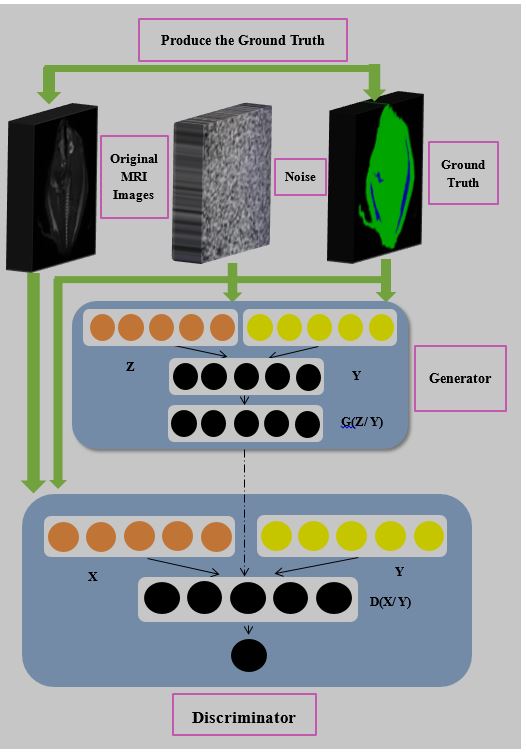
(i) Three species of live economic fish samples with the commercial size were selected in our experiments, including turbot (Scophthalmus maximus, mean weight: 212.05g±33g, N=6), Large yellow croaker (Pseudosciaena crocea, mean weight: 611.1g±11g, N=2), Japanese seabass(Lateolabrax japonicus, mean weight: 663.766675g±28g, N=4). Taking Scophthalmus maximus from the flat fish family as an example, the juvenile fish were also collected through various stages after its birth to adulthood in our experiments. All fish were first anesthetized prior to MR scanning and resuscitated afterwards. (ii) Fish samples were scanned with Philips ingenia 3.0T MRI and Agilent MRBR 9.4T/400(for Juvenile Scophthalmus maximus) imaging system, and a series T2-weighted Turbo-Spin-Echo (T2W-TSE) MRI images and T2-weighted Fast-Spin-Echo (T2W-FSE) MRI images were collected from the two different machines separately for the digital fish physiological atlas. Fig.1 is example MRI physiological slices originally collected from three species of fish samples. (iii) The large thick fat areas of adipose tissues were manually localized and labelled for image decomposition with expert guidance, and 3D-CGANs were further constructed to quantitatively retrieve and integrate the morphological and volumetric estimation of adipose tissues. Fig 2 is our network flow chart. Both the generator and discriminator in our developed 3D-CGAN impose the extended auxiliary manual labelling from the digital physiological atlas for the additional input layers, and feed those conditional knowledge to generate online fat detection and segmentation of adipose tissues for economic fish. In generator, the prior input noise and conditional knowledge are combined into joint hidden representation, and the adversarial training allows for considerable flexibility in the composition of the hidden representation. The objective function of a two-player minimax game for the generator and discriminator is hereby written as, min_{G}max_{D}V(G,D)=E_{xPdata(x)}[log{D(x)}]+E_{xpz(z)}[log(1-D(G(z)))] . (iv) From MRI tissue segmentation in aid of 3D-CGAN, the physiological attributes of adipose tissues in those economic fish, were further calculated with GPU-accelerated execution. Meanwhile, conventional anatomic and chemical analyses in fat deposition measurement were also carried out in comparison with our developed scheme.
Results
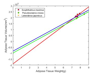
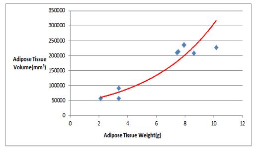
In our experiments, the fat deposit region in was extracted from the physiological tissue segmentation of 3D-CGAN, and the statistics of the total volumes, distribution, size, shape of fat deposit were investigated in detail. Fig. 3 displays the ratio curve of the total weight estimation (the total voxel number) in adipose tissues from three species of fish samples versus the real fat weight measurement. Fig. 4 predicts the growing curve of adipose tissue assessment at different stages for Scophthalmus maximus samples.Discussion
As is demonstrated in our experiment, the proposed auto-evaluation mechanism of adipose tissues for economic fish behaves a scientifically correct manner, and the average relative prediction error between MRI imaging analysis and the chemical analysis for the fat region detection in the adipose tissue of Scophthalmus maximus samples could be 0.35415%.Conclusions
In this paper, we put forward one novel auto-evaluation mechanism of adipose tissues for economic fish with MRI imaging via 3D-CGAN. The invasive means perform a sensitive and feasible online assessment and could be a potential tool in future studies of lipid metabolism.Acknowledgements
No acknowledgement found.References
1. Ren W, Li J, Tan P, et al. Lipid deposition patterns among different sizes of three commercial fish species[J]. Aquaculture Research, 2018, 49(2): 1046-1052. 2. Wu J L, Zhang J L, Du X X, et al. Evaluation of the distribution of adipose tissues in fish using magnetic resonance imaging (MRI)[J]. Aquaculture, 2015, 448: 112-122.Figures