3737
Dysfunction of functional connectivity between default mode network and cerebellar structures in patients with mTBI in acute stage. rsfMRI and DTI study.Maxim Ublinskiy1,2, Natalia Semenova1,2, Tolib Akhadov1, Andrei Manzhurtsev2, Petr Menshchikov3, and Dmitriy Kupriyanov4
1Clinical and Research Institute of Emergency Pediatric Surgery and Trauma, Moscow, Russian Federation, 2Institute of Biochemical Physics Russian Academy of Sciences, Moscow, Russian Federation, 3Semenov institute of Chemical Physics of Russian Academy of Sciences, Moscow, Russian Federation, 4Philips Healthcare Russian Federation, Moscow, Russian Federation
Synopsis
The revealed changes in DMN neuronal connection and cerebellar regions in acute stage of mTBI patients can be an initial step of damages leading to cognitive deficit which can be developed in future in long-term period of injury. We suggest that further research in this direction will demonstrate whether DMN functional integrity analysis can serve as a biomarker for monitoring the recovery of patients with mTBI.
Introduction
Mild traumatic brain injury (mTBI) occupies one of the first places in children injuries. Patients with mTBI may suffer headache, dizziness memory loss and short-term loss of consciousness in acute stage of injury [1]. However, in patients with mTBI there is usually an absence of structural lesions revealed by MRI studies in particular. That’s why mTBI neurobiological mechanisms are not fully understood. Diffusion Tensor Imaging (DTI) makes it possible to estimate the location, orientation, and anisotropy of the brain's white matter tracts. Resting-state functional MRI (rsfMRI) allows us to obtain new information on functional connectivity patterns of human brain. This technique is especially useful to reveal functional network disturbances in cases of MR normal-appearing structures. Among all brain networks at the resting state, the Default Mode Network (DMN) is the most widely studied network [2]. The revealed important role of DMN in cognitive processes and its disruption in different neurocognitive disorders makes the study of DMN functional integrity in patients with mTBI very useful. The aim of this study is to examine functional connectivity in normal-appearing cortex in acute period of mTBI using rsfmRI.Methods
34 MR negative participants were studied in age from 12 to 17 years (mean age – 14.5 years). Group of patients consisted of 17 children with mild traumatic brain injury in acute stage. 17 age-matched healthy volunteers comprised control group. All studies were performed at Phillips Achieva 3.0T MRI scanner using 32-channel head coil. DTI parameters: TR = 9431 ms; TE = 70 ms, matrix 120/144, FOV = 240 mm, voxel 2x2x2 mm3, EPI factor = 63, gap = 0, NSA = 2. fMRI studies were conducted using EPI BOLD (TR = 3000, TE = 30, EPI factor = 240, slice thickness = 4 mm, NSA = 1, 80 dynamics, total duration – 4 minutes). For each participant fMRI performing was repeated twice. fMRI data were processed using functional connectivity toolbox CONN [3]. Seed-based analysis was performed in order to reveal disturbances in functional connectivity. Statistical processing was performed using software package Statistica 12 using Mann-Whitney criterion to evaluate statistical intergroup differences.Results
DTI analysis didn't show any changes in values of apparent diffusion coefficient (ADC) and fractional anisotropy (FA) between two groups (see Fig.1). No statistically significant differences in correlation strength between DMN parts were observed in two groups (see Fig. 2). Seed-based analysis revealed absence of statistically significant neural correlations (p = 0,39) between DMN and cerebellum structural parts: inferior and superior semilunar lobules and flocculus (see Fig. 3). While in control group analysis showed statistically significant correlations (p < 0,05) in connectivity of respective areas.Discussion
One of the most common symptoms of mTBI is dizziness as a result of impaired movements coordination [4]. Cerebellum plays an important role in motor control in human brain. Flocculus as an essential cerebellum part (see Figure 3) plays an important role in the vestibulo-ocular system which is involved in the learning of basic motor skills in the brain [5]. Flocculus aids in the synchronization of eye and motor functions in order for the visual field and the motor skills to function together [6]. Flocculus functional integrity could suffer as a result of brain concussion. Our results show that mTBI appears to be a possible reason of connectivity malfunction in normal-appearing flocculus.Conclusion
Our study demonstrate disrupted functional connectivity between DMN areas and flocculus. This fact may indicate a functioning violation in normal-appearing cerebellum as a result of concussion in patients with mTBI. Resting-state functional MRI could serve as a potential marker for mTBI improved analysis.Acknowledgements
This study is supported by the Russian Fund Basic Research (RFBR) grant 18-315-00165 and the Russian Science Foundation (RSF) grant 18-13-00030References
1. Bigler E.D. Neuropsychology and clinical neuroscience of persistent post-concussive syndrome. J Int Neuropsychol Soc 2008;14(1):1–22. 2. Mayer A.R. Functional connectivity in mild traumatic brain injury. Hum Brain Mapp 2011 32, 1825–1835. 3. Whitfield-Gabrieli S. Conn: a functional connectivity toolbox for correlated and anticorrelated brain networks. Brain Connect. 2012; 2(3): 125-41. 4. Fife, T.D. Persistent vertigo and dizziness after mild traumatic brain injury. Ann N Y Acad Sci. 2015 Apr; 1343: pp 97-105. 5. Ito, M. Cerebellar Control of the Vestibulo-Ocular Reflex--Around the Flocculus Hypothesis. Annual Review of Neuroscience. 1982 5: 275–96. 6. Lisberger, S. The neural basis for learning of simple motor skills. Science. 1988 242 (4879): 728–35.Figures
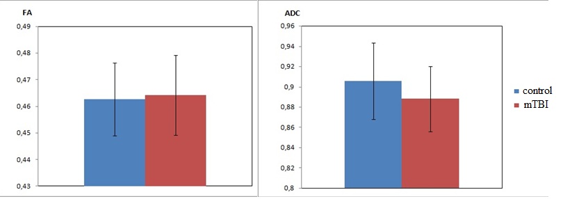
DTI analysis of MPFC-PCC link in two groups
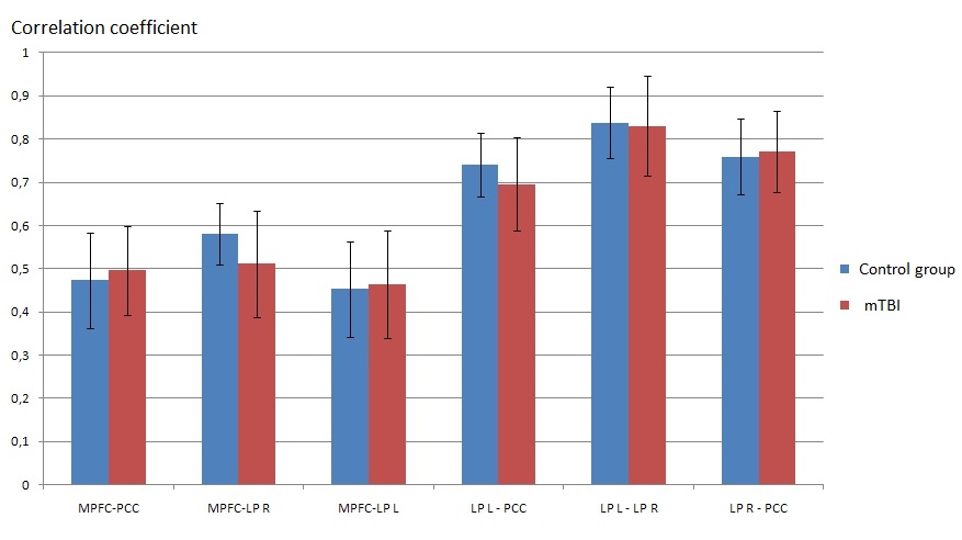
Results of intergroup analysis of the correlations strength of between
DMN regions.
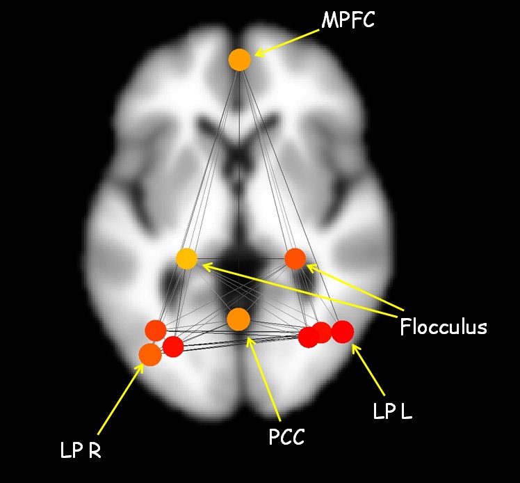
Map of
correlation links between DMN regions and flocculus in CONN software package