3735
Mapping Brain Neurochemical and Functional Coupling Using Dynamic SPICE1Electrical and Computer Engineering, University of Illinois at Urbana-Champaign, Urbana, IL, United States, 2Beckman Institute for Advanced Science and Technology, University of Illinois at Urbana-Champaign, Urbana, IL, United States, 3Biomedical Engineering, Shanghai Jiao Tong University, Shanghai, China
Synopsis
Neuronal metabolite (e.g., glutamate, GABA) concentrations in the brain are known to be correlated with neural activity. Currently, fMRS is the primary tool for measuring neurochemical changes in response to brain activity. However, fMRS has several major practical limitations, including low spatial resolution, low SNR, and very limited brain coverage. In this work, a new dynamic 1H-MRSI technique is used to address these difficulties. This technique can map dynamic metabolic changes from the whole brain at high spatial and temporal resolutions. In addition, it can simultaneously acquire fMRI images to track brain functional activity during the scan. With this unique capability, we have carried out functional MRSI experiments with motor tasks to investigate the coupling between neural metabolism and neural activity. The experimental results clearly show an increase in Glx in the motor cortex during the motor activation.
Introduction
The characterization of neurovascular and metabolic couplings is essential for understanding the complex interactions between neuronal activity and neurochemical changes. Investigating the dynamic changes of neurochemicals in response to brain functional activation could contribute to further understanding brain energy metabolism and metabolic pathways. Currently, fMRS is the primary tool for measuring metabolic changes associated with brain activities1. Using fMRS, a number of studies have been done, which have detected the dynamic changes of neurochemicals, like glutamate, lactate and GABA related to human psychological functions1-4. However, fMRS has low spatial resolution (around centimeters), low SNR, and poor brain coverage (single voxel measurements), which have significantly limited its practical utility. In this work, we investigate neurochemical changes of the brain using a novel dynamic 1H-MRSI acquisition and processing scheme based on SPICE5. This technique can acquire time-resolved MRSI spatiospectral functions with 2.0×2.8×3.0 mm3 nominal spatial resolution and 3.7-min frame rate and a series of fMRI images simultaneously. With this capability, we are able to investigate neurochemical and functional coupling of the whole brain at high spatial resolution. Our technology and experimental study may open up a new opportunity for investigating brain function and metabolism.Methods
The in vivo motor task experiments were performed on a 3T scanner (Siemens Prisma). The dynamic 1H-MRSI scan included 2 fixation blocks and 2 task blocks, lasting for 15 minutes (shown in Fig. 1). In the task block, the subject was instructed to perform repeated cycles of finger tapping for 40 seconds and resting for 16 seconds. The finger tapping task was performed using both hands and the tapping frequency was around 1 Hz. The entire study includes 3 scans and 12 event blocks on healthy subjects.
The dynamic 1H-MRSI sequence is based on a recently proposed imaging technique that extends SPICE (SPectroscopic Imaging by exploiting spatiospectral CorrElation5) for simultaneous MRSI and fMRI acquisition. The technique can acquire high-resolution non-water-suppressed 1H-MRSI data (TR/TE: 160/1.6 ms, FOV: 230×230×48 mm3, Matrix size: 116×80×16, Repetition: 4, Frame rate: 224 s) simultaneously with a time series of fMRI images (Matrix size: 76×76×40, Repetition: 300, Frame rate: 3 seconds).
Our processing pipeline provides several functions to effectively analyze the dynamic MRSI and fMRI data, which include: 1) normalization using the unsuppressed water signals to remove any field drift and T2* modulation in different frames induced by BOLD and physiological effect; 2) identification of the activation region based on the functional networks extracted from the fMRI signals using an ICA-based method6. In this study, the motor cortex is manually identified based on the functional network structure and the temporal changes of the associated fMRI signals; 3) spectral quantification for the whole brain using a subspace approach7; and 4) statistical analysis of the metabolite signals in the motor cortex and compare the difference between task and resting frames.
Results
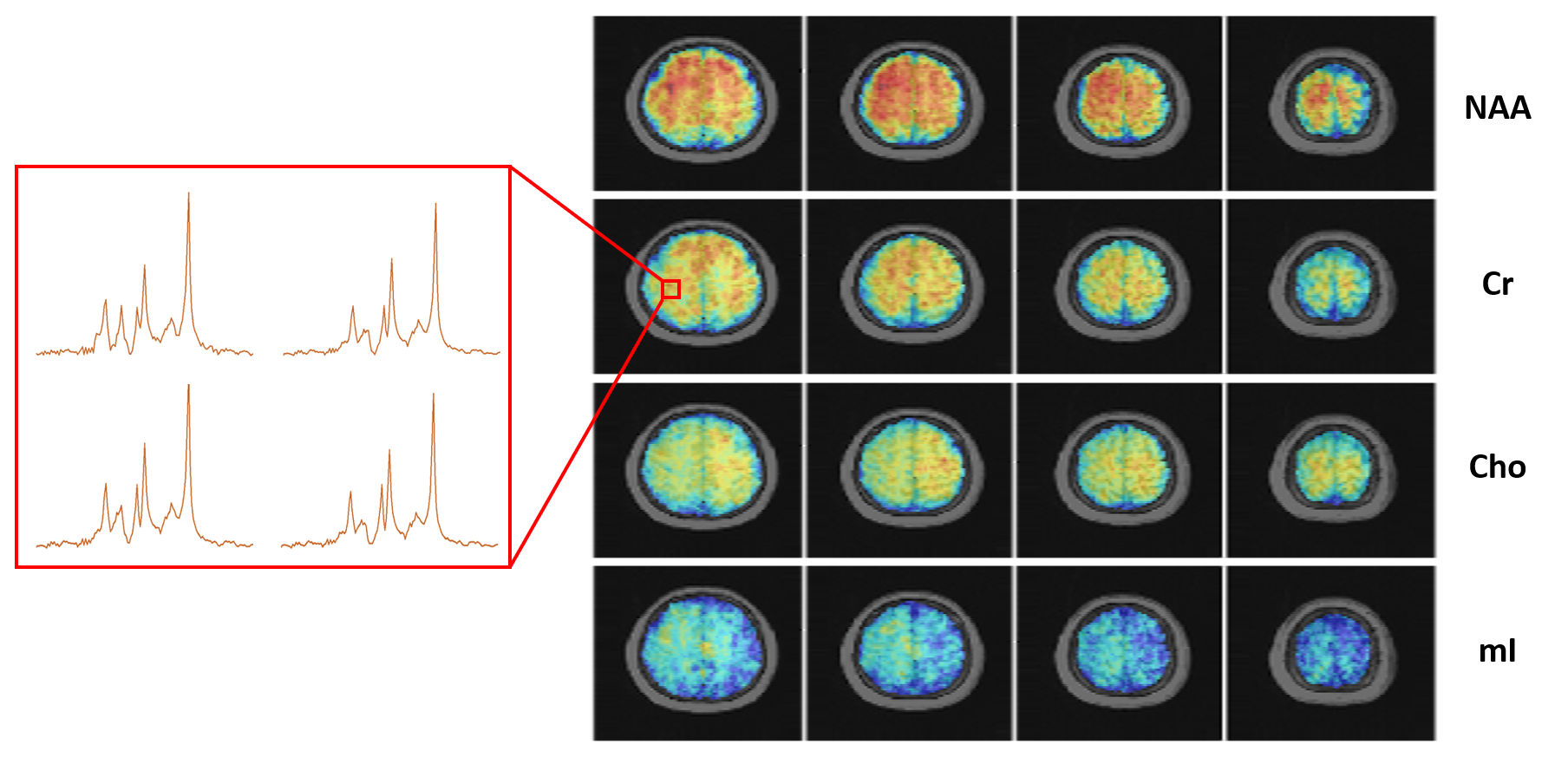
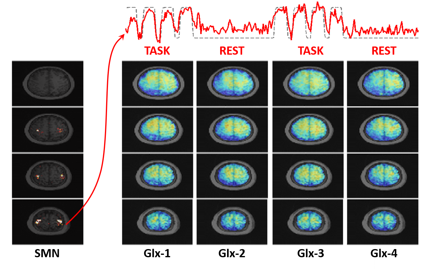
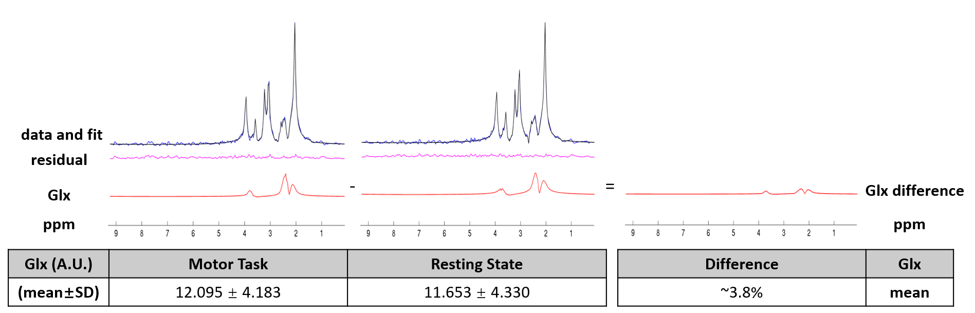
Figure 2 shows the metabolite maps and representative spectra of the first resting frame. Figure 3 shows the somato-motor network (SMN) extracted from the fMRI data and its corresponding task time course. The functional time course matches the block design very well, which implies that the subject followed the designed task well during the 1H-MRSI scan. Figure 3 also shows the dynamic Glx (Glutamate+Glutamine) maps of 4 frames obtained in one of the experiments. These dynamic Glx maps cover the whole brain which make it possible to investigate the dynamic changes of different brain regions. In this study, we were focusing on the neurochemical changes in response to functional activation in the motor cortex. Therefore, the spectra in the motor cortex from three scans (a total of 2132 spectra in the task frames and 2132 spectra in the resting frames) were used to analyze the Glx changes. Figure 4 presents the spectral quantification results on the averaged spectra from both the task frame and resting frame, which show a noticeable difference in the Glx level between the task state and the resting state. This experimental result is consistent with the previous findings8,9.Conclusion
We have successfully carried out a functional MRSI study on neurochemical coupling of brain function. To the best of our knowledge, this is the first functional MRSI study that maps the metabolic changes of a large portion of the brain (FOV: 230×230×48 mm3) at high resolution (2.0×2.8×3.0 mm3) instead of the single-voxel measurements obtained in conventional fMRS studies. Our technology and experimental results may open up a new opportunity for investigating brain function and metabolism.Acknowledgements
This work was supported in part by the National Institutes of Health (NIH-R21-EB021013, NIH-R21-EB023413, NIH-R01-EB023704, and NIH-P41-EB022544)References
1. Mullins PG. Towards a theory of functional magnetic resonance spectroscopy (fMRS): A meta‐analysis and discussion of using MRS to measure changes in neurotransmitters in real time. Scand J Psychol. 2018;59(1):91-103.
2. Prichard J, Rothman D, Novotny E, et al. Lactate rise detected by 1H NMR in human visual cortex during physiologic stimulation. Proc Natl Acad Sci. 1991;88(13):5829-5831.
3. Mangia S, Tkáč I, Gruetter R, et al. Sustained neuronal activation raises oxidative metabolism to a new steady-state level: evidence from 1H NMR spectroscopy in the human visual cortex. J Cereb Blood Flow Metab. 2007;27(5):1055-1063.
4. Jelen LA, King S, Mullins PG, et al. Beyond static measures: A review of functional magnetic resonance spectroscopy and its potential to investigate dynamic glutamatergic abnormalities in schizophrenia. J Psychopharmacol. 2018;32(5):497-508.
5. Lam F, Liang ZP. A subspace approach to high‐resolution spectroscopic imaging. Magn Reson Med. 2014;71(4):1349-1357.
6. McKeown MJ, Makeig S, Brown GG, et al. Analysis of fMRI data by blind separation into independent spatial components. Hum brain mapp. 1998;6(3):160-188.
7. Li Y, Lam F, Clifford B, et al. A subspace approach to spectral quantification for MR spectroscopic imaging. IEEE Trans Biomed Eng. 2017;64(10):2486-2489.
8. Ryan K, Wawrzyn K, Gati JS, et al. 1H MR spectroscopy of the motor cortex immediately following transcranial direct current stimulation at 7 Tesla. PloS one, 2018;13(8): e0198053.
9. Schaller B, Xin L, O'brien, et al. Are glutamate and lactate increases ubiquitous to physiological activation? A 1H functional MR spectroscopy study during motor activation in human brain at 7 Tesla. Neuroimage 2014;93:138-145.
Figures