3718
Elevated activation of a murine limbic network during cue-reward association learning revealed in 15.2 T fMRI1Department of Integrated Biosciences, Graduate School of Frontier Sciences, The University of Tokyo, Tokyo, Japan, 2Center for Neuroscience Imaging Research, Institute for Basic Science, Suwon, Korea, Republic of
Synopsis
An operant learning device for head-fixed mouse was developed for ultra-high field (15.2 Tesla) fMRI. Habituated mice were learned to water reward, associated with cue of light stimulation as a conditioning stimulus. We obtained 1,000 sets of BOLD fMRI (GE-EPI, TR/TE = 1000/11 ms), and found that elevated activation of a limbic learning network, including the entorhinal, perirhinal, and retrosprenial cortices and the hippocampal formation, during cue-reward association in learned mice.
Introduction
fMRI in rodents provides the opportunity for preclinical studies on the effects of pharmacological and genetic manipulation on brain function, which is difficult to perform in humans. However, due to body and head movements, fMRI studies in rodents have been limited to resting state or anesthesia conditions6. In this study, we developed a training and habituating protocol for a cue (light)-reward (water) association operant learning to mice attached with very light plastic head plate (0.2 g), in order to take series of BOLD fMRI and analyze brain activity for association learning.Methods
For awake fMRI, we utilized non-magnetic operant learning device for head fixed mice5 (Fig. 1). Center of the hole of the plastic head bar was adjusted to the bregma of the mouse brain. Numbers of male mice (C57BL/6; 8 weeks old) with plastic head bar was performed an operant learning task procedure consisted of 4 phases; head-post surgery, recovery, licking training and association learning (Fig. 2). Custom-made surface coil for 15.2 T-MRI (650 MHz) was attached to this chamber on top, and the center of the surface coil was fitted to bregma of the mouse brain. Taking advantage of ultra-high field of 15.2 3 we utilized gradient echo-EPI with short TE (TE: 11ms, TR: 1000ms, FOV: 1.5x1.5cm2, 10 slices, slice thickness: 0.5mm). We took series of BOLD fMRI (1,000 fMRI; 50 trials x 20 scans per each trial) from well-habituated head-fixed mice (n=3). For a post-processing of awake fMRI data set, we developed a pipeline by using spm12 platform in the order of slice timing, two-sets of realignment, co-registeration to C57BL/6 template4, normalization, and smoothing, and performed statistical fMRI data analysis. To determine neural network responsible for association learning, we differentiated sets of fMRI data into four categories dependent on the licking behavior of learning mice during task (Fig. 3).Results
To visualize neural network responsible for acquiring cue-reward association memory, we statistically compared sets of fMRI between net correct trials and no-reward trials from three learning mice at day 4 of Go-task (Fig. 4). As a result of the two-sample t-test in spm 12, we detected elevated activation of a limbic-related network, including the entorhinal, perirhinal, and retrosprenial cortices and the hippocampal formation, during net correct trails in cue-reward association learning task from all three mice tested. To determine time course of BOLD response during association learning, we computed fMRI data sets from reward trials in a 1st-level analysis and plotted time course of BOLD signal in the entorhinal cortex (Fig. 5).Discussion
The entorhinal cortex and the perirhinal cortex, as well as the retrosprenial cortex and the hippocampal formation, are known as constituent elements of the medial temporal lobe memory system with the hippocampus as the apex9. These regions supply the sensory information received from the sensory association field to the hippocampus, while providing the recalled memory information to the higher order sensory allied field1. It is also associated with emotional and reward areas such as the orbitofrontal cortex and anterior cingulate cortex8. Change in the BOLD signal of the entorhinal cortex was similar to the visual stimulation BOLD change in V1 area, reported to return to normal level within 10 seconds7. Thus, these limbic areas serve as one of the main pathways of neural circuits related to cue-reward association learning memory. Based on these findings, in this experiment, it is suggested that when the task succeeded, association learning between the light as cue and water as reward is established by the function of the medial temporal lobe a limbic memory system, depicted in this study.Conclusion
This study enabled us to capture the brain activities of mouse during task execution, therefore it seems that it will become possible to measure brain activities of mouse related to association learning. It is also possible to study various neurological disease models, including Alzheimer’s Disease2. We are in progress to perform preclinical studies of various disease by this awake fMRI in the ultra-high field-MRI.Acknowledgements
No acknowledgement found.References
1. Bussey, J. and Saksida, M. Memory, perception, and the ventral visual-perirhinal-hippocampal stream: thinking outside of the boxes. Hippocampus. 2007; 17(9), 898-908
2. Götz, J., et al. Rodent models for Alzheimer disease. Nat Rev Neurosci. 2018; 19, 583-598.
3. Han, S. H., Son J. P., Cho, H.J., Park, J.-Y., and Kim, S.-G. Gradient‐echo and spin‐echo blood oxygenation level–dependent functional MRI at ultrahigh fields of 9.4 and 15.2 Tesla. Magn Reson Med 2018; DOI: 10.1002/mrm.27457.
4. Hikishima, K. et al. In vivo microscopic voxel-based morphometry with a brain template to characterize strain specific structures in the mouse brain. Scientific Reports. 2017; 7, 85.
5. Jomura, N., Shintani, T., Sakurai, K., Kaneko, J., and Hisatsune, T. Mouse BOLD fMRI imaging during operant learning at ultra-high field (14 T). Proc. Intl. Soc. Mag. Reson. Med. 2017; 25, 5365.
6. Jonckers, E., et al. Functional connectivity fMRI of the rodent brain: comparison of functional connectivity networks in rat and mouse. PLoS One. 2011; 6, e18876.
7. Logothetis, N. K. et al. A neurophysiological investigation of the basis of the BOLD signal in fMRI. Nature. 2001; 412(6843), 150-157.
8. Mogami, T. and Tanaka, K. Reward association affects neuronal responses to visual stimuli in macaque te and perirhinal cortices. J. Neurosci. 2006; 26(25), 6761-6770.
9. Squire, L. and Wixted, T. The cognitive neuroscience of human memory since H.M. Annu. Rev. Neurosci. 2011; 34, 259-288.
Figures
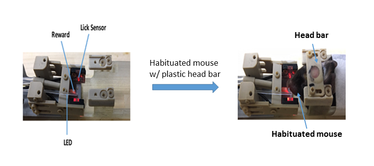
Fig 1. Habituated mouse situated in non-magnetic cue-reward operant learning device
By the use of habituated mouse with a plastic head-bar (0.2 g), we minimized the head movement of the mouse. In this learning device, we utilized light stimulus as a cue and water as a reward. We used an optic sensor that senses the licking action of murine tongue to detect a conditioned response.
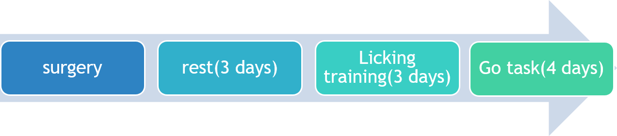
Fig 2. Schedule of cue-reward operant association learning fMRI in mice
Operant learning task for fMRI consisted of 4 phases; head-post surgery, rest for recovery, licking training to take water from plastic nozzle in the dice chamber, and cue-reward association learning (Go task) in the MRI scanner (15.2 T BioSpec 152/11 MRI, Bruker). In the association learning task, mice were provided with water (4 µL) immediately after the licking behavior, sticking out their tongue, during cue (light) stimulus.
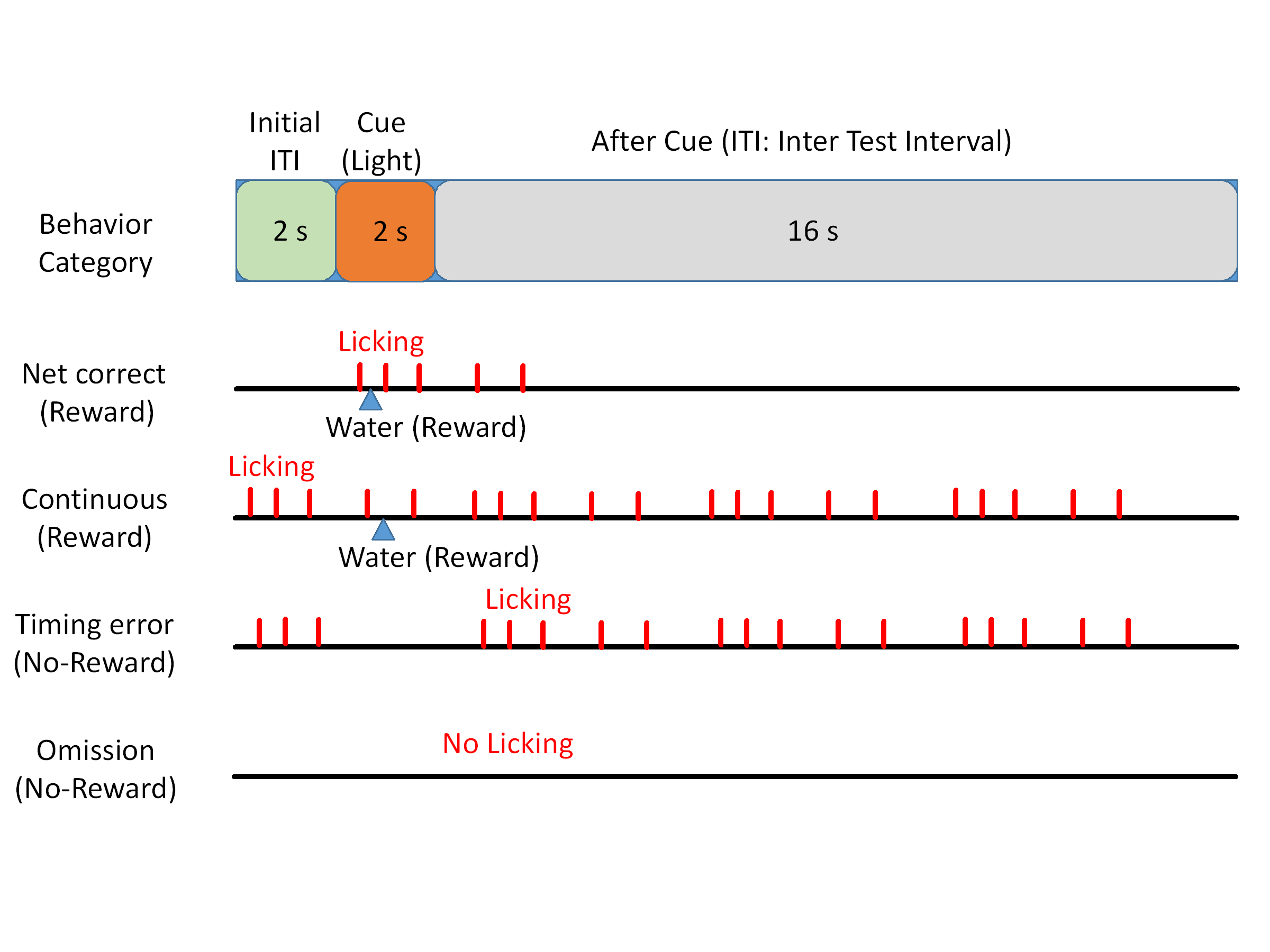
Fig 3. Definition of learning behaviors during cue-reward association task for fMRI
We differentiate learning behavior into four categories: Net correct, Continuous, Timing error, and Omission, in each trial (20 seconds). Mice were performed 50 trials continuously per day (One session).
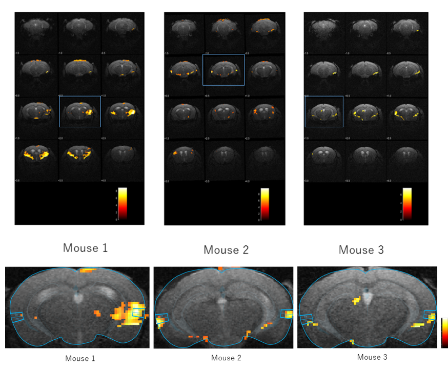
Fig 4. A limbic network responsible for cue-reward association learning in mice
We compared fMRI data during 4.0 – 9.0 second (5 scans) after the initiation of each trial between net correct trials and no-reward trials (timing error and omission) from three mice tested. Upper panel shows all slices, and lower panel shows an enlarged limbic section. PFWE < 0.05, threshold k=20, scale bar represents T-scores.
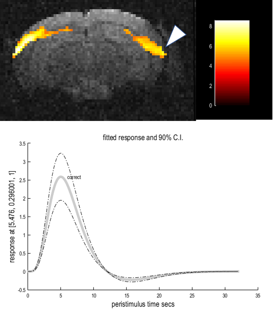
Fig 5. Elevation of BOLD signal in the entorhinal cortex of association learning mice
We computed fMRI data sets from all reward trials (39/50 total trials) from mouse #2, in a spm 12 1st-level analysis. We plotted a time course of BOLD signal (percentage of increase) at the peak voxel in the right entorhinal cortex, immediately after the 2 seconds of light stimulation.