3716
A frequency-domain machine learning (FML) method for dual-calibrated estimation of oxygen extraction fraction (OEF) and cerebral metabolic rate of oxygen metabolism (CMRO2)1Cardiff University, Cardiff, United Kingdom, 2Concordia University, Montreal, QC, Canada
Synopsis
A frequency-domain machine learning method is presented that significantly reduces the bias and variance in dual-calibrated estimation of oxygen extraction fraction, as demonstrated with simulation and in-vivo imaging. In addition, the method substantially reduces the processing time compared to previous robust analysis methods.
Introduction
Non-invasive mapping of the cerebral metabolic rate of oxygen metabolism (CMRO2) with MRI has great potential in the study of the brain in health and in disease. To this end a number of different methods have been proposed to estimate CMRO2 from MRI acquisitions. One such method is based on the inference of the resting oxygen extraction fraction (OEF0) from BOLD and ASL signal fluctuations during hyperoxic and hypercapnic gas challenges.1,2 However, this method suffers instability in OEF0 estimates due to propagation of errors in the analysis pipeline. Here we present a machine learning approach that reduces both the variance and bias in OEF0 estimates. The proposed method takes advantage of the inherent input noise immunity of artificial neural networks (ANN) 3, and the superior feature learning ability of frequency-domain deep learning 4.Methods
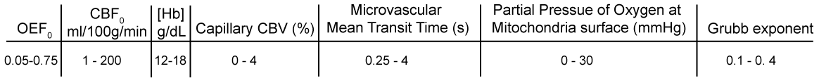
Simulated data were used to train ANNs to estimate resting cerebral blood flow (CBF0) and CMRO2, from which OEF0 was calculated. Two ANNs were cascaded such that the results of the CBF network were fed into the CMRO2 network. The data were simulated using standard physiological models 2 and constrained to be physiologically plausible. Constraints on the cerebral physiology were applied according to a simple model of oxygen transport 5. Physiological limits were chosen to encompass both healthy and pathological brain tissue and are listed in figure 1. Simulated BOLD and ASL time-courses were generated according to an 18-minute gas paradigm 6, with added noise.
Time series data was high-pass filtered (BOLD data only, 320 seconds) and then Fourier transformed. The input feature vector for the CBF ANN consisted of the first 15 points of the magnitude and phase data for both the ASL and BOLD timeseries, [Hb], hyperoxic ΔPaO2, SaO2,0, CaO2,0, and the post-label delay (65 data points in total). The feature vector for the CMRO2 ANN also included the CBF0 estimate (66 data points in total). Simulated datasets were constructed with 1x10⁶ simulations (10% used for early stopping). Networks had 2 hidden layers (50 nodes in each) and a relu activation function. An additional data set (OEF0 range 0.1 to 0.6) was simulated to compare the performance of the FML networks with previously published regularised non-linear least squares (R-NLS) methods 6,7. Each method (FML and R-NLS) was also applied to data acquired (3T Siemens Prisma) in healthy volunteers (n=16, 10 male, mean age 34.5 years) (TR 4.4 seconds, 15 slices, in-plane resolution 3.4 x 3.4 mm and slice thickness 7 mm).7 In-vivo data was processed in the same manner as the simulated data, or as previously described 7 (regularisation was only applied to oxygen diffusivity for the in-vivo analysis as this had the best performance in simulations). No spatial smoothing was applied to the data prior to analysis.
Results
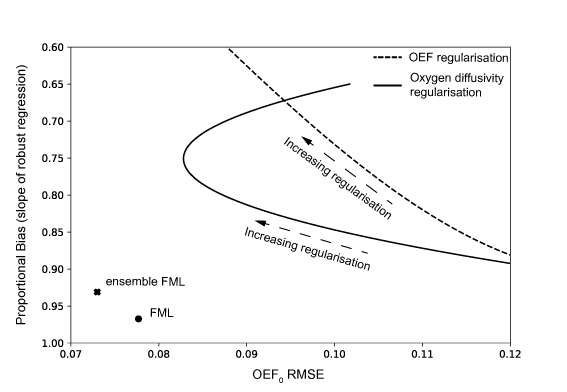
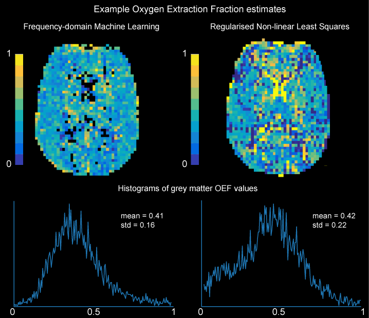
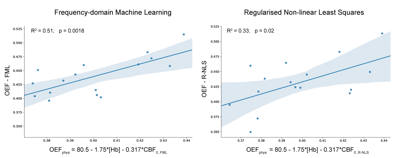
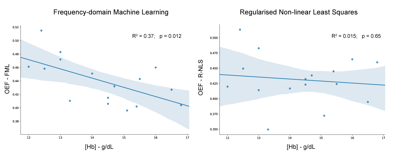
As expected increasing the weight of the regularisation parameter, OEF or oxygen diffusivity, reduced the RMSE of OEF0 estimates but increased the proportional bias (slope of the robust regression line), see figure 2. The FML method produced a significant reduction in both RMSE and bias, with a simulated RMSE of 0.078 and slope of 0.97. Using an ensemble of networks (n=40) further reduced the RMSE to 0.073 while increasing the bias slightly (slope = 0.93). Fitting of in-vivo data was substantially faster using the FML method, approximately 15 - 30 seconds (depending on number of networks used in the ensemble) on a standard laptop versus 17-18 minutes for the R-NLS approaches. The standard deviation in grey matter OEF0 estimates was significantly reduced for the FML method (p<0.001) with a standard deviation of 0.17 ± 0.01 versus 0.21 ± 0.02, see figure 3. Each method showed moderate to strong agreement with empirical observations of OEF0 with variation in CBF0 and [Hb] 8, see figure 4. However, only the FML method demonstrated the expected 8 inverse relationship between OEF0 and [Hb] (figure 5).Discusion
The proposed method demonstrates a significant improvement in speed, RMSE and bias in the analysis of dual-calibrated data for the estimation of OEF0 (the principal source of uncertainty when estimating CMRO2). In-vivo results suggest a significant improvement in sensitivity to physiological variation and reduced variance in voxel-wise estimates. FML appears to be a robust method that should be suitable for online analysis of data, an important step in transitioning dual-calibrated fMRI from a research method to a clinical tool.Acknowledgements
We would like to thank Wellcome for supporting this work: Wellcome Strategic Award, ‘Multi-scale and multi-modal assessment of coupling in the healthy and diseased brain’, grant reference 104943/Z/14/Z (RW, MG, CF and RS). RW also thanks the Higher Education Funding Council for Wales for support. KM is supported by Wellcome grant 200804/Z/16/Z.References
1. Bulte D. et al. Quantitative measurement of cerebral physiology using respiratory-calibrated MRI. NeuroImage. 2012; 60: 582-591
2. Gauthier, C.J. et al Absolute quantification of resting oxygen metabolism and metabolic reactivity during functional activation using QUO2 MRI. Neuroimage. 2012. 63, 1353-1363.
3. Lee Y and Oh S-H. Input Noise Immunity of Multilayer Perceptrons. ETRI Journal. 1994; 16(1): 35-43.
4. Hertel L, Phan H, and Mertins A. Comparing Time and Frequency Domain for Audio Event Recognition Using Deep Learning. International Joint Conference on Neural Networks. 2016: 3407-3411
5. Gjedde A. The pathway for oxygen in brain. APMIS Suppl. 2003; 109: 146-53
6. Germuska M. et al. A forward modelling approach for the estimation of oxygen extraction fraction by calibrated fMRI. NeuroImage. 2016; 139: 313-323
7. Germuska M. et al. Dual-calibrated fMRI measurement of absolute cerebral metabolic rate of oxygen consumption and effective oxygen diffusivity. NeuroImage. 2018. doi:10.1016/j.neuroimage.2018.09.035
8. Ibaraki M. et al. Interindividual variations of cerebral blood flow, oxygen delivery, and metabolism in relation to hemoglobin concentration measured by positron emission tomography in humans. JCBFM 2010; 30(7): 1296-1305
Figures