3707
The correlation between cerebral venous oxygen saturation change and cognitive score in Alzheimer's disease patients by using susceptibility weighted imaging mapping1Department of Radiology, First Affiliated Hospital of Dalian Medical University, Dalian,116011,China, Dalian, China, 2GE Healthcare, Beijing, China
Synopsis
Alzheimer's disease (AD) is a progressive neurodegenerative disease and vascular factors may be important in the development and progression of AD[1]. Magnetic susceptibility weighted imaging (SWI) is widely used in the diagnosis of central nervous system diseases, and venous oxygen content is the basis of SWI angiography. The venous blood oxygen level can be reflected by measuring the phase value of venous blood. This study first made use of SWI mapping (SWIM) to measure the change of magnetic susceptibility and the change of blood oxygen and then explores the correlation between cerebral venous oxygen saturation change and cognitive score in AD patients
Target audience
Researchers related to cognitive impairment of Alzheimer disease(AD).Purpose
To evaluate the change of cerebral venous oxygen saturation of AD patients, and to explore the relevance between venous oxygen saturation and cognitive impairment, based on the quantitative analysis of cerebral venous oxygen saturation by using susceptibility weighted imaging mapping (SWIM).Methods
This study was approved by the hospital ethics committee. The study was divided into the AD patient group (16 males and 28 females, mean age 71.41±9.31 yrs) and the health control (HC) group (5 males and 7 females, age 63.90±6.44 yrs). The mental status of each participant was evaluated through MMSE, MoCA and Clock drawing task (CDT) scores by two neurologists.
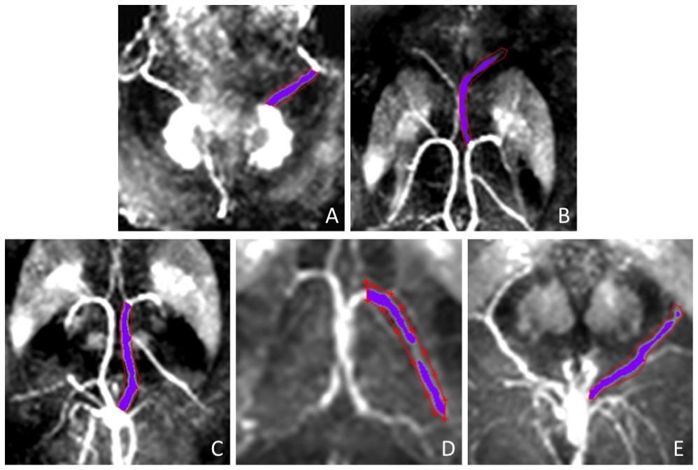
All subjects underwent routine MRI sequences and enhanced gradient echo T2* weighted angiography (ESWAN) on a 3.0T MR scanner. SWIM images were further processed with signal processing in neuro-MR. The regions of interests (ROIs) include the bilateral thalamostriate veins (TV), septal veins (SV), internal cerebral veins(ICV), basal veins (BV) and inferior petrosal sinus (IPS) drawn manually in SWIM images, and magnetic sensitivity value (MSV) was measured (Fig.1).
For the values conformed to normal distribution, two independent samples t-test was used to compare the susceptibility in all regions between the groups. The relations between MSV and MMSE, MoCA scores were analyzed using Pearson's correlation. The relations between MSV and CDT scores was analyzed using Spearman 's correlation.
Result
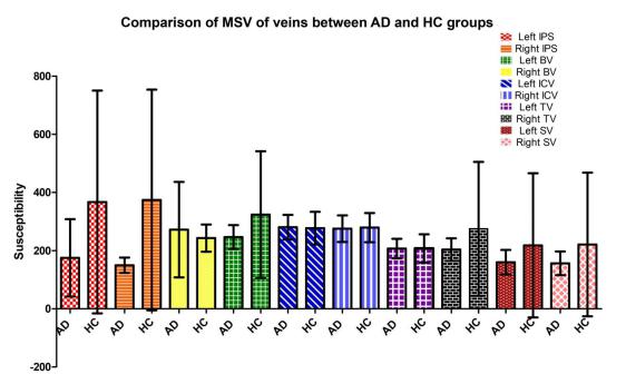
Compared with the HC group, the MSV of all the veins decreased except for the left ICV and the bilateral SV, while no significant difference was found (Fig.2).
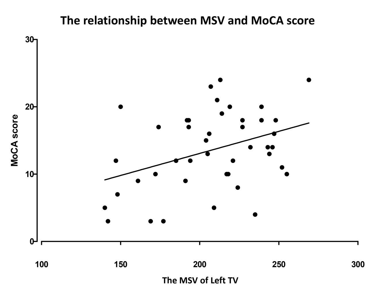
Correlation analysis indicated that there was a significant positive correlation between the susceptibility of left TV and the MoCA score (r = 0.382, P = 0.012) (Fig.3), and the susceptibility of left SV and the CDT score (r = 0.354, P = 0.027).
Discussion and Conclusion
AD is a progressive neurodegenerative disorder resulting from pathological changes[1]. Venous oxygen saturation is an important indicator for evaluating brain function[2]. In this study, the MSV was measured by SWIM method, and the change in blood oxygen was further measured according to Δχvein-tissue=Κ×Δχdo×Hct(1-Y) (Δχ, Magnetic sensitivity; Y, Saturation of blood oxygen). The results show that most of the venous MSV of the AD group were higher than those of the HC group, indicating a lower blood oxygen level of the intracerebral veins, an increased proportion of deoxyhemoglobin and the anoxic state of brain tissues. However, the difference between the two groups was not significant, probably because of partial volume effect.
A significant positive correlation was observed between the MSV of left TV and the MoCA score, the MSV of left SV and the CDT score, suggesting that the decline in blood oxygen levels may lead to a decreased cognitive level of AD patients.
Acknowledgements
No acknowledgement found.References
[1]. Contreras J A, Goñi J, Risacher S L, et al. Cognitive complaints in older adults at risk for Alzheimer's disease are associated with altered resting-state networks[J]. Alzheimers & Dementia Diagnosis Assessment & Disease Monitoring, 2017, 6(7):40-49.
[2]. Fan A P, Benner T, Bolar D S, et al. Phase-based regional oxygen metabolism (PROM) using MRI[J]. Magnetic Resonance in Medicine, 2012, 67(3):669-678.
Figures