3699
Effects of Isoflurane on Brain Functional Connectivity in Common Marmosets1Department of Radiological Sciences, Faculty of Health Sciences, Tokyo Metropolitan University, Tokyo, Japan, 2Regenerative medicine, The Jikei University School of Medicine, Tokyo, Japan, 3Laboratory for Marmoset Neural Architecture, Center for Brain Science, RIKEN, Wako, Japan, 4Department of Radiological Sciences, Human Health Sciences, Tokyo Metropolitan University Graduate School, Tokyo, Japan, 5Veterinary Surgery Laboratory, Graduate School of Agricultural and Life Sciences, The University of Tokyo, Tokyo, Japan
Synopsis
We examined the effects of isoflurane on brain functional connectivity in common marmosets using resting-state fMRI. We found that the thalamus was closely related to the mechanism of action of anesthesia and the default mode network (DMN), which is thought to function at rest. Isoflurane exhibited similar effect on the thalamus and human DMN region in humans but partly different effect onthe marmoset DMN region.In the future, we need to perform further investigations in detail by increasing the amount of data and reviewing analysis methods.
Introduction
Experiments are often performed under anesthesia in animals considering body movements. However, most studies in humans are conducted when they are awake. Thus, differences in brain function between anesthesia and awake conditions should be discussed to allow development from preclinical research to clinical research. There are few reports on brain function under awake and anesthesia conditions in common marmosets, which have physiological and anatomical features similar to those of humans.1 In addition, resting-state fMRI (rsfMRI) is a method that has attracted attention in recent brain function measurements. It focuses on slow voluntary blood oxygen level-dependent(BOLD)activity of less than 0.1 Hz occurring at rest and calculates the functional connectivity (FC) of the brain by identifying the region where the activity is synchronized.2 The default mode network (DMN) is an important network in rsfMRI. However, as the region estimated as the DMN differs between humans and marmosets, it is necessary to examine the region as well.3, 4 The present study aimed to examine the effects of isoflurane on brain FC when compared with that under an awake condition in common marmosets.Methods
This study included one healthy male common marmoset (age, 3 years). A 9.4-T MRI scanner (BrukerBioSpin, Ettlingen, Germany) was used to obtain resting functional images under isoflurane anesthesia and awake conditions. The imaging parameters were as follows: gradient echo (echo-planar imaging); repetition time/echo time, 2,000/16.0 ms; slice thickness, 0.7 mm; field of view, 42 × 28 mm; resolution, 0.7 × 0.7 mm; number of slices, 52; repetition number, 150; and scan time, 60 min. The registration processing and smoothing processing of standard brain images and functional images were performed using Statistical Parametric Mapping (SPM; Wellcome Center for Human Neuroimaging, London, UK) and Artifact Detection Tools (ART; The Gabrieli Lab, McGovern Institute for Brain Research, MIT, US) to detect and eliminate slices with large movement. Thereafter, noise was removed using CONN (The Gabrieli Lab), and BOLD signal was calculated. An FC matrix was created by combining the obtained data and 52 regions of the anatomically segmented side of the brain. Figure 1 shows the analysis process. This study focused on the thalamus, which is potentially closely related to the action mechanism of anesthesia, and the DMN, which is considered to function at rest. With regard to the DMN, we examined each region, as the region considered to be the DMN is known to differ between humans and marmosets. Figure 2 shows ROIs of the DMN.Results
Regions showing reduced correlations with the thalamus owing to isoflurane administration included the olfactory nucleus, auditory cortex, secondary somatosensory cortex, ventral medial prefrontal cortex, posterior cingulate cortex, nucleus accumbens, and other such regions. The FC showed a decrease in human DMN regions, whereas it showed an increase in the DMNregion among marmoset DMN regions (Figure 3 and Figure 4).Discussion
Sensory regions, such as the auditory and the secondary somatosensory cortexes, were part of the area where isoflurane suppressed the correlation. The thalamus is thought to be a relay nucleus of sensory stimulation.5 The correlations between the thalamus and the sensory regions, as observed in this study, are presumed to be the result of communication blockage. These findings suggest that isoflurane blocks the input of sensory stimulation. In addition, the disappearance of consciousness was associated with the division of collaboration between the thalamus and the cerebral cortex,6, 7 and the same result was obtained in this study. Therefore, it was suggested that the region where the correlation with the thalamus was decreased in this study might be involved in the maintenance of consciousness. A previous report mentioned that the FC of the DMN (human DMN region) in rhesus monkeys declined under isoflurane anesthesia,8 and this is consistent with our results. Thus, even in marmosets, isoflurane is believed to have a function-suppressing effect on the human DMN region. However, when examining the marmoset DMN region presumed in a previous study,4 we found that the functional correlation increased between the intraparietal sulcus and the posterior cingulate cortex under anesthesia. This finding suggests that the effects of isoflurane on the DMN may differ between humans and marmosets.Conclusion
As the site of action of isoflurane in the thalamus was generally consistent with the results of previous studies, similarities between humans and marmosets were shown. However, as there are few reports on the marmoset DMN region with regard to the FC and networks themselves, it is necessary to review the amount of data and the analysis methods in the future and perform detailed studies.Acknowledgements
This research is partially supported by the program for Brain Mapping by Integrated Neuro technologies for Disease Studies (Brain/ MINDS) from Japan Agency for Medical Research and development, AMED.References
- Mansfield K.: Comp Med 2003 53(4):383-92.
- Megan H. Lee, et al.: AJNR Am J Neuroradiol. 2013 Oct; 34(10): 1866–1872.
- Marcus E. Raichle: Neuroscience 2015. 38:433–47
- Annabelle M. Belcher, et al.: Neuroscience 2013 33(42):16796 –16804
- Kevin D. Alloway, et al.: Front. Syst. Neurosci., 2017 https://doi.org/10.3389/fnsys.2017.00053
- Alkire MT, Hudetz AG, Tononi G: Science 2008 322:876-880
- Kerssens C, et al.: Anesthesiology 2002 97:382-389
- Li CX, Zhang X: doi: 10.1089/brain.2016.0445. Epub 2017 7(2):98-105
Figures
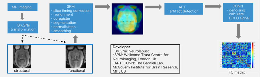
Fig. 1 Analysis Process
After converting the original data to a NifTI file with Bru2Nii, registration processing, smoothing processing, etc. are performed with SPM. Slices with large motion in ART are detected and removed. Noise is removed and the BOLD signal is calculated with CONN. Finally, the obtained data and ROI data of 52 regions of the anatomically segmented side of the brain are combined to create a FC matrix.
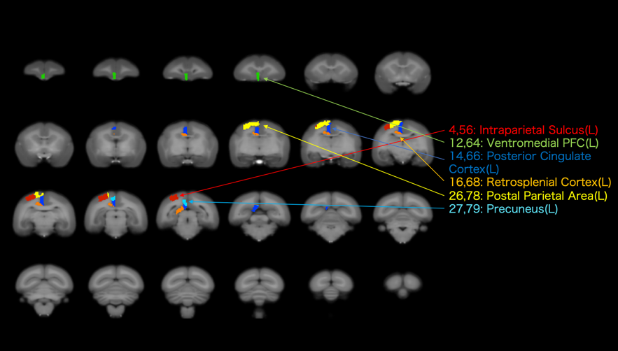
Fig. 2 ROIs of the DMN
The marmoset’s brain in coronal view with several slices and the ROIs of the human and marmoset DMN regions are presented.Human DMN regions: ventromedial prefrontal cortex (12, 64), posterior cingulate cortex (14, 66), and wedge front part (27, 79)Marmoset DMN regions: intercostal groove (4, 56), posterior cingulate cortex (14, 66), encephalic enlarged posterior cortex (16, 68), and parietal cortex (26, 78)The numbers indicate the numbers of the ROIs (1–52 for the left brain and 53–104 for the right brain).
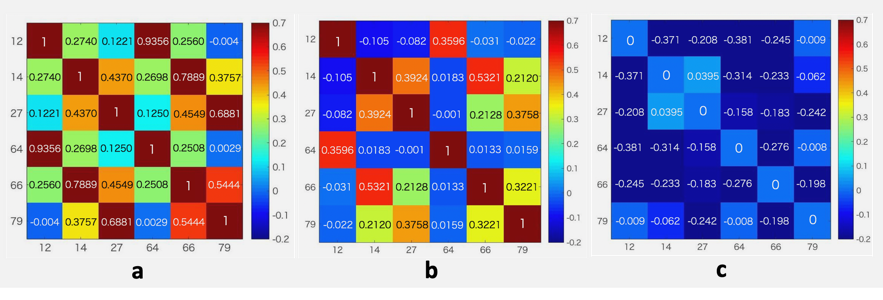
Fig. 3 FC Matrix between ROIs in the Human DMN
The correlation coefficients between ROIs are displayed in a matrix in accordance with the color bar scale, and the numbers correspond to the region numbers in Fig. 2. The human DMN regions are the regions indicated in reference No.3 applied to the ROIs in Fig. 2 of the marmoset brain.(a) Correlation coefficient in a matrix under an awake condition.(b) Correlation coefficient in a matrix under isoflurane anesthesia.(c) Correlation coefficient in a matrix obtained by subtracting the correlation coefficient in a matrix subtracted under an awake condition from the value under isoflurane anesthesia.
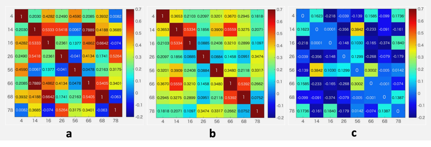
Fig. 4 FC Matrix between ROIs in the Marmoset DMN
The correlation coefficients between ROIs are displayed in a matrix in accordance with the color bar scale, and the numbers correspond to the region numbers in Fig. 2. The marmoset DMN regions are the regions indicated in reference No.4 applied to the ROIs in Fig. 2 of the marmoset brain.(a) Correlation coefficient in a matrix under an awake condition.(b) Correlation coefficient in a matrix under isoflurane anesthesia.(c) Correlation coefficient in a matrix obtained by subtracting the value under an awake condition from the value under isoflurane anesthesia.