3692
Neurological, Physiological, and Behavioral Effects Following Acute Cannabis Nose-Only Exposure in C57BL/6J MiceYasmeen Farra1, Dongyang Yi1, James Coleman2, Praveen Kulkarni2,3, Craig Ferris2,3, Jessica Oakes1, and Chiara Bellini1
1Bioengineering, Northeastern University, Boston, MA, United States, 2Center for Translational Neuroimaging, Northeastern University, Boston, MA, United States, 3Psychology, Northeastern University, Boston, MA, United States
Synopsis
Cannabis use is rising worldwide. Robust standardized methodologies in animal models are needed to further elucidate the impact of cannabis on overall health. Our aim was to identify acute exposure methods that provide a viable model for human cannabis consumption. C57BL/6J mice were exposed to cannabis aerosols using a Volcano® vaporizing device. Cannabis dosage levels were identified that elicited a human-like response. Animals were exposed while undergoing fMRI scans of their neurological activity. BOLD data, coupled with blood pressure and behavioral tests, demonstrated that our exposure methods generated a reproducible response that can be adapted for further studies.
Introduction
Cannabis is used by an estimated 183 million adults annually, representing well over half of all illicit drug use worldwide.1 There is inconclusive scientific evidence regarding the health effects of cannabis and therefore there is a need for established, reproducible methods of acute cannabis exposure in animal studies that model human consumption.2-7 Only then can these methods be expanded to develop a comprehensive understanding of the long-term, systematic effects of cannabis. In this work, we modeled the health effects of acute cannabis administration in recreational-use populations using nose-only, cannabis aerosol inhalation in mice.Methods
Male C57BL/6J mice aged 8-10 weeks and weighing approximately 23-27 grams were used. Two sets of experiments were performed: (1) correlation between cannabis exposure and THC concentration in the blood (n = 18) and (2) assessment of neurological, physiological, and behavioral changes in mice following exposure (n = 24) to an optimal amount of cannabis. For both objectives, acute nose-only cannabis exposure was compared with nose-only exposure to room air. Cannabis containing 13.1% THC and negligible CBD was used in all procedures. The Volcano® vaporizing system (Storz and Bickel, Tuttlingen, Germany) was used to fill a bag with cannabis aerosols.8-9 Particles generated from cannabis were examined for particle mass concentration and sizing. An ELISA assay for serum THC concentration identified the optimal cannabis dosage to match observed human response. Assessment of neurological activity in response to cannabis exposure was conducted using functional magnetic resonance imaging (fMRI). An inline duct fan was modified to pump cannabis directly to an awake restrained mouse via nose-only tubing (Fig. 1). Imaging took place using a Bruker BioSpec 7.0T/20-cm USR horizontal magnet (Bruker, Billerica, Massachusetts) and a 20-G/cm magnetic field gradient insert (ID=12cm). Functional images were acquired using a single-shot RARE (Rapidly Acquired with Refocused Echos) pulse sequence (18 slices; 0.75 mm; FOV 1.8 cm; data matrix 96 X 96; TR 6 sec; TE 4 msec; Effect ET 24 msec; 15 min acquisition time). Images were aligned and registered to a 3D mouse atlas, which is segmented and labeled with 116 discrete anatomical regions. Matrices that transformed each subject’s anatomy were used to embed each slice within the atlas. Images acquired after cannabis exposure were compared to baseline. A non-parametric Kruskal-Wallis test was used to compare the average signal intensity in each of the 15,000 voxels for their first 5 minutes baseline (acquisitions 1-50) to minutes 5 – 15 (acquisitions 50-150). Mice were also tested for physiological and behavioral responses to cannabis exposure using non-invasive blood pressure tail-cuffs and open field behavioral tests. Statistical analyses of all data were performed using a threshold for statistical significance of p < 0.05.Results
Particulate matter quantification showed that the exposure system varies linearly in dose efficiency with amount of cannabis input. A THC serum concentration of approximately 136 ng/mL was produced, which is comparable with reported values of human THC concentration following high-dose cannabis consumption. THC concentrations present in blood samples following acute inhalation experienced a linear effect with cannabis dosages (Fig. 2). fMRI imaging demonstrated that cannabis caused increased BOLD activation in many regions of the brain (Fig. 3). Blood pressure and heart rate were shown to decrease following acute cannabis exposure. Open-field behavioral tests indicated that despite similar levels of overall mobility, cannabis-exposed mice spent significantly more time near walls and away from the center of the testing space than the mice exposed only to room air (Fig. 4).Discussion
The methods conducted in this work produce a consistent effect in mice exposed to aerosolized cannabis. We show that the cannabis particles generated by the Volcano® system are within the respirable range and cause a physiological response within the body (Fig. 5). Clear CNS-mediated effects of cannabis inhalation, are exhibited by fMRI results of significant positive BOLD signal activation in 15 brain regions and negative BOLD activation in 25 regions of the brain, including the brain stem which modulates sympathetic and parasympathetic response. Blood pressure and heart rate results also indicate that there is a bradycardic and hypotensive reaction that agrees with the effects shown in the fMRI data. Finally, behavioral testing data suggests that cannabis-exposed mice experienced significant levels of anxiety following exposure, which agrees with the common human symptoms of anxiety and psychosis following consumption of cannabis.10Conclusion
Acute cannabis exposures in mice were shown to provide a viable model for human cannabis consumption. Particles delivered via nose-only inhalation elicited physiological, neurological and behavioral responses in the mice as supported by the fMRI data, blood pressure, and behavioral activity tests.Acknowledgements
This work was supported, in part, by the Northeastern University Tier 1 Seed Funds. Undergraduates Andrew Szendrey and Chris Le are gratefully acknowledged for their assistance in the experimental methods expressed here.References
- UNDOC. (2017). Fact Sheet on Statistics and Trends in Illicit Drugs. World Drug Report. https://doi.org/10.18356/c595e10f-en
- Fergusson, D. M., & Boden, J. M. (2008). Cannabis use and later life outcomes. Addiction, 103(6). https://doi.org/10.1111/j.1360-0443.2008.02221.x
- Baker, D., Pryce, G., Giovannoni, G., & Thompson, A. J. (2003). Review The therapeutic potential of cannabis. Lancet Neurology, 2(May), 291–298. https://doi.org/10.1016/S1474-4422(03)00381-8
- Whiting, P. F., Wolff, R. F., Deshpande, S., Di Nisio, M., Duffy, S., Hernandez, A. V., … Kleijnen, J. (2015). Cannabinoids for medical use: A systematic review and meta-analysis. JAMA - Journal of the American Medical Association, 313(24), 2456–2473. https://doi.org/10.1001/jama.2015.6358
- Woolridge, E., Barton, S., Samuel, J., Osorio, J., Dougherty, A., & Holdcroft, A. (2005). Cannabis use in HIV for pain and other medical symptoms. Journal of Pain, 29(4). https://doi.org/10.1016/j.jpainsymman.2004.07.011
- Hall, W., & Degenhardt, L. (2009). Adverse health effects of non-medical cannabis use. Lancet, 374(9698), 1383–1391. https://doi.org/10.1016/S0140-6736(09)61037-0
- Patton, G. C., Coffey, C., Carlin, J. B., Degenhardt, L., Lynskey, M., & Hall, W. (2002). Cannabis use and mental health in young people: cohort study. BMJ, 325(7374). https://doi.org/10.1136/bmj.325.7374.1195
- Hazekamp, A., Ruhaak, R., Zuurman, L., Van Gerven, J., & Verpoorte, R. (2005). Evaluation of a Vaporizing Device (Volcano1) for the Pulmonary Administration of Tetrahydrocannabinol. International Journal of Drug Development and Research, 3(2), 26–33. https://doi.org/10.1002/jps
- Abrams, D. I., Vizoso, H. P., Shade, S. B., Jay, C., Kelly, M. E., & Benowitz, N. L. (2007). Vaporization as a smokeless cannabis delivery system: A pilot study. Clinical Pharmacology and Therapeutics, 82(5), 572–578. https://doi.org/10.1038/sj.clpt.6100200
- Morrison, P. D., Zois, V., McKeown, D. A., Lee, T. D., Holt, D. W., Powell, J. F., … Murray, R. M. (2009). The acute effects of synthetic intravenous9- tetrahydrocannabinol on psychosis, mood and cognitive functioning. Psychological Medicine, 39(10), 1607–1616. https://doi.org/10.1017/S0033291709005522
Figures
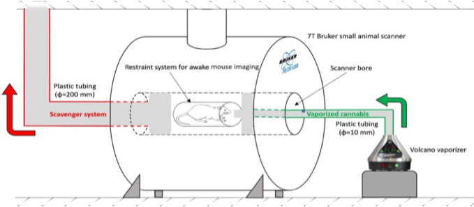
Figure 1. Schematic detailing
the exposure setup for awake animal MRI experiments with cannabis nose-only
exposure.
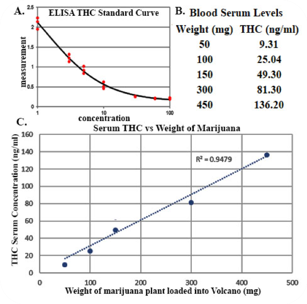
Figure 2. A. THC concentration in the mouse blood serum is estimated
using an ELISA standard curve. B.
THC concentration in the blood is calculated at varying doses of 13.1% THC
cannabis. C. THC concentration in
the serum is linearly related to the dosage of cannabis loaded into the
Volcano®.
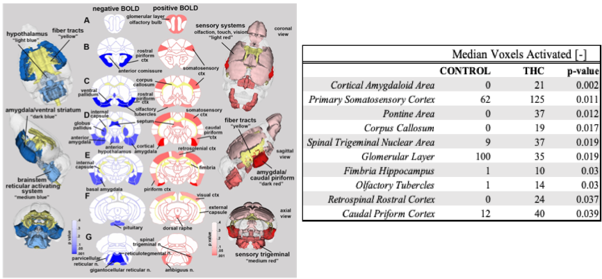
Figure 3. LEFT: 3D and 2D probability maps that show brain regions most
likely to be activated during inhaled exposure to vaporized cannabis in the
MRI. Regions coded in red show areas with significant positive BOLD activation,
while areas in blue show areas with significant negative BOLD activation.
Although all areas shown here have significant activation (p<0.05), darker
shaded regions correspond to lower p-values, while higher p-values are shaded
lighter.
RIGHT: Median number of voxels activated in select brain regions
with significant positive BOLD activity in cannabis exposed vs. control groups.
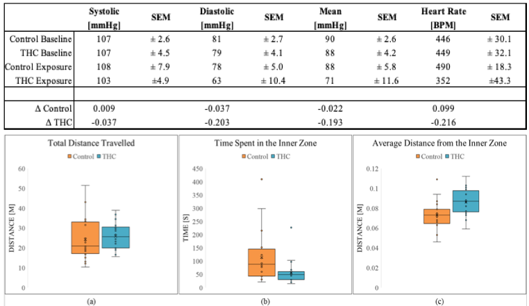
Figure 4. TOP: Physiological blood pressure and heart rate data taken from
control (N=8) and cannabis-exposed (N=7) using non-invasive tail-cuff technique
prior to and immediately following nose-only exposure.
BOTTOM: Key behavioral data demonstrating results of open
field behavioral testing. (a) Total distance travelled is similar between groups, suggesting that overall mobility is not
impaired and should not affect other metrics. (b) Time spent in the inner zone
is significantly (p=0.021) higher for control mice,
suggesting that mice exposed to cannabis demonstrate anxiety-like
behavior. (c) Average distance from the inner zone is significantly (p=0.004)
higher for cannabis-exposed mice, suggesting that cannabis induces anxiety-like behavior.
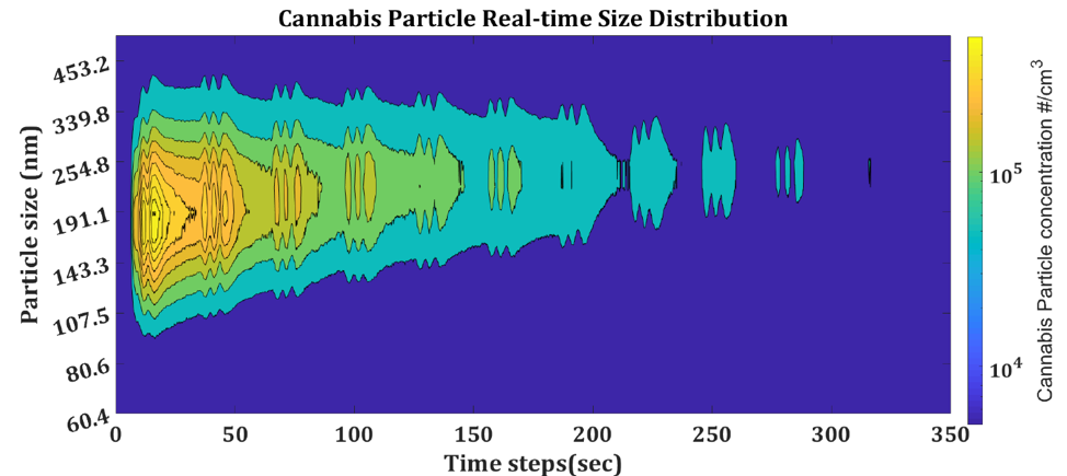
Figure 5. Preliminary particulate
matter data detailing cannabis particle size and concentration distribution.