3690
Awake Macaque MRI Setup on 7T Human Scanner Platform for High-Resolution Anatomical and Functional Imaging1Interdisciplinary Institute of Neuroscience and Technology, Qiushi Academy for Advanced Studies, College of Biomedical Engineering & Instrument Science, Zhejiang University, Hangzhou, China, 2School of Medicine, Zhejiang University, Hangzhou, China, 3MR Collaboration Northeast Asia, Siemens Healthcare, Hangzhou, China
Synopsis
We introduced a customized setup for awake sitting macaque MRI in a 7T horizontal-bore human scanner. Considerations in the specialized design of monkey cage and head fixation device consisting of 1) monkey comfort in a Sphinx position, 2) stable head fixation, 3) easy mounting of the surface array for cortex imaging, and 4) easy presentation of visual stimuli. Our findings demonstrate that, in a well-trained awake monkey, with the increased contrast sensitivity and substantial gains in spatial resolution offered by ultra-high field MRI and a custom-made dense RF array, our setup is capable of achieving improved SNR and high time efficiency.
Introduction
Non-human primates (NHPs) are a valuable model for understanding the neural circuits underlying human cognition, behavior, and disease. Research in awake behaving monkeys may directly bridge non-invasive human studies with the body of knowledge gained from invasive studies in monkeys. In recent years, there have been several awake NHP studies by using high and ultra-high field MRI [1-6], most of which were limited to the 3T human scanner or vertical-bore animal scanner, and with contrast agent injection to pursue higher spatial resolution and the better contrast-to-noise ratio (CNR).
Here we introduce a customized setup for awake sitting macaque MRI in a 7T horizontal-bore human scanner. Considerations in the specialized design of monkey cage and head fixation device consisting of 1) monkey comfort in a Sphinx position, 2) stable head fixation, 3) easy mounting of a dense surface array for cortex imaging, and 4) easy presentation of visual stimuli. Anatomical and functional imaging has been collected to evaluate its performance.
Method
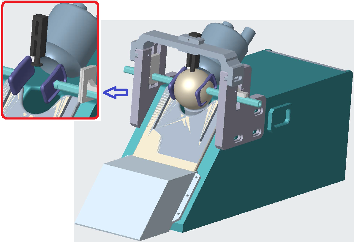
After a few months’ adaption and training, a male monkey (weight: 9kg, age: 8-year old) with a headpost installed was trained to sit calmly inside the scanner bore in a Sphinx position and undergo MRI scans. All procedures were in accordance with NIH standards and approved by our Institutional Animal Care Committee. A custom primate chair (height: 33cm, width: 23cm, made by polycarbonate) was designed to accommodate the monkey during the scan, with additional support holders attached to fix the headpost, as shown in Fig.1. Cushions supported by two additional bars s covered the ears to reduced scan noise. A custom-built surface array, which consists of a transmit-only loop (diameter: 9cm) and 16 peripherally located receive-only loops (diameter:1.5cm) [7], was mounted over the macaque head covering visual cortical areas as indicated in Fig.1.
The awake macaque experiment was performed on a 7T research scanner (Siemens Healthcare, Erlangen, Germany) equipped with a whole-body gradient set (70mT/m and 200T/m/s). High-spatial-resolution (0.2mm in-plane) anatomical images (T2*-weighted 2D GRE) were acquired with an acceleration rate of 2 along F/H phase encoding direction [TR/TE/α: 1,600ms/20ms/60°, matrix size 384×384, FOV 76×76mm2, slice thickness 1mm, 1 average, scan time 5'34'']. For functional brain imaging, visual stimulus (red/blue checkboard, temporal frequency of 8Hz) was presented to the monkey on a screen facing the monkey’s eyes, and a block design with 144 images in 8 blocks of 9 images OFF/ON was used. Single-shot gradient-echo EPI was used to capture the BOLD signal with an acceleration rate of 3 along F/H phase encoding direction [TR/TE/α: 2,000ms/20ms/70°, matrix size 96×96, FOV 96×96mm2, slice thickness 1mm, bandwidth 1,212Hz/px, echo spacing 1ms]. The significant response to visual stimuli was calculated by identifying voxels with significant T-test p-values with generalized linear models (GLM).
Results
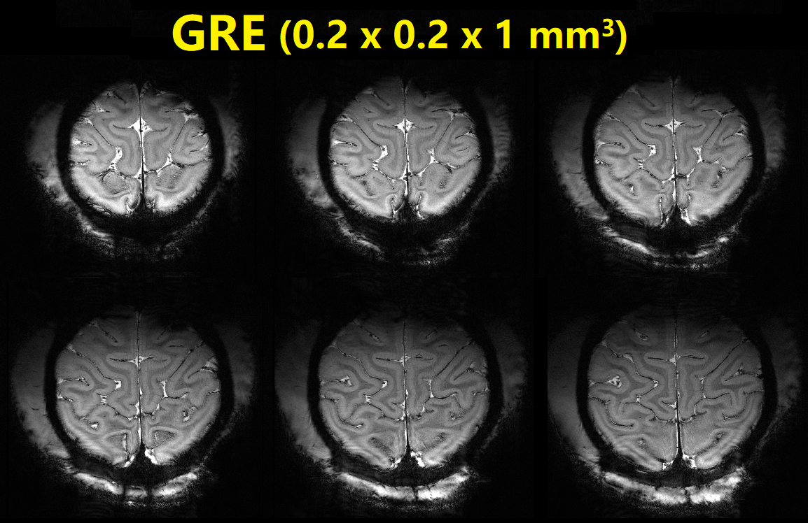
Fig.2 shows, high-resolution MRI microscopy images have been successfully obtained over monkey cortical areas under the awake condition in ~5 minutes. Detailed structural information, revealing diving cortical pial vessels and cortical laminar patterns, can be visualized in anatomical scans as discerned in the T2*-weighted GRE images.
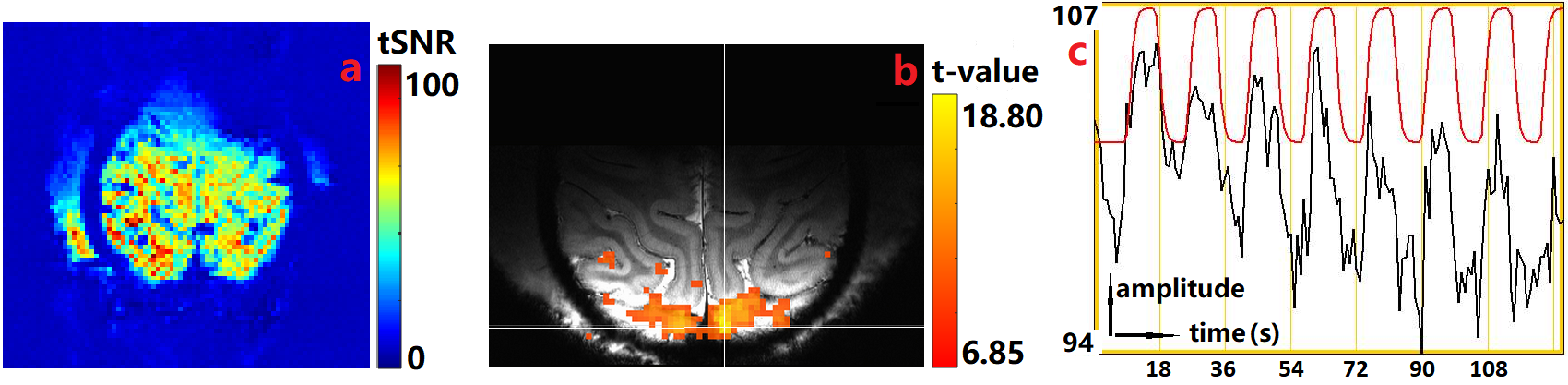
Fig.3a shows an assessment of tSNR for BOLD imaging. The profile indicates the stability of the monkey head, the good performance in receive sensitivity and data acquisition acceleration (GRAPPA factor 3) of the RF coil, and the good overall tSNR (50~90) for functional recording at 1mm isotropic resolution. Fig.3b shows functional activation maps overlaid onto 2D GRE images. Bilaterally, visual areas (including V1. V2 and MST) were significantly activated by high frequency flashing checkerboard stimuli presented on a monitor (uncorrected p<3×10⁻¹⁰, corrected p<1×10⁻⁸). Fig.3c shows the strong correlation between BOLD time course (black: peak voxel) and ideal general linear model (red: signal change range 5%-7%).
Discussion and Conclusion
We have evaluated the performance of our setup for obtaining anatomical and functional imaging in an awake monkey seated in a 7T scanner with a horizontal bore. A customized 16-channel multiarray RF coil aided in achieving high-resolution imaging in an awake head-fixed macaque monkey trained to sit calmly in the MRI. 200um in-plane-resolution T2* images acquired in ~5min showed clear, stable structural images, and 1mm-isotropic EPI images recorded robust BOLD activation in visual cortical areas. Our findings clearly demonstrate that, in a well-trained awake monkey, with the increased contrast sensitivity and substantial gains in spatial resolution offered by ultra-high field MRI and a custom-made dense RF array, our setup is capable of achieving improved image SNR and high time efficiency.Acknowledgements
National Natural Science Foundation 81701774 and 61771423. We thank Jiaming Hu, Jianbao Wang, and Jie Zhao for helpful discussion and technical support, and Wim Vanduffel and Doris Tsao for tremendous helpful advice in the early phases of this project.References
[1] N.K. Logothetis, H. Guggenberger, S. Peled, J. Pauls, Nat. Neurosci. 2 (1999) 555–562.
[2] L. Stefanacci, P. Reber, J. Costanza, E. Wong, R. Buxton, S. Zola, L. Squire, T. Albright, Neuron 20 (1998) 1051–1057.
[3] D.J. Dubowitz, D.Y. Chen, D.J. Atkinson, K.L. Grieve, B. Gillikin, W.G. Bradley Jr., R.A. Andersen, Neuroreport 9 (1998) 2213–2218.
[4] K. Nakahara, T. Hayashi, S. Konishi, Y. Miyashita, Science 295 (2002) 1532– 1536.
[5] D.Y. Tsao, W. Vanduffel, Y. Sasaki, D. Fize, T.A. Knutsen, J.B. Mandeville, L.L. Wald, A.M. Dale, B.R. Rosen, D.C. Van Essen, M.S. Livingstone, G.A. Orban, R.B. Tootell, Neuron 39 (2003) 555–568.
[6] G. Chen, F. Wang, J.C. Gore, A.W. Roe, NeuroImage 59(2012) 3441-3449.
[7] X. Zhang, Y. Gao, M. Qian, Y. Sun, A.W. Roe, ISMRM 2018 4278.
Figures