3689
An awake mouse MRI method using mouse clothes for fMRI applicationsSosuke Yoshinaga1, Satoshi Fujiwara1, Shunsuke Kusanagi1, Kazunari Kimura1, Rikita Araki2, Mitsuhiro Takeda1, and Hiroaki Terasawa1
1Faculty of Life Sciences, Kumamoto University, Kumamoto, Japan, 2Bruker Japan K.K., Yokohama, Japan
Synopsis
In current awake MRI methods, fixing apparatuses implanted in the brain by surgery and acclimation procedures by training have been explored, to suppress head movement in the scanner. Previously, we reported an awake MRI method using mouse clothes designed for a cryogenic coil system, without surgery and training. We now report the successful optimization of the awake mouse MRI method for fMRI applications, using the newly designed mouse clothes. Resting-state analyses showed bilateral functional connectivities in the cortical and limbic system networks in an awake mouse. Our method will contribute to the acquisition of awake-specific neuronal responses.
Introduction
Functional MRI studies for awake animals are attracting increased attention, for their potential to capture awake-specific neuronal responses. In the current awake MRI methods, fixing apparatuses implanted in the brain by surgery [1] and acclimation procedures by training [2] have been explored, to suppress head movement in the scanner. However, these methods require technical proficiency. If the animal accommodation space in the MRI coils is small, a bulky apparatus cannot be used, which is the case for the latest mouse brain cryogenic probe (Fig. 1A). An awake MRI method with the cryogenic coil system was recently reported, but it utilizes head–fixation implements [3]. Previously, we reported an awake MRI method using mouse clothes designed for the cryogenic coil system, without surgery and training [4]. This approach enables the head motion to be suppressed in a non-invasive manner. Although the previous clothes suppressed the mouse brain movement to some extent, the quality of the MR images was lower than that of anesthetized mice without clothes. The aim of this study is to acquire higher Signal-to-Noise Ratio (SNR) functional MR images, by optimizing the mouse clothes.Methods
MRI experiments were performed with a 7.0 Tesla Bruker BioSpec 70/20 scanner and a mouse brain 2-channel phased array surface cryogenic coil. The mouse clothes were newly designed to fit the mouse head accommodation space of the coil (Fig. 1). The experiments were performed with adult male C57BL/6 mice (22–25 g). Throughout the experiments, the mice were maintained at 37±1°C by a heating pad, and the respiration and heart rates were monitored. For their first experience of wearing the clothes, the mice were anesthetized with 2% isoflurane, placed in the clothes, and fixed within the MRI cradle. At 60 minutes after cessation of the anesthesia, EPI images (FOV = 18×18 mm2, matrix = 90×90, resolution = 0.2×0.2 mm2, slice thickness = 0.6 mm, number of slices = 16, TR/TE = 2,000/15 ms, flip angle = 75°) and FISP images (FOV = 18×18 mm2, matrix = 90×90, resolution = 0.2×0.2 mm2, slice thickness = 1.5 mm, number of slices = 5, TR/TE = 2.3/1.15 ms, flip angle = 60°) were acquired. The standard deviations (S.D.) of the movements of the mouse brains in the three directions of Left–Right (L–R), Superior–Inferior (S–I), and Anterior–Posterior (A–P) were calculated using SPM8, based on the acquired FISP images. For resting-state functional connectivities, an Independent Component Analysis (ICA) was applied to the EPI images with the FSL software, MELODIC.Results
At first, FISP images of awake mice wearing the clothes previously designed for the room temperature coil were acquired using the cryogenic coil, and we found that the mice tended to escape backward from the clothes. Therefore, we newly designed clothes intended to prevent the escape by squeezing the body, sewn in advance together with a neck belt (Design A). As a result, better brain immobilization effects were obtained using the Design A clothes, but the SNR of the MR images was lower than that of anesthetized mice without clothes. Thus, we improved the clothes by removing the hood cover material, to obtain higher SNR images (Design B) (Fig. 1B(b)). During the awake MRI experiments, the respiration and heart rates of the mice stably remained within the ranges normally observed in awake mice. The S.D. of the mouse brain movements in the Design B clothes were larger than those of the anesthetized mice, but smaller than those in the Design A clothes and remained within the target S.D. value range (< 0.1 mm) (Fig. 2). The SNR of the EPI images in the Design B clothes was better than that of the Design A clothes, and it was about 80% of that of the anesthetized mice without clothes (Fig. 3). Next, resting-state analyses were performed for awake mice wearing the Design B clothes (Fig. 4). As a result, the bilateral cortical and limbic system networks were observed.Discussion
The mouse brain immobilization effect of the Design B clothes was better than that of Design A. In addition, higher SNR MR images were obtained with Design B than with Design A. The removal of the hood cover part in Design B shortened the brain–coil distance, resulting in the higher SNR and improved head movement suppression. The observation of bilateral functional connectivities in the cortical and limbic system networks agrees well with previous studies [3], demonstrating that our method is practical and more tractable.Conclusion
We successfully optimized the awake mouse MRI method for the cryogenic coil system, and observed functional connectivities in the resting state. Our method will greatly contribute to the acquisition of awake-specific neuronal responses.Acknowledgements
No acknowledgement found.References
[1] Desai, M. et al., J. Neurophysiol. 105, 1393–1405 (2011), [2] King, J.A. et al., J. Neurosci. Methods 148, 154–160 (2005), [3] Yoshida, K. et al., J. Neurosci. Methods 274, 38–48 (2016), [4] Nakata, E. et al., Proc. Intl. Soc. Mag. Reson. Med. 24, 1757 (2016)Figures

Fig. 1 Mouse head accommodation space
of a cryogenic coil and procedure for the awake mouse MRI. (A) The dome-shaped mouse
head accommodation space of a cryogenic coil-imitating position gauge (Bruker)
is shown. (B) A mouse is being dressed in Design B clothes (a), placed on the MRI
cradle with the clothes (b), and fitted with the cryogenic coil system (c).

Fig. 2 Immobilization effects of the designed
clothes on the mouse brain movement.
The S.D. of the brain movements of
mice wearing the Design A and B clothes were
compared (N=3). As a reference, those of the anesthetized mice without clothes
(Anesthetized) are shown (N=3). The S.D. in the three directions of Left–Right (L–R,
red), Superior–Inferior (S–I, green), and Anterior–Posterior (A–P,
blue) were calculated, based on the acquired FISP
images. The target S.D. value (0.1 mm) is shown by the red dashed line.
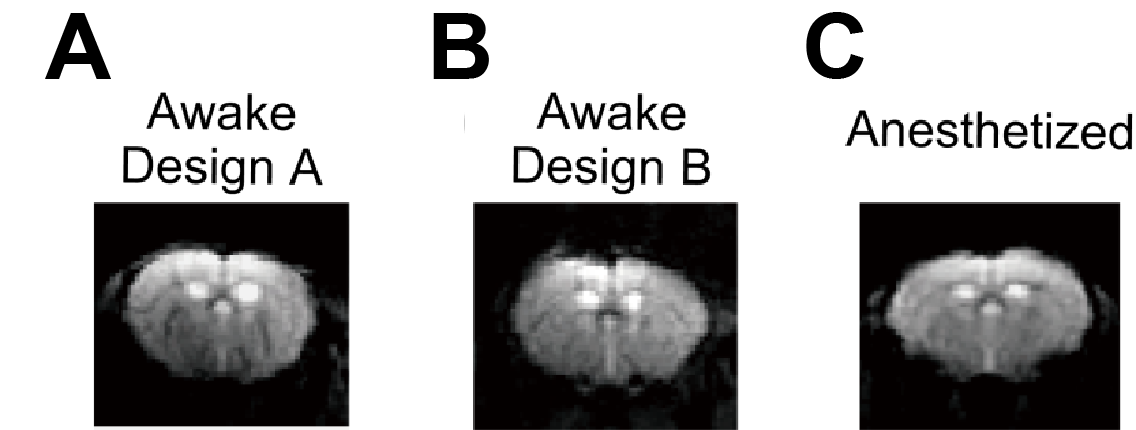
Fig.
3 Comparison of EPI brain images of
awake mice wearing the differently designed clothes.
Representative
EPI images of awake mice wearing the Design A and B clothes are
shown in (A) and (B), respectively. As a reference, a representative EPI image
of an anesthetized mouse without clothes (Anesthetized) is shown in (C). All
EPI images were acquired in axial cross sections of the mice.
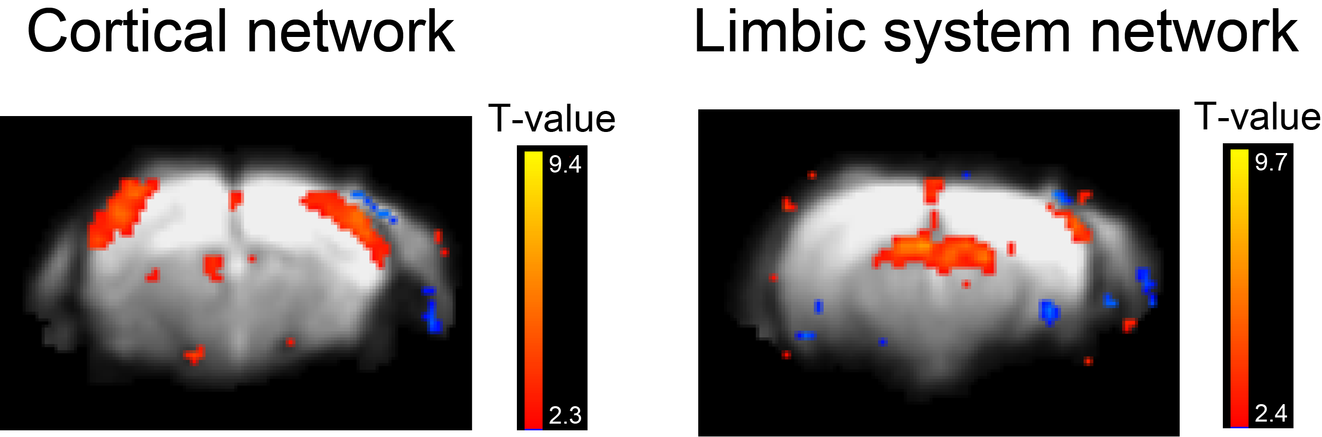
Fig. 4 Functional networks in the brain of an awake
mouse.
Representative
functional connectivity maps in the brain of an awake mouse wearing the Design B clothes,
resulting from ICA, are presented. The color-coded T-maps are overlaid on
average EPI functional images.