3687
BOLD-fMRI evaluation of analgesic candidates with different analgesic mechanisms against allodynia-specific pain in a chronic pain animal modelMikio Sameshima1, Sosuke Yoshinaga1, Naoya Yuzuriha1, Mitsuhiro Takeda1, and Hiroaki Terasawa1
1Faculty of Life Sciences, Kumamoto University, Kumamoto, Japan
Synopsis
The aim of this study is to evaluate the pain-relieving effect of a novel chemokine signal-inhibiting compound on allodynia-specific responses in a chronic pain animal model, using our BOLD-fMRI-based pain evaluation system with a green laser. We observed activations in four brain regions related to pain. We found that the compound suppresses green laser-evoked allodynia-specific responses in three regions. We expect that the compound has synergistic pain-relieving effects with existing analgesic agents. Our system is useful to evaluate the effects of new analgesic candidates in non-clinical and, expectantly, clinical studies.
Introduction
Fibromyalgia and neuropathic disorders characterized by chronic pain induce the pathological condition “allodynia”, in which a stimulus that is normally not painful causes pain sensations (Fig. 1A). Previously, we successfully observed allodynia-specific brain responses evoked by green laser stimulation in fibromyalgia model animals, using BOLD-fMRI. Recently, it was reported that chemokine signals enhance the excitatory synaptic transmission of neurons in chronic pain patients (Fig. 1B) [1]. We screened compounds that inhibit chemokine signals, and obtained Compound-A. The aim of this study is to evaluate the analgesic effect of Compound-A on the allodynia-specific responses in a rat chronic pain model, with our BOLD-fMRI-based pain evaluation system using a green laser [2].Methods
The reserpine-induced myalgia rats with the symptoms of allodynia were prepared [3]. The rats were ventilated and treated with gallamine and urethane (1.25 g/kg i.p.). Saline was administered (10 mL/kg i.v.), and 30 minutes later the BOLD experiment was performed. All MRI experiments were performed with a 7.0 Tesla Bruker BioSpec scanner and a rat brain 4-channel phased array surface coil (Bruker). Functional data were acquired with a 4-shot GRE-EPI sequence (TR: 2,000 ms, TE: 15 ms, FA: 45°, matrix: 64 × 64, FOV: 2.56 × 2.56 cm2, 13 slices, slice thickness: 0.6 mm). The green laser was used to irradiate the left hind paws 5 times for 2 s every 2 minutes, during the scans (Fig. 2). After the first BOLD experiment, the rats were treated with Compound-A (20 mg/kg i.p.), and 30 minutes later the same BOLD experiments were performed. The Independent Component Analysis (ICA) was performed with the FSL software. We searched for the brain regions that showed periodic BOLD responses with the frequency of the laser stimulation (8.3 mHz). The BOLD signal intensity was analyzed with the SPM8 software.Results
After the administration of saline, the primary somatosensory cortex (S1), insular cortex (IC), thalamus (Th), and anterior cingulate cortex (ACC), which are brain regions related to pain sensations, exhibited periodic signal intensity changes with the frequency (8.3 mHz) of the laser stimulation. Therefore, they were detected by ICA (Fig. 3A). Upon the laser stimulations, the signal intensities in S1, IC, Th, and ACC significantly increased. After the administration of Compound-A, the periodic signal intensity changes in S1, IC, and Th were detected by ICA, but the signal intensity increases were suppressed. Unlike the other three regions, the periodic signal intensity change in ACC was still significantly detected by ICA. Furthermore, the increases of the signal intensities in ACC were similar to those before the administration of Compound-A, and thus no suppression effect was observed (Fig. 3C).Discussion
Green laser-evoked signal increases are supposed to reflect the activation of the brain, based on the BOLD responses. The activation of the brain regions related to pain sensations was suppressed in S1, IC, and Th after the Compound-A administration. In contrast, the suppression of activation in ACC could not be confirmed after the Compound-A administration. In patients with fibromyalgia, the ACC region is easily activated, even during the anticipation of pain [4]. If this phenomenon is also applicable to the chronic pain model rat, then the ACC region may tend to be easily activated by weak stimulations. The ACC activation may be suppressed by increasing the dose of Compound-A.Conclusion
We found that a chemokine signal inhibitor, Compound-A, has the effect suppressing part of the allodynia-specific pain responses related to the chronic pain. Our experimental system is useful for the evaluation of new analgesic candidates in nonclinical and, expectantly, clinical studies.Acknowledgements
No acknowledgement found.References
[1] Zhang, Z. et al., Cell. Mol. Life Sci. 74, 3275–3291 (2017), [2] Yuzuriha, N. et al., Proc. Intl. Soc. Mag. Reson. Med. 24, 1722 (2016), [3] Nagakura, Y. et al., PAIN 146, 26–33 (2009), [4] Burgmer, M. et al., NeuroImage 44, 502–508 (2009)Figures
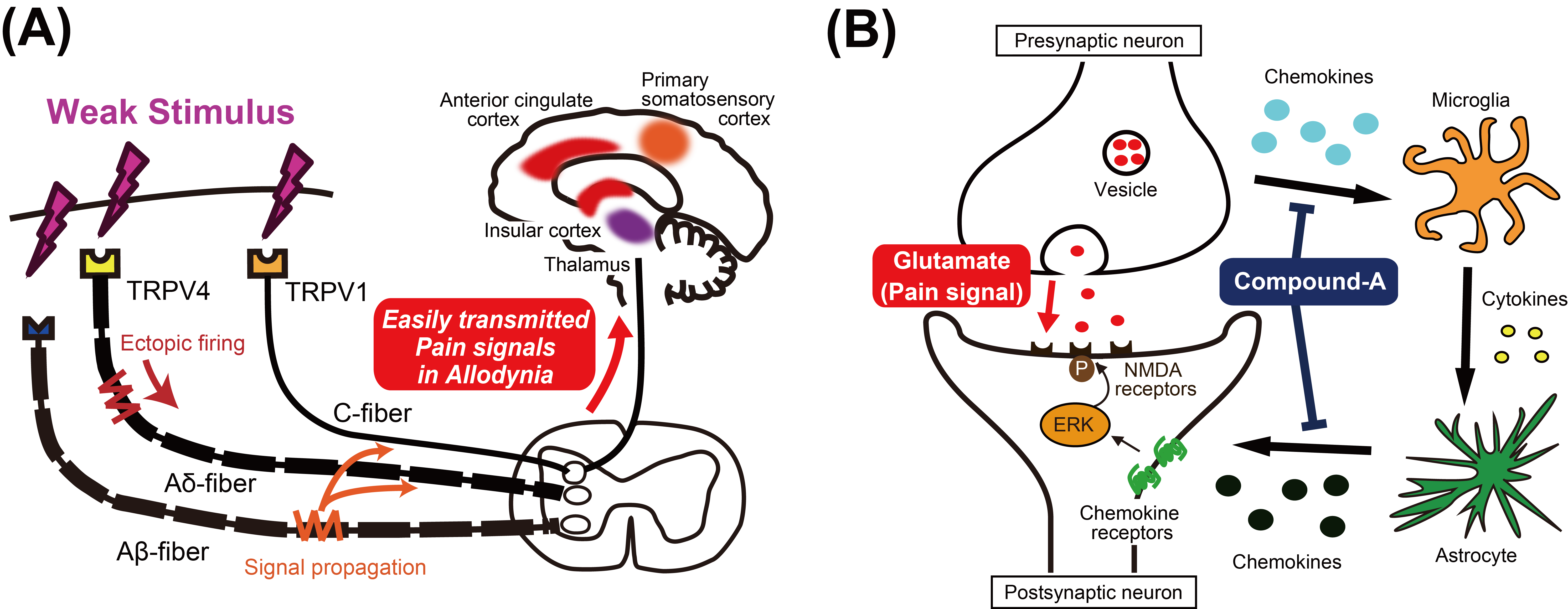
Fig. 1 Pain signals in chronic pain diseases and the
mechanism of action of the analgesic candidate Compound-A.
(A) Neuropathic disorders
induce the pathological condition “allodynia”, in which a weak stimulus that is
normally not painful causes pain sensations, due to signal propagation from
Aβ-fibers and ectopic firing of Aδ- and C-fibers. (B) Under chronic pain
conditions, the pain signal is upregulated by activated ERK. Presynaptic cells release
chemokines, which activate microglia and astrocytes. The chemokines released
from astrocytes activate ERK in postsynaptic cells. Compound-A is considered to
suppress the pain signal by blocking the chemokine signals.
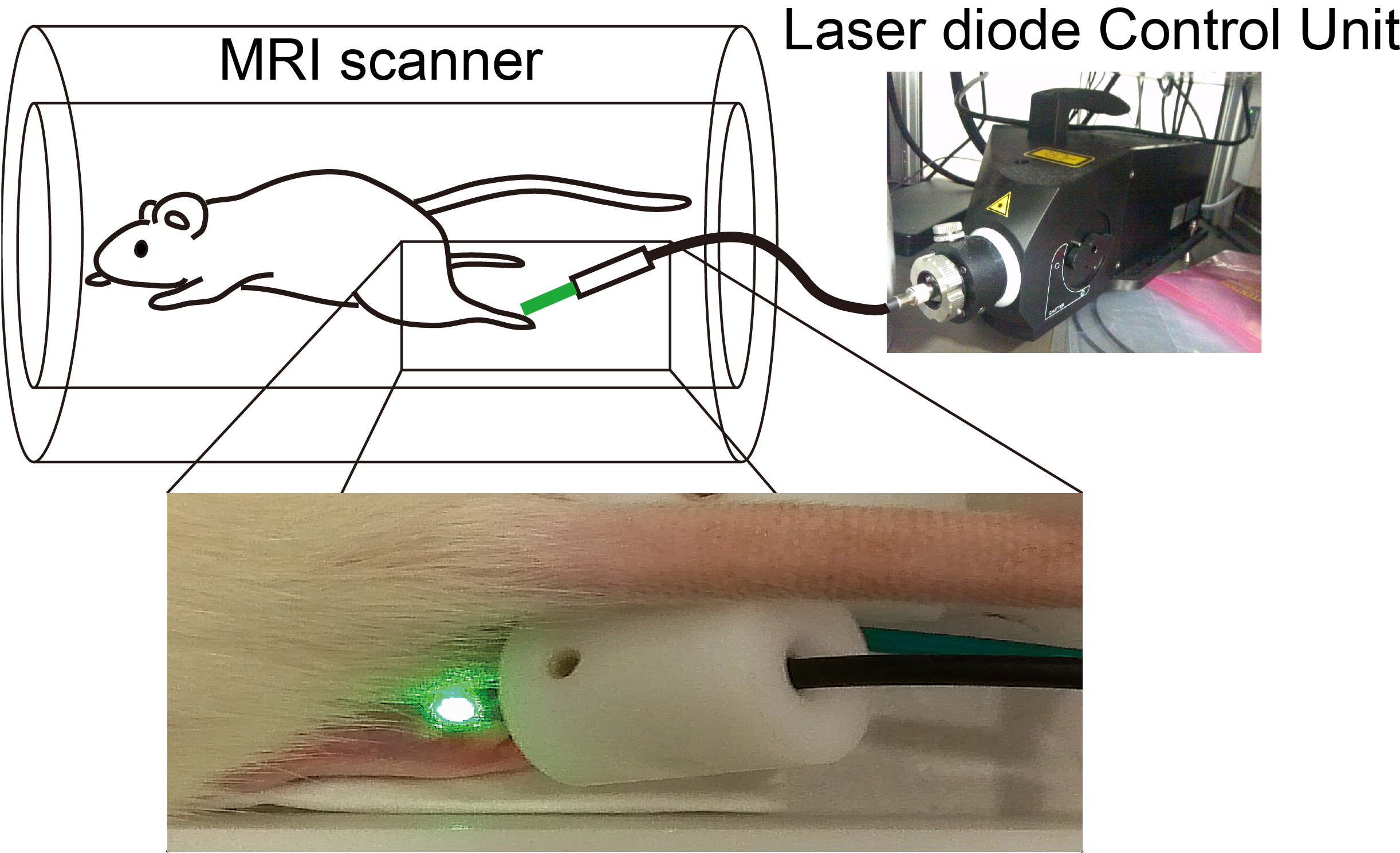
Fig. 2
On-site stimulation device for green laser-evoked BOLD MRI experiments.
An optical fiber from the laser diode control
unit, located outside the MRI room, was set in an MRI scanner. The left hind
paw of the rat was irradiated by the green laser light emitted from the optical
fiber.
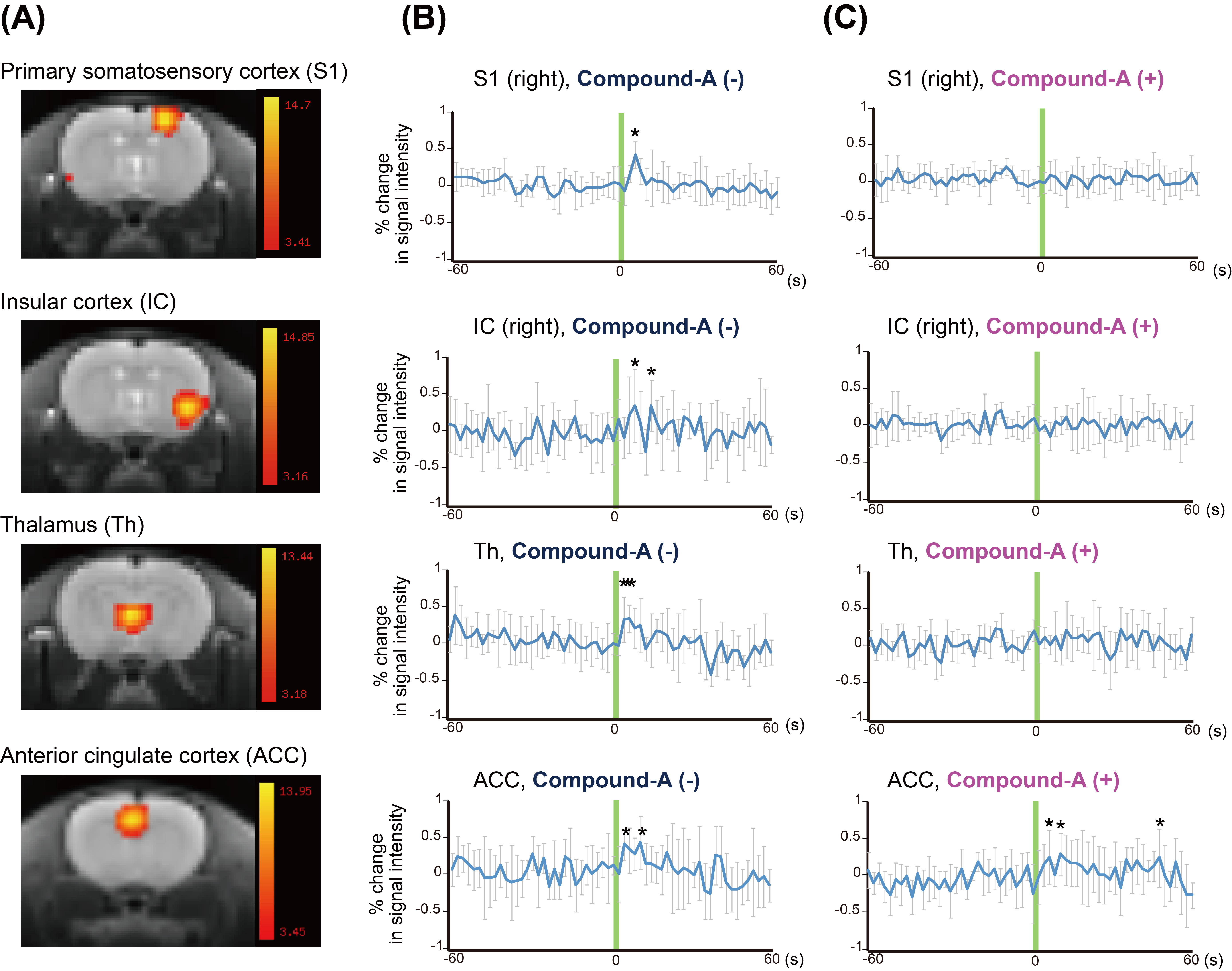
Fig. 3 Green laser-evoked brain
activation maps and event-related averages of the signal changes.
(A) Green laser-evoked brain activation maps are represented for Compound-A
(-) (n = 5). The color-coded T-maps based on ICA are overlaid on the T2w anatomical
standard images. (B) and (C) Event-related averages of the signal changes in
the activated regions (mean signal intensity ± S.D., *: p < 0.05) before (B) and after (C) the treatment with Compound A
(n = 5) are represented. The duration of green laser irradiation was 2 sec, as
shown by the green lines.