3686
Structural and Resting-State Functional Connectivity Changes of the MPS I Mouse Brain1Center for Magnetic Resonance Research, Department of Radiology, University of Minnesota, Minneapolis, MN, United States, 2Department of Biomedical Engineering, Penn State University, State College, PA, United States, 3Department of Neurosurgery, University of Minnesota, Minneapolis, MN, United States, 4College of Biological Sciences, University of Minnesota, Minneapolis, MN, United States
Synopsis
Mucopolysaccharidoses (MPSs) is a group of inherited lysosomal storage disorders that could cause multiple organ failure, cognitive impairment, and shortened life span. In spite of the recognizable clinical morphological and physiological features associated with MPSs, brain connectivity changes and physiopathologic mechanisms responsible for these alterations in the central nervous system are rarely studied but might be a reliable biomarker for disease severity and treatment efficacy. A new study of brain connectome on MPS I mouse model using resting-state functional MRI (rs-fMRI) technique was conducted and a dramatic deterioration on functional connections involving multiple brain regions were observed.
INTRODUCTION
Mucopolysaccharidoses (MPSs) is a group of rare genetic disorders characterized by a reduced or stopped production of lysosomal enzymes which are essential for glycosaminoglycan (GAG) catabolism in multiple organs1. The undegraded GAGs accumulate in the lysosomes, cause chronic cell swelling and progressive organ enlargement which further, and could lead to organ failure, cognitive impairment, and reduced life span. Given the clinical heterogeneity of MPSs, early and accurate diagnosis and severity ranking are challenging especially for central nervous system. Although morphological and physiological anomaly can be detected using various assay and imaging methods1-3, brain connectivity changes and physiopathologic mechanisms responsible for these alterations are rarely studied. The lack of reliable functional evaluation methods also impedes the development and test of new treatments for MPS patients with mental retardations and deterioration4. To bridge this gap, we hypothesize that resting-state functional MRI (rs-fMRI) can provide a safe, noninvasive, and sensitive method for MPS diagnosis and new treatment assessment. In this work, by utilizing a MPS I mouse model, we observed a dramatic functional connectivity deterioration on MPS I mouse brain.METHODS
Animals and scan conditions: Six MPS I-mutant mice (3 male/3 female) and the corresponding controls (n=5) with similar age and body weights were scanned under a protocol approved by the University of Minnesota. All animals were inducted with 5% isoflurane mixed in O2:N2O (30:70) gas and anesthetized with 1.6% isoflurane during the preparation. After mice were in the magnet, the anesthesia was switched to dexmedetomidine (i.p. 0.3 mg/kg bolus followed by 0.6 mg/kg/hr infusion). Data acquisition started once the respiration rate stabilized at > 140 BPM, and the animals’ physiology was monitored and well controlled throughout the study.
MRI experiments and data analysis: The MRI experiments were conducted on a 9.4T/31cm animal scanner (Varian/VNMRJ) using a single loop (1.5cm diameter) surface coil. T2 weighted anatomical images were acquired with TR/TE=4000/10 ms; matrix = 256 × 128; FOV=2.4 × 1.2 cm2; and 12 0.5 mm slices. Gradient echo (GE)-echo planar imaging (EPI) based rs-fMRI images were obtained with TR/TE = 1000/20 ms; matrix = 96 × 48; FOV = 2.4 × 1.2 cm; 12 slice with 0.5 mm thickness. For each mouse, 2-5 rs-fMRI datasets with 310 volumes were obtained. The rs-fMRI data were preprocessed with the standard pipeline and 43 RSNs were generated via seed analysis where brain regions outlined based on the mouse atlas (Allen Institute)5 were used as seeds.
RESULTS
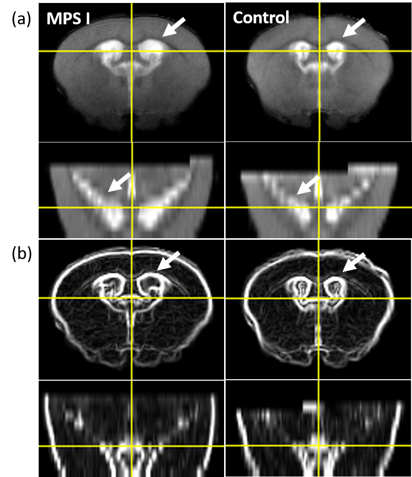
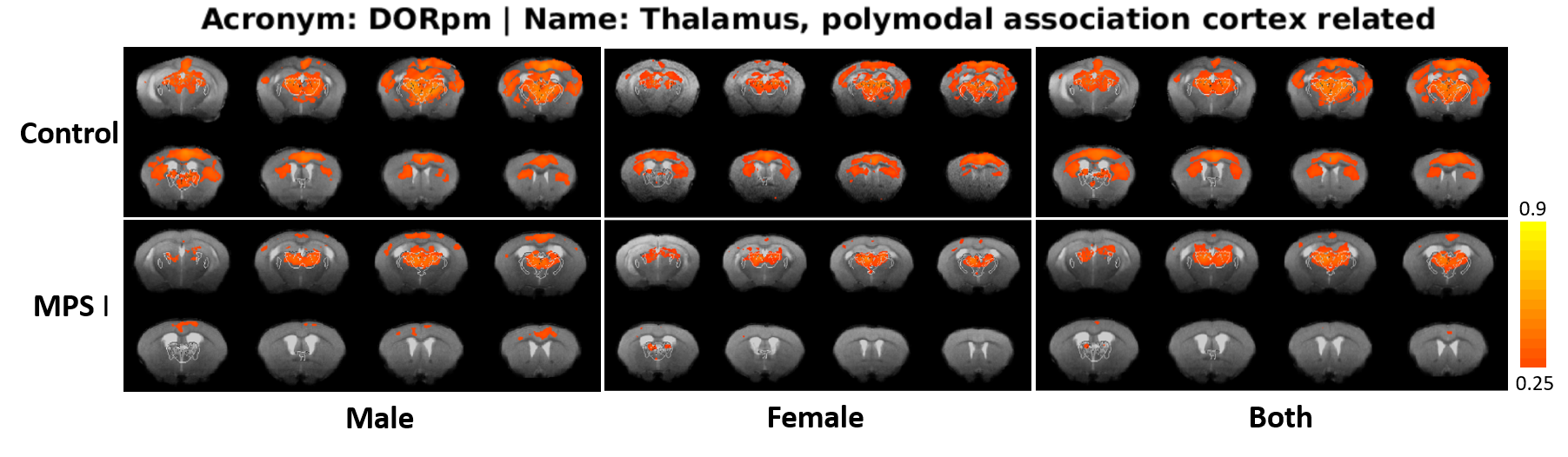
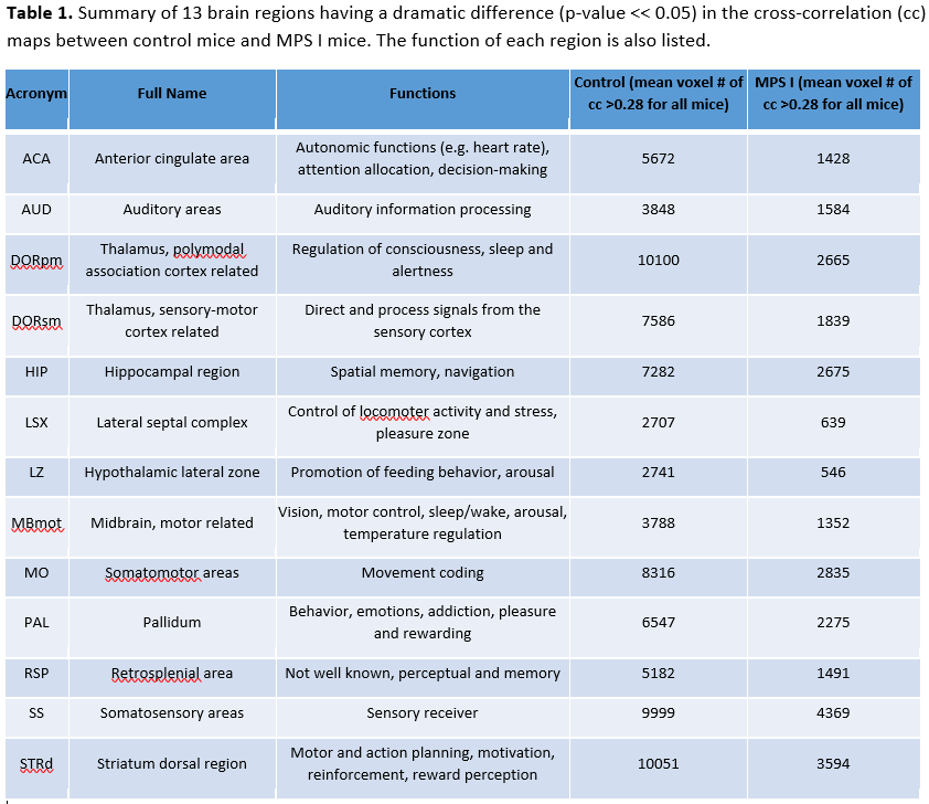
Figure 1 shows (a) anatomical structure and (b) brain region contour of the brain for control and MPS I mice with similar age and physiology. The co-registered transversal and coronal images are displayed in the same scale. MPS I mouse brain, especially in the perivascular spaces indicated by the white arrows, is significantly larger than the controls. Figure 2 illustrates an example of the resting-state functional connectivity differences between control and MPS I mice. In this case, the thalamus (polymodal association cortex related region), identified from atlas was used as a seed and the corresponding cross-correlation (cc) map indicates that the connectivity between thalamus and neocortex is stronger in healthy mice than that of MPS I mice for both male and female. Further analysis with a total of 43 seeds was also performed and the resting state networks associated with 13 brain regions were found to have a dramatic difference between MPS I and control mice. The mean voxel number of the cc map with a cc value > 0.28 were counted for both groups of mice. A summary of these 13 regions and their corresponding functions are listed in Table 1.DISCUSSION and CONCLUSION
In this study, the influence of MPS I disorder on brain structure and functional connectivity were explored using anatomical MRI and rs-fMRI under dexmedetomidine sedation, respectively. Based on the T2-weighted image, brain structure, especially the perivascular spaces, was found enlarged in MPS I mice which is consistent with the literature report3. More interestingly, rs-fMRI revealed a dramatic deterioration in functional connectivity in MPS I mice brain. The areas of weakened connectivity involve various cortical and subcortical regions that are critical to alert, arousal, memory, sensory and behavior. Also, we found that MPS I was not gender specific because it is an autosomal recessive inheritance disease. In summary, we showed for the first time that rs-fMRI could potentially be a promising tool for clinical diagnosis and treatment evaluation of abnormal brain connectivity in MPS I disease.Acknowledgements
Grants P41 EB015894, P30 NS076408, Keck foundation, U01 EB026978, R01 MH111447, R24 MH106049, Hackett Fund.References
1. Muenzer, J. Overview of the mucopolysaccharidoses. Rheumatology (Oxford) 50 Suppl 5, v4-12, doi:10.1093/rheumatology/ker394 (2011).
2. Scaramuzzo, L. et al. Skeletal modifications in mucopolysaccharidoses: an overview. J Biol Regul Homeost Agents 26, 139-144 (2012).
3. Nicolas-Jilwan, M. & AlSayed, M. Mucopolysaccharidoses: overview of neuroimaging manifestations. Pediatr Radiol 48, 1503-1520, doi:10.1007/s00247-018-4139-3 (2018).
4. Muenzer, J., Wraith, J. E., Clarke, L. A., International Consensus Panel on, M. & Treatment of Mucopolysaccharidosis, I. Mucopolysaccharidosis I: management and treatment guidelines. Pediatrics 123, 19-29, doi:10.1542/peds.2008-0416 (2009).
5. Lein, E. S. et al. Genome-wide atlas of gene expression in the adult mouse brain. Nature 445, 168-176, doi:10.1038/nature05453 (2007).
Figures