3684
Stress and the brain: Perceived stress mediates the impact of the superior frontal gyrus spontaneous activity on depression in late adolescence1Huaxi MR Research Center (HMRRC), Department of Radiology, West China Hospital of Sichuan University, Chengdu, China
Synopsis
Perceived stress (PS), which reflects the tendency to appraise one’s life situations as stressful and overwhelmed, is a stable predictor for depression. Here, we used resting-state functional magnetic resonance imaging to investigate the neural basis of PS and the underlying brain mechanism linking PS and depression in 217 adolescents. We found that PS was positively related to the spontaneous activity in the left superior frontal gyrus (SFG). Furthermore, PS mediated the link between the left SFG activity and depression. Altogether, our study might present a neurofunctional marker of PS and reveal an underlying brain-stress mechanism for predicting depression.
Introduction
Identifying factors for the prediction of depression is a long-standing research topic in psychiatry and psychology. Many previous studies have suggested that perceived stress, which refers to the degree to which events in a person’s life are assessed as stressful and uncontrollable 1, can predict subsequent depressive symptoms in different populations 2-4. However, the neurobiological bases of perceived stress and the underlying mechanisms for how perceived stress influences depression in the brain remained largely unknown. Here, we investigated these issues in 217 healthy adolescents by estimating fractional amplitude of low-frequency fluctuations (fALFF) via resting-state functional magnetic resonance imaging (RS-fMRI).Methods
A total of 217 healthy adolescent students (110 women; mean age = 18.50 ± 0.55 years) from several local public high schools participated in this study. We used the 10-item Perceived Stress Scale (PSS) 1 to assess perceived stress and used the Beck Depression Inventory 5 to evaluate individual’s depressive symptoms. Additionally, to exclude the potential impact of anxiety on the relationships among perceived stress, resting-state brain activity and depression, we used the State Anxiety Inventory 6 to assess participants’ anxious symptoms. The image data were collected using a 3.0 T Siemens-Trio Erlangen MRI scanner. Each participant took part in an 8-minute RS-fMRI scan including 240 echo-planar imaging volumes (repetition time/echo time = 2000/30 ms; flip angle = 90°; slices = 30; matrix = 64 × 64; thickness = 5 mm; field of view = 24 × 24 cm2; voxel size = 3.75 × 3.75 × 5 mm3). During the image acquisition, we asked each participant to close his/her eyes and remain awake without thinking about anything purposely. The DPARSF toolbox 7 was employed to preprocess the RS-fMRI data, with the following steps: discarding the first ten images, slice timing and head motion correction, realignment, normalization with 3 × 3 × 3 mm3 resolution, smoothing using an 8-mm FWHM Gaussian kernel, and removing linear trends. Next, the fALFF of each participant was computed according to the procedure developed by Zou et al. (2008) 8. To detect the neurofunctional substrates of perceived stress, a whole-brain correlation analysis was conducted between PSS scores and voxel-vise fALFF. The Gaussian random field approach 9 was employed to determine the regions of significance, with a threshold of p < 0.005 at voxel level and p < 0.05 at cluster level. Then, we performed a prediction analysis 10 to test the stability of the association between perceived stress and regional fALFF. Finally, we conducted mediation analyses 11 to explore the association between regional fALFF, perceived stress and depression.Results
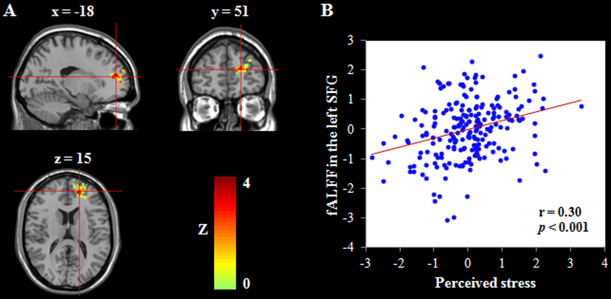
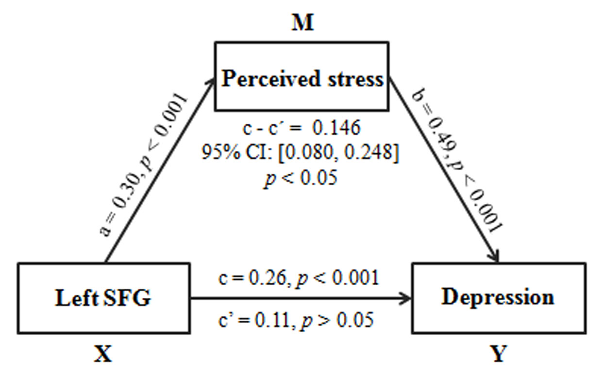
The whole-brain correlation analyses found that after adjusting for gender, age and head motion, perceived stress was positively associated with the fALFF in the left superior frontal gyrus (SFG; MNI coordinates: -18, 51, 15; cluster size = 212 voxels; Z = 3.92; r = 0.30, p < 0.001; Figure 1). The prediction analyses revealed that perceived stress could be stably predicted by the fALFF in the left SFG [r(predicted, observed) = 0.27, p < 0.001], after controlling for gender, age and head motion. Furthermore, mediation analyses showed that perceived stress played a mediating role in the relation between the fALFF in the left SFG and depression (indirect effect = 0.146, 95% CI = [0.080, 0.248], p < 0.05; Figure 2), after controlling for gender, age and head motion. Finally, these results persisted even after excluding the impact of anxiety, suggesting the specificity of our findings.Discussion
The current research was conducted to investigate the functional brain basis of perceived stress and the underlying brain mechanism linking perceived stress and depression. First, higher levels of perceived stress were linked to greater fALFF in the left SFG, which is consistent with previous findings showing an association between SFG function and structure and stress-related processing 12-16. Additionally, the positive association of the fALFF in the left SFG with perceived stress may reflect a compensatory mechanism to counteract functional or structural brain abnormalities 17,18. Second, perceived stress served as a mediator in the link between SFG spontaneous activity and depression. This finding may present further evidence for the predictive role of perceived stress in depression and highlight that perceived stress may be a potential mechanism linking spontaneous brain activity with depression.Conclusion
In conclusion, this research provides initial evidence for neurofunctional makers underlying perceived stress and reveals a potential brain-stress mechanism for predicting depression.Acknowledgements
No acknowledgements.References
1. Cohen S, Kamarck T, Mermelstein R. A global measure of perceived stress. J Health Soc Behav. 1983; 24:385-396.
2. Chao SF. Functional disability and depressive symptoms: Longitudinal effects of activity restriction, perceived stress, and social support. Aging Ment Health. 2014; 18:767-776.
3. Lorenzo-Blanco EI, Unger JB. Ethnic discrimination, acculturative stress, and family conflict as predictors of depressive symptoms and cigarette smoking among Latina/o youth: The mediating role of perceived stress. J Youth Adolescence. 2015; 44:1984-1997.
4. Tsai HJ, Chang FK. Associations of various perceived-stress situations with depressive symptoms in >= 50-year old Taiwanese men and women: Results from the Taiwan Longitudinal Study on Aging. Arch Gerontol Geriat. 2016; 67:113-119.
5. Beck AT, Ward CH, Mendelson M, Mock J, Erbaugh J. An inventory for measuring depression. Arch Gen Psychiatry. 1961; 4:561-571.
6. Spielberger CD, Gorsuch RL, Lushene RE. STAI-manual for the State Trait Anxiety Inventory. Palo Alto (CA): Consulting Psychologists Press, 1988.
7. Yan CG, Wang XD, Zuo XN, Zang YF. DPABI: Data processing & analysis for (resting-state) brain imaging. Neuroinformatics. 2016; 14:339-351.
8. Zou QH, Zhu CZ, Yang YH, Zuo XN, Long XY, Cao QJ, Wang YF, Zang YF . An improved approach to detection of amplitude of low-frequency fluctuation (ALFF) for resting-state fMRI: Fractional ALFF. J Neurosci Meth. 2008; 172:137-141.
9. Worsley KJ, Evans AC, Marrett S, Neelin P. A three-dimensional statistical analysis for CBF activation studies in human brain. Journal of Cerebral Blood Flow and Metabolism. 1992; 12:900-918.
10. Supekar K, Swigart AG, Tenison C, Jolles DD, Rosenberg-Lee M, Fuchs L, Menon V. Neural predictors of individual differences in response to math tutoring in primary-grade school children. Proc Natl Acad Sci USA. 2013; 110:8230-8235.
11. Hayes AF. Introduction to mediation, moderation, and conditional process analysis: A regression-based approach. New York, NY: The Guilford Press, 2013.
12. Treadway MT, Buckholtz JW, Zald DH. Perceived stress predicts altered reward and loss feedback processing in medial prefrontal cortex. Front Hum Neurosci. 2013; 7:180.
13. Pruessner JC, Declovic K, Khalili-Mahani N, Engert V, Pruessner M, Buss C, Renwick R, Dagher A, Meaney MJ, Lupien S. Deactivation of the limbic system during acute psychosocial stress: Evidence from positron emission tomography and functional magnetic resonance Imaging studies. Biol Psychiat. 2008; 63:234-240.
14. Li GY, Ma XY, Bian HM, Sun XH, Zhai N, Yao MY, Qu HR, Ji SZ, Tian HJ, Zhuo CJ. A pilot fMRI study of the effect of stressful factors on the onset of depression in female patients. Brain Imaging Behav. 2016; 10:195-202.
15. Tyborowska A, Volman I, Niermann HCM, Pouwels JL, Smeekens S, Cillessen AHN, Toni I, Roelofs K. Early-life and pubertal stress differentially modulate grey matter development in human adolescents. Sci Rep-Uk. 2018; 8:9201.
16. Ansell EB, Rando K, Tuit K, Guarnaccia J, Sinha R. Cumulative adversity and smaller gray matter volume in medial prefrontal, anterior cingulate, and insula regions. Biol Psychiat. 2012; 72:57-64.
17. Bing X, Ming-Guo Q, Ye Z, Jing-Na Z, Min L, Han C, Yu Z, Jia-Jia Z, Jian W, Wei C, Han-Jian D, Shao-Xiang Z. Alterations in the cortical thickness and the amplitude of low-frequency fluctuation in patients with post-traumatic stress disorder. Brain research. 2013; 1490:225-232.
18. Orr C, Morioka R, Behan B, Datwani S, Doucet M, Ivanovic J, Kelly C, Weierstall K, Watts R, Smyth B, Garavan H. Altered resting-state connectivity in adolescent cannabis users. Am J Drug Alcohol Ab. 2013; 39:372-381.
Figures