3676
Decreased Relative Cerebral Blood Flow In Unmedicated Heroin Use Disorders1Department of Radiology, Second Xiangya Hospital of Central South University, Changsha, China, 2Department, Hunan Judicial Police Academy, Changsha, China
Synopsis
Sixty-eight patients with heroin use disorders(HUD) and forty-seven matched healthy controls underwent a high resolution T1 and whole-brain pulse arterial spin labeled (PASL) perfusion magnetic resonanceMRI scanning. We found that compared to control subjects, HUD showed worse neuropsychological performance and significantly decreased regional relative CBF ((rCBF)) in HUD patients had hypoperfusion in limbic, frontal, and temporol areas,and the correlation between middle frontal gyrus(MFG) and theneurocognitive measures.
Introduction:
Methods:
Sixty-eight (42 males and 26 females, ages:40.9±7.3) HUD patients with heroin use disorders and forty-seven (34 males and 13 females, ages:39.3±9.2) matched healthy controls underwent a high resolution T1 and whole-brain pulse arterial spin labeled (PASL) perfusion magnetic resonanceMRI scanning. Clinical characteristics were collected to assess the neurocognitive function as well.Results:
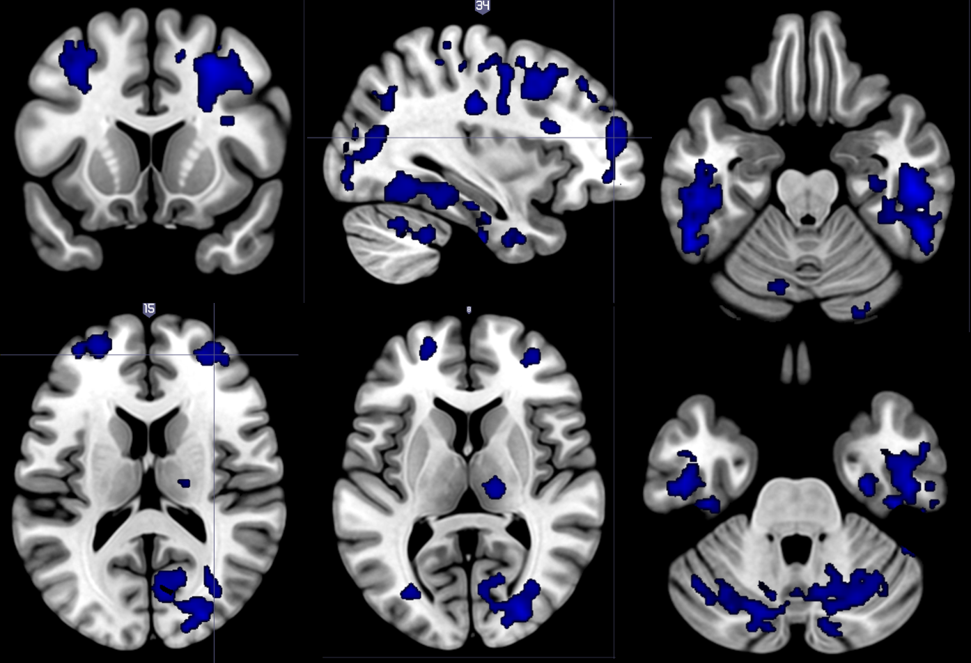
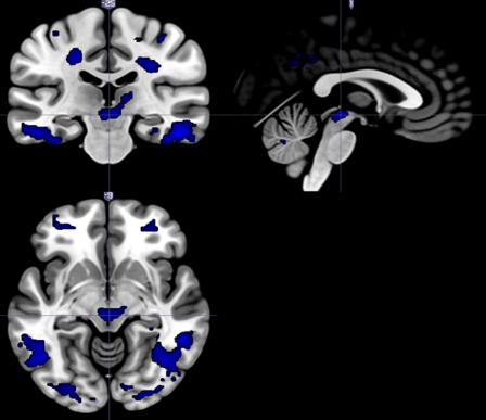
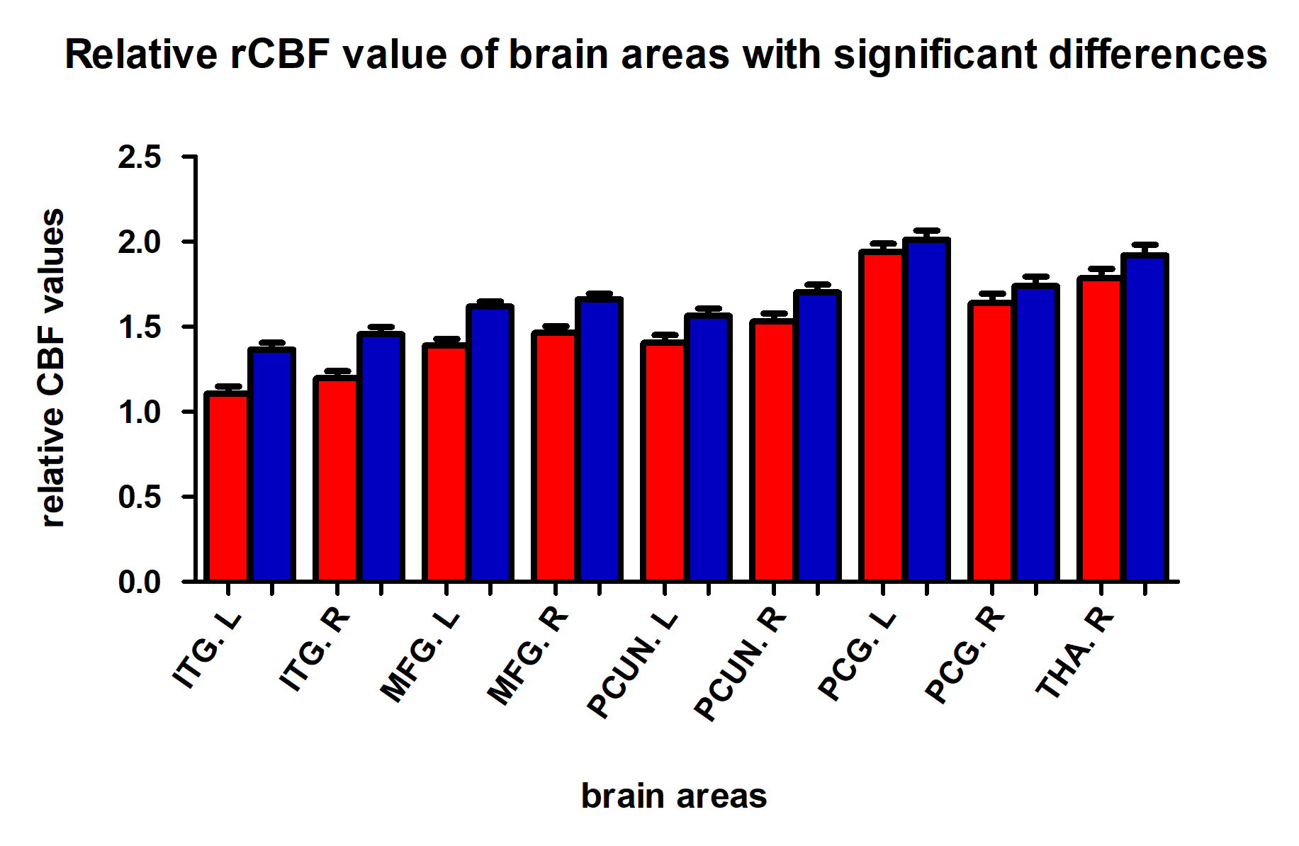
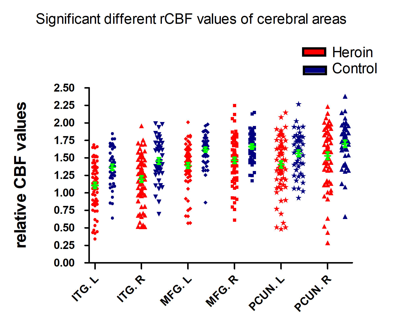
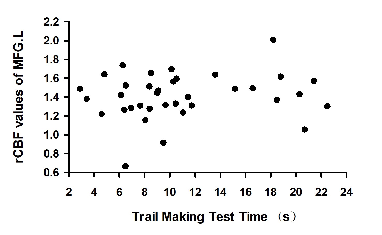
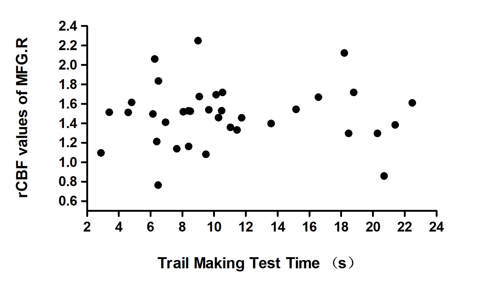
Compared to control subjects, HUDHeroin patients showed worse neuropsychological performance and significantly decreased regional relative CBF ((rCBF)) in bilateral inferior temporal gyrus(ITG),middle frontal gyrus(MFG), cerebellar vermis and posterior cerebellar lobe, precuneus, right posterior cingulate cortex(PCC), thalamus, and the midbrain adjacent to the ventral tegmental area (VTA);. N no significant rCBF increase was observed in Heroin patients. Heroin patients showed significantly better performance in the following neuropsychological testing: Wechsler Adult Intelligence (WAIS-3) Test Symbol Test 2nd-minute score (t=-2.663, p=0.011) and backward digital memory span (t=-2.557, p=0.013), and Trail Making Test Time (t=3.556, p=0.001). Both the metrics of rCBF in left MFG and right MFG were negatively correlated with the Trail Making Test Time (left: r = -0.409, p = 0.013; right: r = -0.456, p = 0.005) in HUD patients.Conclusion:
HUD patients had hypoperfusion in limbic, frontal, and temporal areas. Lower CBF in MFG predicts more cognitive impairment of HUD patientsOur findings suggest the prevalent decreased CBF of the brain areas in Heroin use disorders than that in controls, and that the levels in bilateral MFG are significantly associated with the Trail Making Test Time. Together, these findings suggest MFG as a critical region in heroin dependence and suggest ASL CBF as a potential marker for heroin addiction studySuch findings suggest that there is a connection between the decreased CBF and the impaired cognitive function .Acknowledgements
The authors express their appreciation to their patients and volunteers for participating in this study.References
[1]. Gerra, G., et al., Regional cerebral blood flow and comorbid diagnosis in abstinent opioid addicts. Psychiatry Res, 1998. 83(2): p. 117-26.
[2]. Pezawas, L., et al., Opioid addiction changes cerebral blood flow symmetry. Neuropsychobiology, 2002. 45(2): p. 67-73.
[3]. Denier, N., et al., Association of frontal gray matter volume and cerebral perfusion in heroin addiction: a multimodal neuroimaging study. Front Psychiatry, 2013. 4: p. 135.
[4]. Qiao, P.G., et al., Clinical assessment of cerebral hemodynamics in Moyamoya disease via multiple inversion time arterial spin labeling and dynamic susceptibility contrast-magnetic resonance imaging: A comparative study. J Neuroradiol, 2017. 44(4): p. 273-280.
[5]. Xu, Q., et al., Tumor recurrence versus treatment effects in glioma: A comparative study of three dimensional pseudo-continuous arterial spin labeling and dynamic susceptibility contrast imaging. Medicine (Baltimore), 2017. 96(50): p. e9332.
[6]. Riederer, I., et al., Alzheimer Disease and Mild Cognitive Impairment: Integrated Pulsed Arterial Spin-Labeling MRI and F-FDG PET. Radiology, 2018. 288(1): p. 198-206.
[7].Feltenstein, M.W. and R.E. See, The neurocircuitry of addiction: an overview. Br. J. Pharmacol., 2008. 154(2): p. 261-74.
[8]. Clay, S.W., J. Allen and T. Parran, A review of addiction. Postgrad Med, 2008. 120(2): p. E01-7.
[9]. Narita, M., M. Funada and T. Suzuki, Regulations of opioid dependence by opioid receptor types. Pharmacol. Ther., 2001. 89(1): p. 1-15.
[10]. Wang, Z., et al., Empirical optimization of ASL data analysis using an ASL data processing toolbox: ASLtbx. Magn Reson Imaging, 2008. 26(2): p. 261-9. [11]. Loprinzi, P.D., Epidemiological investigation of muscle-strengthening activities and cognitive function among older adults. Chronic Illn, 2016. 12(2): p. 157-62.
[12]. Nakahachi, T., et al., Frontal activity during the digit symbol substitution test determined by multichannel near-infrared spectroscopy. Neuropsychobiology, 2008. 57(4): p. 151-8.
[13]. Frith, E. and P.D. Loprinzi, Physical Activity and Cognitive Function Among Older Adults with an Elevated Gamma Gap. Med Princ Pract, 2018.
[14]. Jensen, A.R. and R.A. Figueroa, Forward and backward digit span interaction with race and IQ: predictions from Jensen's theory. J Educ Psychol, 1975. 67(6): p. 882-93.
[15]. Hester, R.L., G.J. Kinsella and B. Ong, Effect of age on forward and backward span tasks. J Int Neuropsychol Soc, 2004. 10(4): p. 475-81.
[16]. Wechsler, D., Technical manual for the Wechsler Adult Intelligence and Memory Scale–Third Edition. New York: The Psychological Corporation., 1997.
[17]. Hester, R.L., G.J. Kinsella and B. Ong, Effect of age on forward and backward span tasks. J Int Neuropsychol Soc, 2004. 10(4): p. 475-81.
[18]. Wilckens, K.A., et al., Sleep moderates the relationship between amyloid beta and memory recall. Neurobiol. Aging, 2018. 71: p. 142-148.
[19]. Estimation of the cool executive function using frontal electroencephalogram signals in first ‑ episode schizophrenia patients. BioMedical Engineering, 2016.
[20]. Port, A.P., et al., Cognition and brain function in elderly Tai Chi practitioners: A case-control study. Explore (NY), 2018.
[21]. Spreen O, S.E., A compendium of neuropsychological tests : administration, norms, and commentary. 2nd ed. New York: Oxford University Press, 1998.
[22]. Langston, R.G. and T. Virmani, Use of a Modified STROOP Test to Assess Color Discrimination Deficit in Parkinson's Disease. Front Neurol, 2018. 9: p. 765
[23]. Volkow, N.D. and M. Boyle, Neuroscience of Addiction: Relevance to Prevention and Treatment. Am J Psychiatry, 2018. 175(8): p. 729-740.
[24]. Karila, L., et al., New synthetic opioids: Part of a new addiction landscape. Neurosci Biobehav Rev, 2018.
[25]. Valentino, R.J. and N. Volkow, Untangling the complexity of opioid receptor function. Neuropsychopharmacology, 2018.
[26]. Jay, G.W. and R.L. Barkin, Perspectives on the opioid crisis from pain medicine clinicians. Dis Mon, 2018. 64(10): p. 451-466.
[27]. Darcq, E. and B.L. Kieffer, Opioid receptors: drivers to addiction? Nat. Rev. Neurosci., 2018. 19(8): p. 499-514.
[28]. Cornejo, M.P., et al., Ghrelin recruits specific subsets of dopamine and gaba neurons of different ventral tegmental area sub-nuclei. Neuroscience, 2018.
[29]. Kopra, J., et al., Constitutive Ret signaling leads to long-lasting expression of amphetamine-induced place conditioning via elevation of mesolimbic dopamine. Neuropharmacology, 2018. 128: p. 221-230.
[30]. Artiges, E., et al., Striatal and Extrastriatal Dopamine Transporter Availability in Schizophrenia and Its Clinical Correlates: A Voxel-Based and High-Resolution PET Study. Schizophr Bull, 2017. 43(5): p. 1134-1142.
[31]. Morel, C., S. Montgomery and M.H. Han, Nicotine and alcohol: the role of midbrain dopaminergic neurons in drug reinforcement. Eur. J. Neurosci., 2018.
[32]. Luo, Z., et al., Acute cocaine induces fast activation of D1 receptor and progressive deactivation of D2 receptor striatal neurons: in vivo optical microprobe [Ca2+]i imaging. J. Neurosci., 2011. 31(37): p. 13180-90.
[33]. Volkow, N.D., et al., Profound decreases in dopamine release in striatum in detoxified alcoholics: possible orbitofrontal involvement. J. Neurosci., 2007. 27(46): p. 12700-6.
[34]. Sadat-Shirazi, M.S., et al., Alteration of Dopamine Receptors subtypes in the Brain of Opioid Abusers: a postmortem study in Iran. Neurosci. Lett., 2018.
[35]. Francis, T.C., et al., Synaptic and intrinsic plasticity in the ventral tegmental area after chronic cocaine. Curr. Opin. Neurobiol., 2018. 54: p. 66-72.
[36]. Alaghband, Y., et al., CREST in the nucleus accumbens core regulates cocaine conditioned place preference, cocaine-seeking behavior, and synaptic plasticity. J. Neurosci., 2018.
[37]. Ostroumov, A. and J.A. Dani, Inhibitory Plasticity of Mesocorticolimbic Circuits in Addiction and Mental Illness. Trends Neurosci., 2018.
[38]. Goldstein RZ, V.N., Dysfunction of the prefrontal cortex in addiction: neuroimaging findings and clinical implications. Nat Rev Neurosci., 2011.
[39]. Koob, G.F.A.V., Neurocircuitry of addiction. Neuropsychopharmacology, 2013(35, 217–38).
[40]. Schluter, R.S., et al., Effects of Non-invasive Neuromodulation on Executive and Other Cognitive Functions in Addictive Disorders: A Systematic Review. Front Neurosci, 2018. 12: p. 642.
[41]. Terentjeviene, A., et al., Prefrontal Cortex Activity Predicts Mental Fatigue in Young and Elderly Men During a 2 h "Go/NoGo" Task. Front Neurosci, 2018. 12: p. 620.
[42]. Schluter, R.S., et al., Effects of Non-invasive Neuromodulation on Executive and Other Cognitive Functions in Addictive Disorders: A Systematic Review. Front Neurosci, 2018. 12: p. 642.
[43]. Gardner, M.P., et al., Medial orbitofrontal inactivation does not affect economic choice. Elife, 2018. 7.
[44]. Dien, J., et al., Combined ERP/fMRI evidence for early word recognition effects in the posterior inferior temporal gyrus. Cortex, 2013. 49(9): p. 2307-21.
[45]. Geng, X., et al., Salience and default mode network dysregulation in chronic cocaine users predict treatment outcome. Brain, 2017. 140(5): p. 1513-1524.
[46]. Luo, X., et al., Decreased Bilateral FDG-PET Uptake and Inter-Hemispheric Connectivity in Multi-Domain Amnestic Mild Cognitive Impairment Patients: A Preliminary Study. Front Aging Neurosci, 2018. 10: p. 161.
[47]. Schmahmann, J.D., Disorders of the cerebellum: ataxia, dysmetria of thought, and the cerebellar cognitive affective syndrome. J Neuropsychiatry Clin Neurosci, 2004. 16(3): p. 367-78.
[48]. Schmahmann, J.D., The cerebellum and cognition. Neurosci. Lett., 2018.
[49]. Yucel, K., et al., Cerebellar vermis volume in major depressive disorder. Brain Struct Funct, 2013. 218(4): p. 851-8.
[50]. Du, C., et al., Cocaine-induced ischemia in prefrontal cortex is associated with escalation of cocaine intake in rodents. Mol. Psychiatry, 2018.
Figures