3675
3D T1-weighted Images with Scan Time of Less than 2 Minutes by Using Wave-CAIPI1Department of Diagnostic Imaging and Nuclear Medicine, Kyoto University Graduate School of Medicine, Kyoto, Japan, 2Human Brain Research Center, Kyoto University Graduate School of Medicine, Kyoto, Japan, 3Integrated Clinical Education Center, Kyoto University Hospital, Kyoto, Japan, 4Siemens Shenzhen Magnetic Resonance Ltd., Shenzhen, China, 5Siemens Healthineers, San Francisco, CA, United States, 6Siemens Healthineers, Erlangen, Germany
Synopsis
A preliminary imaging study for MPRAGE with Wave-CAIPI has been introduced at the clinical MR scanner. MPRAGE with Wave-CAIPI 3×3 shows relatively better contrast despite its short scan time. Less imaging noise and segmentation error was found in MPRAGE with Wave-CAIPI 3×3. MPRAGE with GRAPPA 3×3 showed prominent imaging noise and segmentation errors, however, MPRAGE with CAIPI 3×3 showed few segmentation errors and almost equivalent to Wave-CAIPI 3×3.
Introduction
Wave-CAIPI acquisition enables highly accelerated volumetric imaging with low artifact and signal-to-noise ratio (SNR) penalties by playing sinusoidal 𝐺y and 𝐺z gradients during the readout of each phase encoding line. By taking advantage of coil sensitivity encoding from multi-channel receiver coils, reduction of phase-encoding steps has been available with the parallel imaging technique such as SENSE and GRAPPA. Controlled aliasing in parallel imaging results in higher acceleration (2D-CAIPIRINHA) has enabled image reconstruction with further acceleration. More acceleration has been expected with the introduction of Wave-CAIPI, however, image quality has not been evaluated well in the clinical practices.
The purpose of this study is to evaluate Wave-CAIPI MPRAGE in clinical scanners, and to establish the preliminary comparison study among MPRAGE images with various kinds of acceleration mode.
Methods
- Subjects
Healthy volunteers were enrolled in this study. This study was approved by the institutional review board and written informed consent was obtained from each subject.
- MR imaging
Subjects underwent MR imaging at 3T MR scanners (MAGNETOM Prisma, Siemens Healthcare, Erlangen, Germany) with a 64-channel head/neck coil. The imaging protocols of MPRAGE and a prototype Wave-CAIPI MPRAGE were as follows: (i) MPRAGE with GRAPPA 2×1, ref lines PE, 24; 208 slices; scan time, 5 m 21 s. (ii) MPRAGE with Wave-CAIPI 3×3; ref lines PE, 24; ref lines 3D, 24; CAIPI shift factor, 1; 240 slices; scan time, 1 m 42 s.(iii) MPRAGE with CAIPIRINHA 3×3; ref lines PE, 24; ref lines 3D, 24; CAIPI shift factor, 1; 240 slices; scan time, 1 m 42 s. (iv) MPRAGE with GRAPPA 3×3; ref lines PE, 24; ref lines 3D, 24; 240 slices; scan time, 1 m 42 s. TR of 2300 ms, TE of 4.67ms, flip angle of 9, and bandwidth of 130 Hz/pixel were adapted for all MPRAGEs.
- Visual inspection
All MPRAGEs were visually inspected by focusing on noise distribution.
- Post-imaging process
Recon-all function of FreeSurfer (v5.3.0) was applied for all MPRAGE images. The Parcellation status was visually checked and voxel sizes of VOIs were compared.
Results
Representative images of original MPRAGE images were shown in Figure 1.
MPR with axial reconstruction images of all MPRAGEs are shown in Figure 2. MPRAGE with GRAPPA 2×1 shows best image quality, however, MPRAGE with Wave-CAIPI 3×3 shows relatively better contrast between gray and white matters. Image noise was conspicuous in MPRAGE with GRAPPA 3×3 as well as CAIPI 3×3.
By using FreeSurfer, segmentation and cortical parcellation were done fairly well for all MPRAGE images, however mild segmentation errors are shown especially in MPRAGE with GRAPPA 3×3 (Figure 3).
The volume of each VOI was shown consistent among MPRAGE with GRAPPA 2×1, Wave-CAIPI 3×3, and CAIPIRINHA 3×3. Prominent differences were seen in MPRAGE with GRAPPA 3×3 (Figure 4).
Discussion
Wave-CAIPI MPRAGE was evaluated in comparison with other accelerated techniques of MPRAGE. MPRAGE with Wave-CAIPI 3×3 shows relatively better contrast despite its short scan time. Less imaging noise and segmentation error was found in MPRAGE with Wave-CAIPI 3×3. As expected, MPRAGE with GRAPPA 3×3 showed prominent imaging noise and segmentation errors, however, MPRAGE with CAIPIRINHA 3×3 showed little segmentation errors as similarly shown in Wave-CAIPI 3×3.Conclusion
Performance Wave-CAIPI MPRAGE was evaluated in several criteria including segmentation error and imaging noise. MPRAGE with Wave-CAIPI may be a viable tool for clinically acceptable imaging with scan time of < 2 min. It may be also used for voxel base morphometry.Acknowledgements
We are grateful to Mr. Yuta Urushibata, Siemens Healthineer K. K. for his kindest help.References
1. Bilgic B, Gagoski BA, Cauley SF, Fan AP, Polimeni JR, Grant PE, Wald LL, Setsompop K. Magn Reson Med. 2015 Jun;73(6):2152-62. Wave-Caipi for highly accelerated 3D imaging.
2. Moriguchi H, Duerk JL. Magn Reson Med. 2006 Mar;55(3):633-48. Bunched phase encoding (BPE): a new fast data acquisition method in MRI.
3. Breuer FA, Moriguchi H, Seiberlich N, Blaimer M, Jakob PM, Duerk JL, Griswold MA. Magn Reson Med. 2008 Aug;60(2):474-8. Zigzag sampling for improved parallel imaging.
4. Seiberlich N1, Breuer F, Blaimer M, Jakob P, Griswold M. Magn Reson Med. 2008 Apr;59(4):930-5. Self-calibrating GRAPPA operator gridding for radial and spiral trajectories.
5. Dale, A.M., Fischl, B., Sereno, M.I. Neuroimage 1999. 9, 179-194. Cortical surface-based analysis. I. Segmentation and surface reconstruction.
6. Reuter, M., Rosas, H.D., Fischl, B. Neuroimage 2010. 53 (4), 1181–1196. Highly Accurate Inverse Consistent Registration: A Robust Approach.
Figures
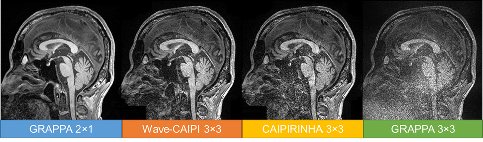
Figure 1.
Representative MPARGE images are shown. Although MPRAGE with GRAPPA 2×1 shows best image quality, however, MPRAGE with Wave-CAIPI 3×3 shows relatively less image noise compared with MPRAGE with CAIPIRINHA 3×3 or GRAPPA 3×3.
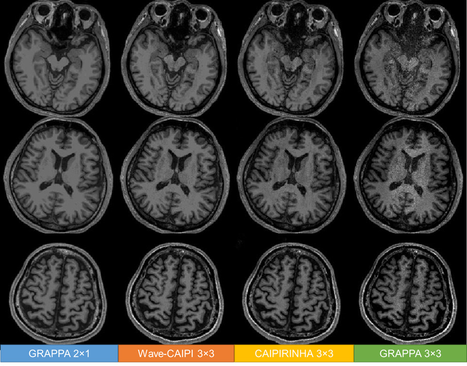
Figure 2.
MPR with axial reconstruction images of all MPRAGEs are shown. MPRAGE with GRAPPA 2×1 shows best image quality, however, MPRAGE with Wave-CAIPI 3×3 shows relatively better contrast between gray and white matters. Image noise was conspicuous in MPRAGE with GRAPPA 3×3 as well as CAIPIRINHA 3×3.
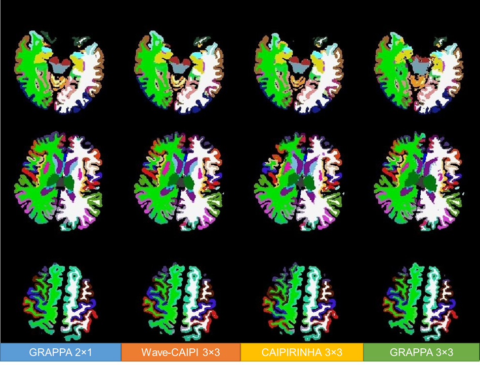
Figure 3.
Segmentation and cortical parcellation were almost well done for all MPRAGE images, however mild segmentation errors are shown especially in MPRAGE with GRAPPA 3×3.
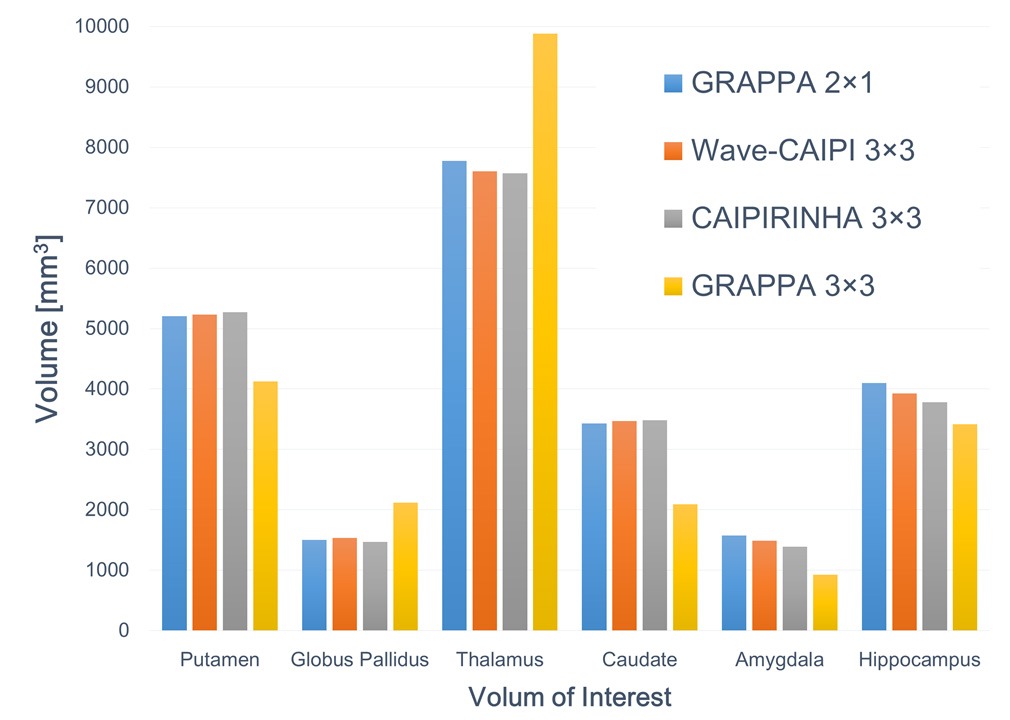
Figure 4.
The volume of each VOI are almost similar among MPRAGE with GRAPPA 2×1, Wave-CAIPI 3×3, and CAIPIRINHA 3×3. Prominent differences were seen in MPRAGE with GRAPPA 3×3.