3673
3D CUBE MRI of facial nerve lesions in temporal bone: a preliminary study1Shandong Provincial Hospital Affiliated to Shandong University, Jinan, China, 2Department of Radiology, Shandong Provincial Hospital Affiliated to Shandong University, Jinan, China, 3GE Healthcare MR Research, Beijing, China
Synopsis
This study mainly investigated the feasibility of three-dimensional (3D) CUBE MRI for high resolution facial nerve imaging in patients with peripheral paralysis. We thus systematically measured the corresponding patients with three different syndromes using 3D fat suppressed CUBE technique. While significant image enhancement between the affected and unaffected sides was almost shown in all segments for the patients, different effects were found in mastoid and internal auditory canalicular regions,We thus can demonstrate the feasibility of 3D CUBE MRI in the diagnosis of patients with facial paralysis relevant diseases.
Synopsis
This study mainly investigated the feasibility of three-dimensional (3D) CUBE MRI for high resolution facial nerve imaging in patients with peripheral paralysis. We systematically measured the corresponding patients with three different syndromes using 3D fat suppressed CUBE technique. While significant image enhancement between the affected and unaffected sides was almost shown in all segments for the patients, different effects were found in mastoid and internal auditory canalicular regions,We thus can demonstrate the feasibility of 3D CUBE MRI in the diagnosis of patients with facial paralysis relevant diseases.Introduction
The incidence of facial paralysis, as one clinical common disease affecting patients’ life quality dramatically, has shown an ascending trend in the recent years1. Peripheral facial paralysis, as a main kind of facial paralysis, has been usually occurred in the temporal bone based on its complex pathogenesis2-5. Due to the requirements in the clinical treatment, accurate localization for lesions plays an important role in the diagnosis for peripheral facial paralysis2-5.A three-dimensional (3D) CUBE magnetic resonance imaging (MRI) technique, developed mainly for high resolution imaging with high signal-to-noise-ratio, was assumed to have the potential for facial nerve imaging. Therefore, in this study, the main goal was to investigate the feasibility of 3D CUBE MRI for the internal facial nerve in the temporal bone and thus to explore the corresponding clinical values of this technique in different temporal bone diseases.Materials and methods
Subjects:In total forth-four patients (mean age: 47.8±4.3 years old), confirmed with peripheral facial paralysis clinically, were recruited in this study for MRI experiments. Of them, 15 were Bell paralysis, 20 were Ramsay Hunt syndrome, and 9 were temporal bone trauma.
MRI experiments:All experiments were performed on a 3T clinical scanner (Discovery 750w, GE Healthcare, Milwaukee, WI, USA) equipped with a 16-channel head and neck connecting coil. A 3D CUBE imaging technique with lipid suppression was employed to acquire T1-weighted facial nerve imaging before and after contrast agent (gadolinium) injection. A field-of-view of 20mm x 20mm was applied to fully cover the area from the lower margin of the temporal lobe to the upper margin of the parotid gland. Other scan parameters included repetition time=1420ms, echo time=16ms, echo train length=60, NEX=2, matrix size=256×256, and slice thickness=1.0mm. The scan time was 3 minutes 30 seconds.
Data analysis:All data were analyzed at a GE MR workstation (Advantage workstation 4.6; GE Medical Systems). Following the anatomical structures of facial nerve, five sub-regions were segmented: (1) internal auditory canalicular, (2) labyrinthine, (3) geniculate ganglion, (4) tympanic and (5) mastoid or vertical.Contrast enhancement of facial nerve imaging after Gd administration was first qualitatively evaluated according to a three-point rule (0: no contrast enhancement; 1: mild contrast enhancement, weaker than that in the surrounding vessels; 2: strong contrast enhancement, comparable to that in the surrounding vessels), and also quantitatively assessed by using a relative signal enhancement ratio lRFV, defined as follows:IRFV = IFV / IBS, where IFV and IBS represent the respective signal intensity in different regions of facial nerve and brainstem after Gd administration. The statistical analysis was performed using SPSS software version 20.0. The embedded paired-t-test was used to evaluate the difference of the relative signal enhancement ratio IRFV and The Wilcoxon-rank-sum-test was applied to estimate the enhancement degrees between different segments at the affected and unaffected sides for all the patients . The significant threshold was set as p=0.05.
Results
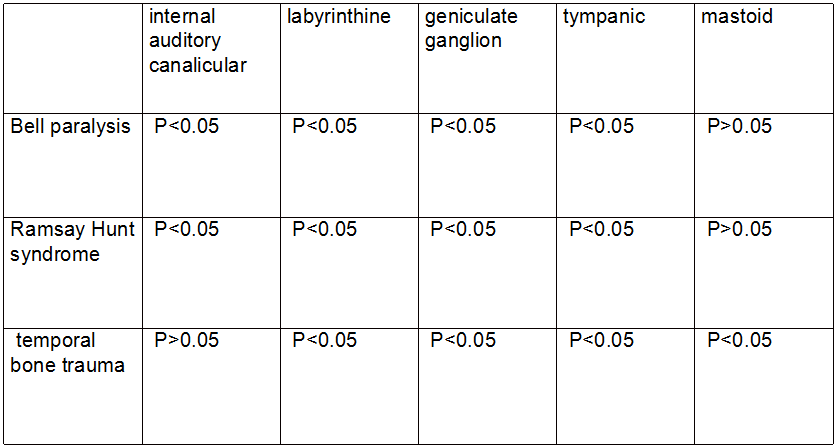
For all the patients with three different syndromes, , the affected nerves became thickened and also showed abnormal enhancement (Fig.1).The image enhancement between the affected and unaffected sides was qualitatively assessed by a 3-point rule and quantitatively evaluated with a defined relative signal enhancement ratio IRFV. As shown in Tables 1 and 2, significant difference was found in almost all segments of the patients (P<0.05). However, in mastoid region comparable qualitative and quantitative results were shown for the patients with Bell paralysis and Ramsay Hunt syndrome, and only comparable qualitative values were observed for patients with temporal bone trauma. In addition, in internal auditory canalicular region only comparable results was revealed for the patients with temporal bone trauma after qualitative and quantitative analyisis.Discussion and Conclusion
In this study, 3D CUBE MRI was applied to systematically measure the facially paralyzed patients separated based on three different syndromes. Ensuring the clear visualization of facial nerve with contrast enhancement, their own characteristics of different syndromes were revealed in CUBE images, respectively. While significant image enhancement between the affected and unaffected sides was almost shown in all segments for the patients, different effects were found in mastoid and internal auditory canalicular regions Therefore, it can demonstrate the clinical value of CUBE MRI in the diagnosis of facial nerve relevant diseases.Acknowledgements
No acknowledgement found.References
1.Finsterer J.Management of peripheral facial nerve palsy[J]. Eur ArchOtorhinolaryngol. 2008,265(7):743-52.
2.Kress BP,Griesbeck F,Efinger K,et al.Bell's palsy: what is the prognostic value of measurements of signal intensity increases with contrast enhancement on MRI?Neuroradiology 2002;44:428-433.
3.Kim IS,Shin SH,Kim J,et al.Correlation between MRI and operative findings in Bell's palsy and Ramsay Hunt syndrome[J].Yonsei medical journal,2007,48(6):963-968.
4.Lee LN, Lyford-Pike S, Boahene KD.Traumatic facial nerve injury[J]. OtolaryngolClin North Am. 2013,46(5):825-39.
5.Shirazi MA, Leonetti JP, Marzo SJ, et al.Surgical management of facial neuromas:lessons learned[J]. Otol Neurotol. 2007,28(7):958-63.
Figures