3672
A test-retest multi-site reproducibility of neurovascular 4D flow MRI1MR, the First Affiliated Hospital of Zhengzhou University, zhengzhou, China, 2the First Affiliated Hospital of Zhengzhou University, zhengzhou, China, 3GE Healthcare, beijing, China
Synopsis
4D
flow shows great potential in neurovascular disorders such as stenosis,
atherosclerotic disease, aneurysms, and vascular malformations; its widespread
application in neurovascular system requires evidence of good test-retest
multi-site reproducibility. This study would like to assess the multi-center
reproducibility, test-retest variability and inter-observer dependence of 4D
flow MRI in neurovascular system.
Introduction
Abnormal cerebral blood flow (CBF) patterns have been shown in a variety of diseases1.2,3. Quantitative assessment of the blood flow and velocity is important for the understanding and management of these diseases 4. 4D flow MRI is a non-invasive technique with the capability of imaging complex vessel geometries and measuring blood flow velocities. This study would like to assess the multi-center reproducibility, test-retest reliability and inter-observer dependence of 4D flow MRI in measuring flow related parameters, such as the blood flow and peak velocity of main vessels in the neurovascular system.Methods
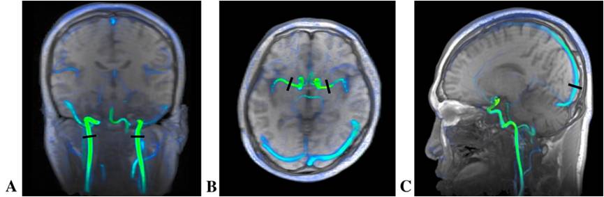
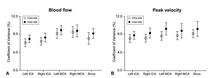
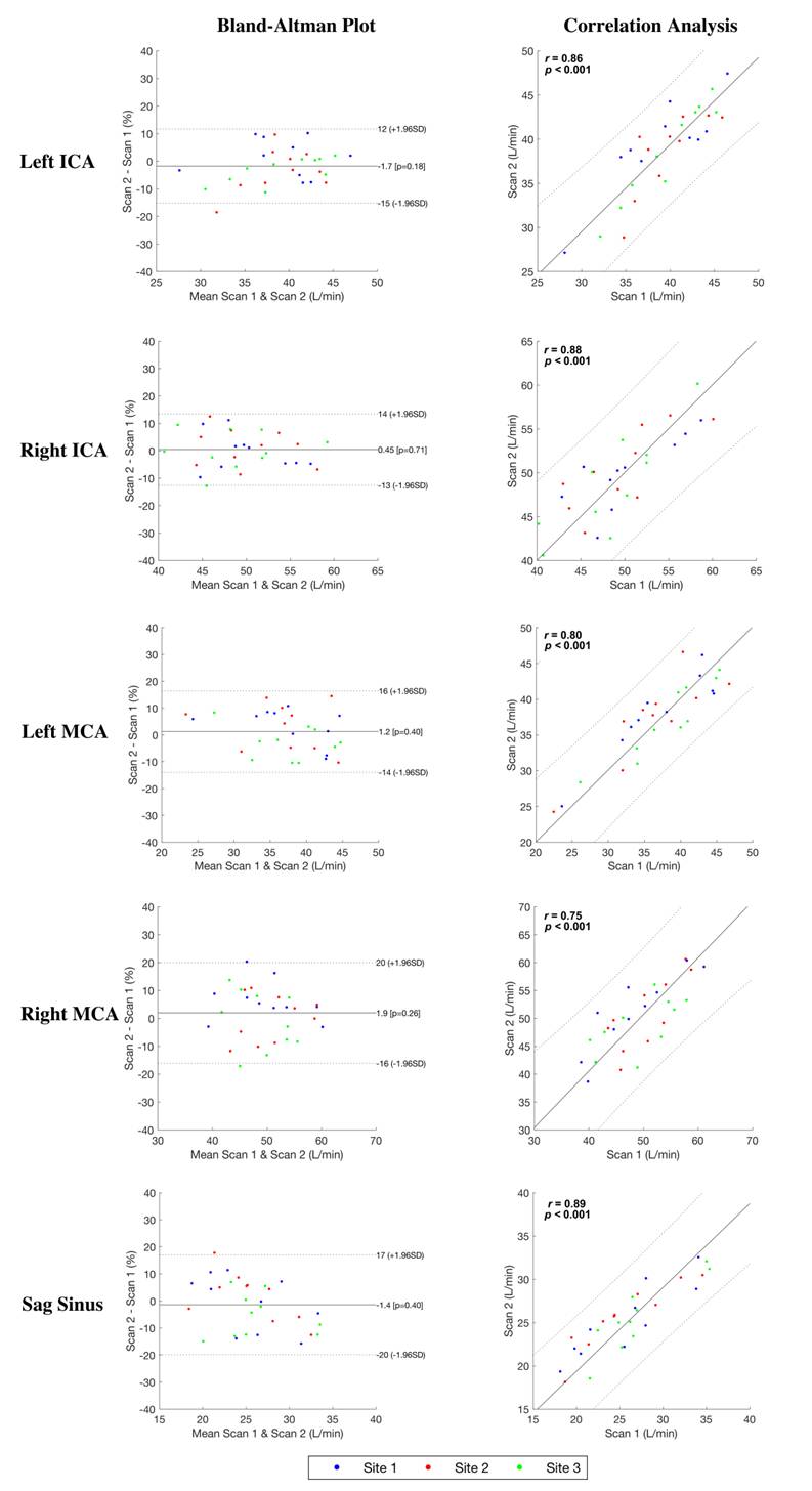
Ten healthy subjects (4 female) underwent 4D flow scans in three different centers, consent forms were obtained prior to the scan. All subjects were scanned twice on a 3T MR system (Discovery MR750, GE Healthcare, Waukesha, USA) equipped with an 8-channel head coil at two different days each center. The Arterys software 5 was used for 4D flow post-processing to derive flow related parameters. Multi-center reproducibility, test-retest reliability and inter-observer agreement of measurements of the blood flow and peak velocity of five regions of interest were assessed (bilateral internal carotid arteries, bilateral medial cerebral arteries and sagittal sinus). Shapiro-Walk test was conducted to assess normality of measurements in each scan. Coefficient of variances (CV) were computed to evaluate intra- and inter-site variances of measurements. The multi-center reproducibility of 4D flow was assessed by two-way mixed intra-class correlation coefficient (ICC). Bland-Altman plot and Pearson correlation were used to evaluate test-retest reliability. ICC was calculated to assess inter-observer agreements.Results
The placement of the ROIs is illustrated in Figure 1. All p-values for Shapiro-Walk test were greater than 0.05, which indicated normality of all measurements from all scans. Both intra- and inter-site CVs were lower than 12%, and the CVs plots are shown in Figure 2. There was good test-retest reliability for both blood flow and peak velocity of all ROIs (r = 0.75 – 0.94). In addition, high multi-center reproducibility was detected (ICC = 0.77 – 0.96, all p-values < 0.001). The results of these measurements also showed great inter-observer agreement (all ICC > 0.9 and all p-value < 0.001). Bland-Altman plot and correlation analysis are shown in Figure 3.Discussion
The 4D flow MRI has been widely used in cardiovascular systems to assess dynamic characteristics of blood flow. The present study assesses the test-retest reliability, multi-center reproducibility and inter-observer agreement of 4D flow in measuring blood flow of intracranial arteries. Good test-retest reliability was observed in the present study. The varying level of reliability and reproducibility might be related to different arteries assessed. The resultsConclusion
High multi-center reproducibility and test-retest reliability was shown for 4D flow in the measurements of blood flow and peak velocity of intracranial vessels, Besides, these measurements featured great inter-observer agreement.Acknowledgements
No acknowledgement found.References
1. Turski P, Scarano A, Hartman E, et al. Neurovascular 4DFlow MRI (Phase Contrast MRA): emerging clinical applications. Neurovascular Imaging 2016;2(1).
2. Futami K, Sano H, Misaki K, Nakada M, Ueda F, Hamada J. Identification of the inflow zone of unruptured cerebral aneurysms: comparison of 4D flow MRI and 3D TOF MRA data. American Journal of Neuroradiology 2014;35(7):1363-1370.
3. Wu C, Ansari S, Honarmand A, et al. Evaluation of 4D vascular flow and tissue perfusion in cerebral arteriovenous malformations: influence of Spetzler-Martin grade, clinical presentation, and AVM risk factors. American Journal of Neuroradiology 2015;36(6):1142-1149.
4. Pereira VM, Delattre B, Brina O, Bouillot P, Vargas MI. 4D flow MRI in neuroradiology: techniques and applications. Topics in Magnetic Resonance Imaging 2016;25(2):81-87.
5. Vasanawala SS, Hanneman K, Alley MT, Hsiao A. Congenital heart disease assessment with 4D flow MRI. Journal of Magnetic Resonance Imaging 2015;42(4):870-886.
Figures