3667
Improving Patient Comfort by Shortening MR Scan Duration with the Help of Compressed SENSE1Philips Health Systems, Philips India Limited, Chennai, India, 2Deaprtment of Radiology, Fortis Memorial Research Institute, Gurugram, India, 3Philips Health Systems, Philips India Limited, Gurugram, India
Synopsis
Compressed SENSE i.e parallel imaging combined with Compressed Sensing, plays a vital role in reducing the overall imaging time without compromising the image quality. Objective of the study is to analyze the impact of using Compressed SENSE to improve the patient comfort by reducing the scan time. This is achieved by implementing Compressed SENSE in T1, T2, PD Weighted TSE, T1 & T2 FFE based sequences in 2D & 3D mode in routine MR Examinations.
Introduction
Since the introduction of MRI, one of the biggest challenge is the time taken per study. Thus, key development in the field of MRI related to patient comfort is the focus on acceleration techniques1, 2, 3. One of the vastly use technique to accelerate MR acquisition is parallel imaging technique like (SENSE). Due to limitations with the parallel imaging techniques, there was always a need to more robust technique to further reduce the scan time. Recently developed novel Compressed SENSE4 technique takes the advantage of both compressed sensing & parallel imaging (SENSE) by adopting balanced sampling method. In this study, we have applied compressed SENSE in 90% of the routine sequences in different anatomical regions (except DWI, Multivane enabled sequence which contributes to 10% of the complete sequences) to improve the patient comfort by reducing the sequence time, i.e., total time for the patient to be inside the magnet and also the breath hold duration for certain scans for critically ill patients. Results from accelerated scanning protocol were evaluated in terms of time reduction with respect to routine non-Compressed SENSE protocols for each anatomy.Methodology
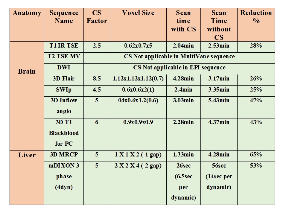
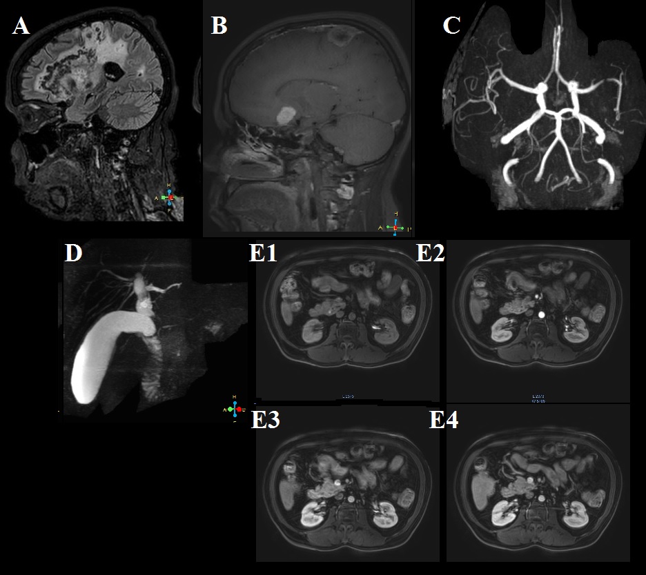
In this study, we have acquired MRI data of total 50 human subjects: 5 subjects each in 10 different anatomies such as: routine brain, pituitary, orbit, neck, cervical spine, lumbar spine, 3-station-angio, breast, MRCP, Knee, Ankle, Hip, Neck at 3.0 T Ingenia (Philips, The Netherlands). Coils used for each of these anatomies were: 15 channel head coil for brain, pituitary, orbit, Ankle, 44 channel spine coil for cervical spine, and lumbar spine, 32 channel torso coil for MRCP, 8 channel coil for knee joint. Necessary sequences for a comprehensive MR study for each anatomy were acquired using Compressed SENSE. Sequences were optimized to keep the same geometrical parameters (slice thickness, gap, field of view and resolution). Compressed SENSE factors used for each sequence were designed and optimized to achieve possible scan duration reduction, while keeping the visual image quality comparable with previously used sequences (non-Compressed SENSE protocols). For an example, our comprehensive brain examination protocol includes T1W IR TSE, T2W TSE, DWI, 3D Flair, SWI, Inflow angio and T1 3D Black-blood sequence when contrast is used. Resulting images from the compressed SENSE enabled fast scans were evaluated by senior radiologist to ensure similar visual appearance without losing any critical clinical information.Results
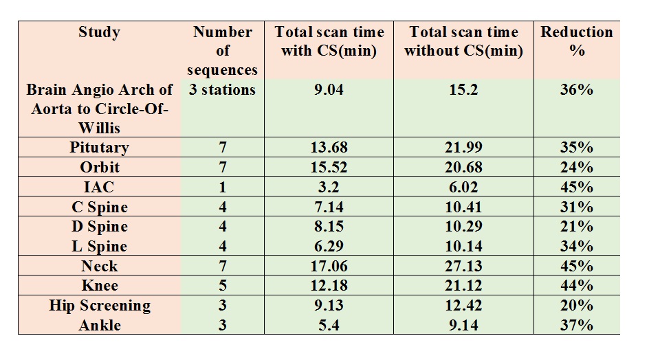
In this example, we have reported comprehensive exams for brain & MRCP. Table 1 shows the sequences used for a comprehensive brain, MRCP and liver 3 phase study with the geometric parameters, scan time and breath hold time with and without compressed SNESE. For our comprehensive brain exam 31% reduction in scan duration is achieved and when contrast is given 34% reduction is achieved, for 3D MRCP 65% reduction in scan duration and for Liver 3 Phase 53% reduction in breath hold duration is achieved. Table 2 shows the list of studies and scan time reduction achieved using Compressed Sense.Discussion
The results achieved from our study showed, patient comfort can be improved with Compressed SENSE by reducing the scan duration more than 30% for one MRI study without compromising the image quality. This is possible with Compressed SENSE because this method uses variable density incoherently under-sampling of k-space which has full freedom to optimize k-space sampling for best possible signal and image sharpness.
Implementing Compressed SENSE in routine clinical practice can bring down the number of repeat scans due to patient moment, sedation time and can increase the patient throughput. It can also improve the patient comfort by reducing the breath hold time by more than 50% for such sequences. In an alternate way, using Compressed SENSE can add more value by increasing the resolution of an existing sequence or by adding extra sequences without increasing the scan time.
Conclusion
Compressed SENSE can be a solution for the increasing demand in MR to perform imaging in a shorter time without compromising image quality to improve patient comfort.Acknowledgements
No acknowledgement found.References
[1] Lustig M, Donoho D, Pauly JM. Sparse MRI: The application of compressed sensing for rapid MR imaging. Magn Reson Med. 2007 Dec;58(6):1182-95.
[2] Donoho DL. Compressed sensing. IEEE Transactions on Information Theory. 2006;52(4):1289-306.
[3] Jaspan ON, Fleysher R, Lipton ML. Compressed sensing MRI: a review of the clinical literature. Br J Radiol. 2015;88 (1056):20150487
[4] Geerts-Ossevoort, L., de Weerdt, E., Duijndam, A., van IJperen, G., Peeters, H., Doneva, M., Nijenhuis, M. and Huang, A., Speed done right. Every time.
Figures