3656
Free water mapping in diffusion MRI: How do two common approaches compare?1Rotman Research Institute, Baycrest Health Sciences, Toronto, ON, Canada, 2Medical Biophysics, University of Toronto, Toronto, ON, Canada, 3Brigham and Women's Hospital, Harvard Medical School, Boston, MA, United States
Synopsis
Free water eliminated diffusion tensor imaging (fwDTI) and neurite orientation dispersion and density imaging (NODDI) are two increasingly established techniques that measure free water (FW) in diffusion MRI. Yet, despite the utility of each approach, their corresponding FW estimates have yet to be compared. In this work, we find that FW measurements near cerebrospinal fluid are highly similar between the two approaches, but within tissue, NODDI tends to compute slightly higher FW values in the white matter and lower FW values in gray matter than fwDTI. Potential sources of this discrepancy are discussed.
Introduction
Extracellular free water content, as measured by diffusion MRI, has recently gained attention as a sensitive biomarker of brain aging and pathology, and its elimination from the diffusion MRI signal can increase specificity of other diffusion-derived biomarkers to processes within the tissue. One method of free water mapping is to extend the conventional diffusion tensor model to incorporate a free water compartment via free water DTI (fwDTI)1. This model can be used to estimate free water from standard single-shell acquisitions1.
Alternatively, rather than modeling the tissue compartment as a single tensor, multiple compartments attributed to different tissue types can be modeled using multi-shell data. A popular tissue-modeling approach is NODDI, which includes a free-water compartment2.
While fwDTI is commonly used on single-shell data and NODDI is commonly used on multi-shell data, cross-validation between free water measurements in each of these approaches has yet to be shown. This is the motivation for the current study.
Methods
Three healthy volunteers were scanned on a 3T Siemens TIM Trio system with diffusion MRI at TR=11.3s, TE=118ms, FOV=25.6cm, 2mm3 resolution, 72 slices. Two shells were acquired: 60 directions with b=1000mm2/s + 6 b=0; and 60 directions with b=2000mm2/s + 6 b=0. Free water maps in fwDTI were obtained1 by fitting the fwDTI model using only the b=1000 shell. Free water maps in NODDI were obtained2 using both shells via the NODDI Matlab Toolbox (UCL). Fractional anisotropy (FA) maps were also computed from the b=1000 shell.Results
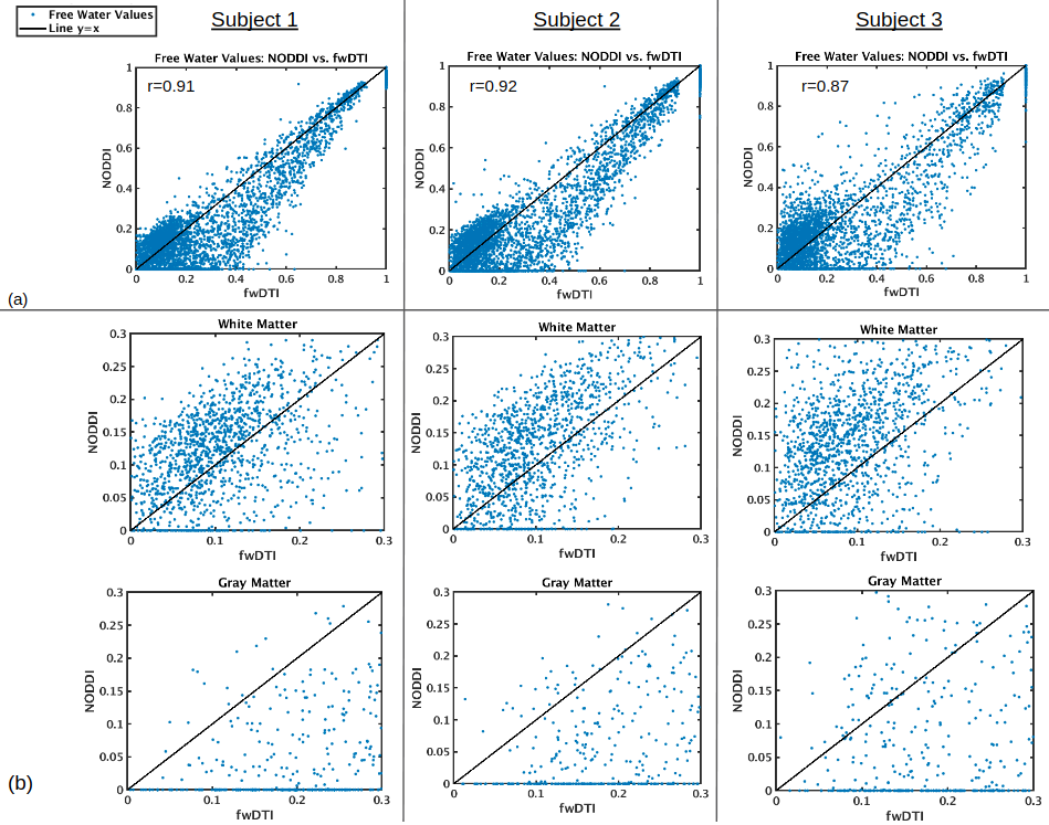
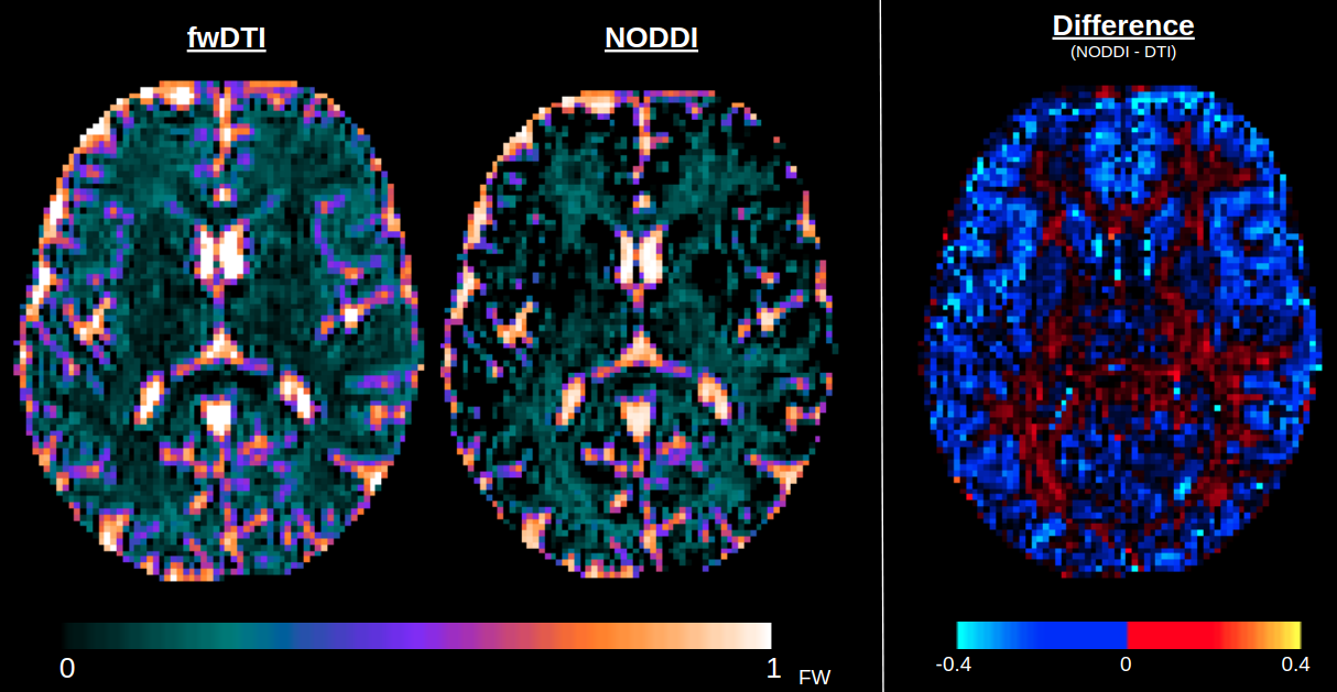
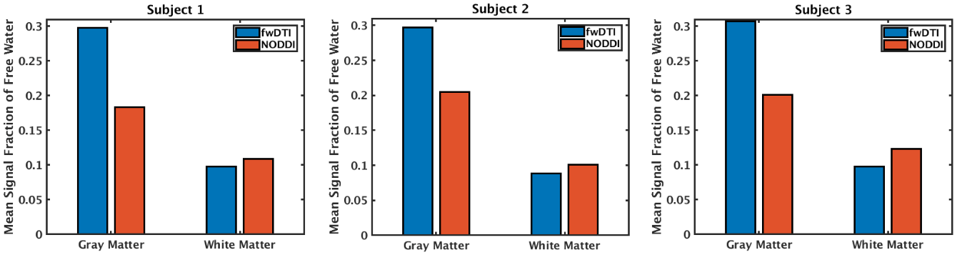
In all three subjects, FW values computed by fwDTI and NODDI are correlated throughout the brain, with correlation coefficient r ranging between 0.8 and 0.9 within slices. Correlations in a central slice is shown in Figure 1. The variance in free water values between the two approaches is smaller at higher FW values, i.e., the approaches are more strongly correlated in regions of partial-voluming with cerebrospinal fluid. Within tissue, NODDI tends to compute lower FW values in the gray matter and higher FW values in the white matter relative to fwDTI (Figure 2), a trend consistent across all three subjects (Figure 3).
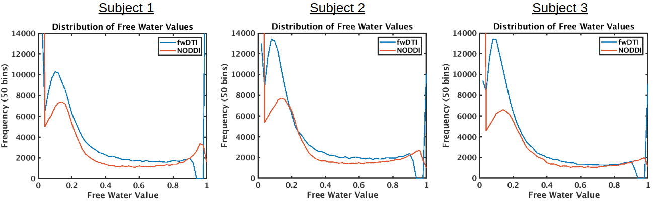
Figure 4 shows that the frequency of FW values in the brain peaks between 0.1 and 0.2 when computed from both methods. NODDI detects a significant amount of voxels with zero FW content, particularly in the gray matter, whereas the lowest FW values computed by fwDTI tend to be nonzero.
Discussion
This is the first in-depth study to compare FW measurements computed by two common methods, fwDTI and NODDI. It was found that (1) FW values are highly correlated between the two approaches, computing particularly similar values of FW in regions of partial-voluming with cerebrospinal fluid (CSF), and (2) in regions secluded from CSF, while FW values are still correlated between each approach, the absolute-values of FW display a consistent pattern of discrepancies across all three subjects. Sources of the discrepancies can potentially include issues with goodness-of-fit for fwDTI, as a regularizer is required for fitting single-shell data1, and issues with modeling assumptions for NODDI, which have been disputed3. Preliminary work suggests that regularizing the fwDTI model to fit single-shell data can lead to comparable goodness-of-fit to a multi-shell approach4,5, although a full validation of the fit of fwDTI to single-shell data remains. It has also been found that invalid modeling assumptions for NODDI result in NODDI underestimating FW in gray matter regions and overestimating FW in white matter regions compared to a gold-standard multidimensional diffusion MRI approach3. Therefore, given that FW values in tissue are expected to be small, the overestimation of FW in white matter in NODDI relative to fwDTI can likely be attributed to invalid modeling assumptions in NODDI. It is not immediately clear whether zero FW content (as computed by NODDI) or low, nonzero FW content (as computed by fwDTI) is more accurate within GM regions. Ground-truth validation of FW measurements in fwDTI and NODDI is part of future work.Conclusion
Free water measurements computed from two common methods, fwDTI and NODDI, are highly correlated. That said, there is a consistent pattern of discrepancies in the absolute-values of FW between the methods, so this work serves as a reminder that absolute values of FW should be viewed in relation to the method of computation rather than being trusted unconditionally. Nonetheless, this work demonstrates that while fwDTI only requires single-shell data, it computes comparable FW values with NODDI, a multi-shell technique.Acknowledgements
We thank the Canadian Institute for Health Research for financial support.References
1. Pasternak O, Sochen N, Gur Y, et al. Free water elimination and mapping from diffusion MRI. Magn Reson Med. 2009;62(3):717-730.
2. Zhang H, Schneider T, Wheeler-Kingshott CA, Alexander DC. NODDI: practical in vivo neurite orientation dispersion and density imaging of the human brain. Neuroimage 2012;61:1000-1016.
3. Lampinen B, Szczepankiewicz F, Mårtensson J, van Westen D, Sundgren PC, Nilsson M. Neurite density imaging versus imaging of microscopic anisotropy in diffusion MRI: a model comparison using spherical tensor encoding. Neuroimage 2017;147:517-531.
4. Pasternak O, Shenton ME, Westin CF. Estimation of extracellular volume from regularized multi-shell diffusion MRI. Med Image Comput Comput Assist Interv 2012;15:305-312.
5. Chad JA, Chen JJ, Pasternak O. Regularization stabilizes the fit of the two-compartment free water diffusion MRI model. Submitted to ISMRM 2019
Figures