3650
Examining Links Between Free Water and a TSPO-PET Marker of Neuroinflammation1Psychiatry Neuroimaging Laboratory, Brigham and Women's Hospital, Boston, MA, United States, 2Department of Psychiatry and Behavioral Sciences, Johns Hopkins Medical Institutions, Baltimore, MD, United States, 3Russell H. Morgan Department of Radiology and Radiological Science, Johns Hopkins Medical Institutions, Baltimore, MD, United States, 4Department of Radiology, Brigham and Women's Hospital, Boston, MA, United States, 5Research and Development, VA Boston Healthcare System, Boston, MA, United States
Synopsis
Free-water (FW) is a diffusion MRI marker of freely diffusing water in the extracellular space, which is expected to increase in the presence of neuroinflammation. Here, we test associations between FW and positron emission tomography (PET) imaging of the translocator protein (TSPO), which is a putative neuroinflammatory marker. We show that increased FW relates to higher TSPO binding in the hippocampi of healthy controls, but not of individuals with sports-related, repetitive traumatic brain injury. Thus, while FW relates to TSPO under healthy conditions, pathological variance in TSPO may complicate associations between FW and TSPO-indexed neuroinflammation.
Introduction
Free-water (FW) is a diffusion MRI (dMRI) marker, modeling water molecules that are not hindered or restricted. In the brain, FW is found in the extracellular space, which includes cerebrospinal fluid (CSF), interstitial water and plasma. Inflammation is expected to increase interstitial water volume, and increases in FW are therefore hypothesized to coincide with inflammatory processes. Previous observations support this hypothesis, showing increased FW in vasogenic edema,1 and in other disorders with inflammatory components.2 Here, we explicitly test the relationship between FW and a putative marker of neuroinflammation, the 18 kDa translocator protein (TSPO), using positron emission tomography (PET) with [11C]DPA-713 that binds TSPO. This relationship was examined in healthy control subjects, and in a group of National Football League (NFL) players with a history of repetitive, sports-related traumatic brain injury (TBI).Methods
9 healthy controls and 11 active or former NFL players underwent dMRI and [11C]DPA-713 PET. A previous analysis revealed higher regional [11C]DPA-713 binding in the players compared to the controls, including in the hippocampus.3
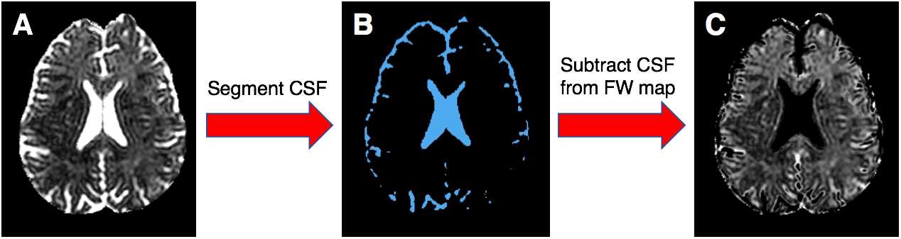
dMRI had 32 gradient directions at b=700mm2/s, 1 b=0, 70 slices reconstructed to 0.8X0.8X2.2mm3 and two repetitions. All scans were visually inspected and corrected for motion and eddy current artifacts. FW maps (Figure 1A) were generated using a regularized minimization.1 To eliminate CSF from FW maps, CSF probability was estimated by segmentation (SPM), and subtracted from FW maps (Figure 1).
[11C]DPA-713 PET scans were acquired using a high-resolution research tomograph PET system with 2.5mm3 spatial resolution. Images were reconstructed using the iterative ordered subsets expectation maximization algorithm (six iterations and 16 subsets), with correction for radioactive decay, dead time, attenuation, scatter, and randoms.4 Pre-processing with PMOD v3.7 included inter-frame motion correction and PET-MRI co-registration. Logan graphical method with metabolite-corrected arterial input function was applied to [11C]DPA-713 time activity curves to generate regional total distribution volume (VT).
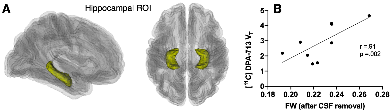
The hippocampus was selected as a region of interest (ROI), as it is functionally relevant to cognitive sequelae following TBI.3 Bilateral hippocampi were automatically segmented using FreeSurfer, based on T1-weighted MRI. Regions were visually inspected, transformed to subject-specific dMRI and PET space respectively, and eroded to minimize partial volume effects from potential misregistrations. VT and FW were then bilaterally averaged within the hippocampi. FW was compared between the study groups, and partial correlations examined the relationships between hippocampal [11C]DPA-713 VT and FW before and after CSF subtraction. All statistical tests controlled for the rs6971 polymorphism (C/C or C/T genotype), which affects the binding affinity of [11C]DPA-713.
Results
Hippocampal FW was significantly higher in healthy controls compared to ex-NFL players (p=0.03), but this higher level only trended toward significance after subtracting CSF (p=0.06). In the control group, a significant positive correlation was observed between hippocampal [11C]DPA-713 VT and FW before subtracting CSF (r=.89, p=0.003), which was stronger after subtracting CSF (r=.91, p=0.002; Figure 2). There was no correlation between hippocampal FW and [11C]DPA-713 VT in the players. Furthermore, there was no correlation between hippocampal fractional anisotropy and [11C]DPA-713 VT in players or in controls.Discussion
We found that variability in hippocampal FW relates to levels of TSPO in a sample of healthy control subjects. Subtracting CSF from FW strengthened this association, suggesting that interstitial or plasma-related changes accompany changes in TSPO. CSF subtraction is important in a cross-modality context to reduce macroscopic effects, such as atrophy and CSF-associated noise. This approach may be improved in future studies to better eliminate CSF signal. Conversely, there was no relationship between hippocampal FW and [11C]DPA-713 VT in the active or former NFL players, who displayed higher hippocampal [11C]DPA-713 VT relative to controls. These findings may suggest that a chronic neuroimmune response to repeated TBI complicates associations between FW and TSPO-indexed neuroinflammation. Inflammatory responses are dynamic and involve multiple biological processes, which may change within disease-specific contexts.5 Specifically, inflammation after repeated TBI may be associated with glial changes that reduce the extracellular space, such as increased density, scars, or swollen cell bodies.6Conclusion
This preliminary analysis suggests an intricate relationship between FW and TSPO binding. Complexity may stem from multiple pathological processes within the inflammatory response, such that some inflammatory events similarly effect TSPO binding and extracellular changes, while others produce discordant effects. Future work in different clinical populations and in animal models are needed to further establish the cellular mechanisms that underlie changes in the FW fraction. Establishing this link has important implications for indexing inflammation with FW, which is more feasible than PET for clinical studies.Acknowledgements
This project was funded in part by NIH grants R01MH108574, U01NS093334, P41EB015902 and a Veterans Affairs Merit Award. This study was also funded in part by financial support from the following NIH grants and foundations: NIH 5R21MH082277, NIH 5R01MH092443, NIH R01EB012547, NIEHS ES007062, NIH 5T32EB006351, NIH P50AG005146, the Lupus Foundation for America, NFL Charities and the GE NFL Head Health challenge.
References
1. Pasternak, O., Assaf, Y., Intrator, N., and Sochen, N. (2008). Variational multiple-tensor fitting of fiber-ambiguous diffusion-weighted magnetic resonance imaging voxels. Magn. Reson. Imaging 26, 1133–1144.
2. Pasternak, O., Kubicki, M., and Shenton, M.E. (2016). In vivo imaging of neuroinflammation in schizophrenia. Schizophr. Res. 173, 200–212.
3. Coughlin, J.M., Wang, Y., Munro, C.A., Ma, S., Yue, C., Chen, S., Airan, R., Kim, P.K., Adams, A.V., Garcia, C., et al. (2015). Neuroinflammation and brain atrophy in former NFL players: An in vivo multimodal imaging pilot study. Neurobiol. Dis. 74, 58–65.
4. Rahmim, A., Lenox, M., Reader, A.J., Michel, C., Burbar, Z., Ruth, T.J., and Sossi, V. (2004). Statistical list-mode image reconstruction for the high resolution research tomograph. Phys. Med. Biol. 49, 4239–4258.
5. Di Biase, M.A., Zalesky, A., O’keefe, G., Laskaris, L., Baune, B.T., Weickert, C.S., Olver, J., McGorry, P.D., Amminger, G.P., Nelson, B., et al. (2017). PET imaging of putative microglial activation in individuals at ultra-high risk for psychosis, recently diagnosed and chronically ill with schizophrenia. Transl. Psychiatry 7, e1225.
6. Bigler, E.D. (2013). Neuroinflammation and the dynamic lesion in traumatic brain injury. Brain J. Neurol. 136, 9–11.
Figures