3648
Diffusion Time Dependence of Diffusion Tensor Parameters in the Evaluation of Meningioma Subtype: a Preliminary Study1Radiology, Juntendo University Hospital, Tokyo, Japan, 2Radiology, The University of Tokyo Hospital, Tokyo, Japan, 3Siemens Healthcare Japan KK, Tokyo, Japan
Synopsis
We investigated the DTI eigenvalues and MD of five types of meningioma acquired with a shorter diffusion time using an OGSE sequence. Eleven meningiomas that consisted of 4 meningothelial meningiomas, 2 fibrous meningiomas, 2 transitional meningiomas, 1 psammomatous meningioma, and 2 atypical meningiomas were analyzed. Our results showed that the psammomatous and atypical meningiomas had a relatively strong diffusion time-dependence of diffusion tensor metrics. The use of shorter diffusion time with DTI provide additional information about the microstructure of each meningioma subtypes.
Introduction
Oscillating gradient spin-echo (OGSE) prototype sequences can shorten diffusion times by replacing the long-lasting diffusion-sensitizing gradients used in pulsed gradient spin-echo (PGSE) method with rapidly oscillating gradients1-5. Previously, the changes of ADC values obtained with shorter diffusion time has been shown to add information regarding the internal structures of lesion6,7. Meningioma is an extra-axial tumor that mostly classified as benign (World Health Organization [WHO] grade I). However, some meningiomas are classified as atypical (WHO grade Ⅱ) or malignant (WHO grade III). The WHO classified 15 subtypes of meningioma ranging from grade I to III according to their cell type by histopathology. All types of meningiomas usually appear hyperintense on diffusion-weighted imaging (DWI), therefore it is difficult to differentiate between subtypes. We hypothesized that changes in diffusion tensor imaging (DTI) eigenvalues and mean diffusivity (MD) acquired with a shorter diffusion time using OGSE might estimate the more precise internal structure of each meningioma subtype. To test this hypothesis, we evaluated the DTI eigenvalues and MD in 5 types of meningiomas using OGSE and compared it with PGSE methods.Methods
Eleven meningiomas that consisted of 4 meningothelial meningiomas, 2 fibrous meningiomas, 2 transitional meningiomas, 1 psammomatous meningioma, and 2 atypical meningiomas were scanned using a 3T MR scanner. DTI was performed with prototype sequences using b-values of 0 and 1000 s/mm2and six uniformly distributed directions for both OGSE and PGSE acquisitions. OGSE using a trapezoid-cosine waveform8 and PGSE sequences were performed with an effective diffusion time (Δeff) of 6.5 ms (frequency = 30 Hz) and 35.2 ms, respectively. Other parameters for the OGSE and PGSE sequences were as follows: repetition time, 4800 ms; echo time, 101 ms; field of view, 200 × 200 mm2; matrix size, 82 × 82; slice thickness, 5 mm; and acquisition time, approximately 2 minutes. The DTI eigenvalues (λ1, λ2, and λ3) and MD for each meningioma were measured by manually defined a range of interest (ROI). The relative percentage change between shorter and longer diffusion times was then calculated.Results
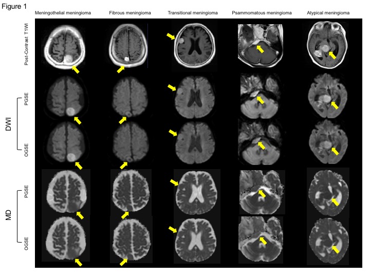
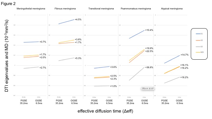
Representative cases of meningiomas are shown in Figure 1. After contrast administration, all meningiomas demonstrate homogeneous enhancement. The meningiomas showed high intensity on DWI using PGSE. The meningiomas showed slightly decreased visualization on DWI using OGSE. MD of meningiomas showed slightly higher on OGSE, compared with those on PGSE. The mean values of DTI eigenvalues (λ1, λ2, and λ3) and MD with the relative percentage changes for meningiomas are shown in Figure 2. Among all types of meningioma, the psammomatous meningioma had the strongest diffusion time-dependence of DTI eigenvalues and MD, followed by and the atypical meningiomas.Discussion
The psammomatous meningioma (WHO grade Ⅰ) and atypical meningiomas (WHO grade Ⅱ) showed relatively stronger diffusion time-dependence of DTI eigenvalues and MD compared to the meningothelial, fibrous, and transitional meningiomas (WHO grade Ⅰ). These results might indicate that the time-dependent of DTI eigenvalues and MD provide unique information that is sensitive to differences among subtypes of meningiomas. Mean square distances of water molecule movement for diffusion times of Δeff = 6.5 ms and 35.2 ms at body temperature are 10.8 μm and 25.1 μm, respectively9. Though the diffusion time that we can probe with a clinical MR scanner is still too long for direct quantification of cell sizes10, the differences of relative percentage changes among tumor subtypes presumably reflect the differences in structural complexity of meningioma11,12.Conclusion
We observed a relatively stronger diffusion time-dependence of DTI eigenvalues and MD in psammomatous and atypical meningiomas. Our results showed that the use of DTI with shorter diffusion time provide additional information about the microstructure of each meningioma subtypes.Acknowledgements
No acknowledgement found.References
1. Martin M. Measuring restriction sizes using diffusion weighted magnetic resonance imaging: a review. Magn Reson Insights 2013;6:59-64.2.
2. Does MD, Parsons EC, Gore JC. Oscillating gradient measurements of water diffusion in normal and globally ischemic rat brain. Magn Reson Med 2003;49(2):206-15.3.
3. Aggarwal M, Jones MV, Calabresi PA, Mori S, Zhang J. Probing mouse brain microstructure using oscillating gradient diffusion MRI. Magn Reson Med 2012;67(1):98-109.4.
4. Wu D, Martin LJ, Northington FJ, Zhang J. Oscillating gradient diffusion MRI reveals unique microstructural information in normal and hypoxia-ischemia injured mouse brains. Magn Reson Med 2014;72(5):1366-74.5.
5. Novikov DS, Jensen JH, Helpern JA, Fieremans E. Revealing mesoscopic structural universality with diffusion. Proc Natl Acad Sci U S A 2014;111(14):5088-93.6.
6. Baron CA, Kate M, Gioia L, Butcher K, Emery D, Budde M, et al. Reduction of Diffusion-Weighted Imaging Contrast of Acute Ischemic Stroke at Short Diffusion Times. Stroke 2015;46(8):2136-41.7.
7. Andica C, Hori M, Kamiya K, Koshino S, Hagiwara A, Kamagata K, et al. Spatial Restriction within Intracranial Epidermoid Cysts Observed Using Short Diffusion-time Diffusion-weighted Imaging. Magn Reson Med Sci 2017.8.
8. Van AT, Holdsworth SJ, Bammer R. In vivo investigation of restricted diffusion in the human brain with optimized oscillating diffusion gradient encoding. Magn Reson Med 2014;71(1):83-94.9.
9. Einstein A. Zur Theorie der Brownschen Bewegung. Ann Phys 1906;324(2):371.10.
10. Reynaud R. Time-Dependent Diffusion MRI in Cancer: Tissue Modeling and Applications. FrontPhys. 2017;5(58):1-16.11.
11. Novikov DS, Jensen JH, Helpern JA, Fieremans E. Revealing mesoscopic structural universality with diffusion. Proc Natl Acad Sci U S A. 2014;111(14):5088-93.12.
12. Xu J, Attia A, Arlinghaus LR, Kirschner AN, Osmundson EC, Kang H, et al. Selective Size Imaging using Filters via diffusion Times (SSIFT): A new contrast-free highly-specific MR cancer imaging method. Proceedings of the International Society for Magnetic Resonance in Medicine; 2018 June 16-21; Paris, France. 0953.
Figures