3637
Multisite evaluation of repeatability and stability for a novel quantitative diffusion kurtosis phantom1Radiology, University of Michigan Health System, Ann Arbor, MI, United States, 2Medical Physics, Memorial Sloan Kettering Cancer Center, New York, NY, United States, 3Columbia University Irving Medical Center, New York, NY, United States, 4Medical Physics and Radiology, Memorial Sloan Kettering Cancer Center, New York, NY, United States
Synopsis
Multi-center clinical trials utilizing quantitative diffusion kurtosis imaging (DKI) protocols require accurate, precise, and stable phantoms for validation of derived imaging metrics. This study examines the precision and reproducibility of isotropic (i)DKI parameters obtained from a phantom based on nanostructured vesicles that restrict diffusion and mimic tissue cellularity. Ten test-retest iDKI studies were performed on four scanners at three imaging centers over a six-month period. The tested prototype phantoms exhibited physiologically-relevant and highly-repeatable apparent diffusion and kurtosis parameters. Achieved precision was sufficient to characterize thermal and temporal stability trends to guide robust quantitative iDKI phantom production.
Introduction
To establish precision (repeatability) and accuracy (bias)1 of a potential diffusion kurtosis imaging (DKI) derived biomarker2,3, multi-center trials need quantitative phantoms that supply true DKI model parameter values. The DKI phantoms developed to date have either been single-parameter perishable (natural2,4,5) or provided limited range of kurtosis parameters with low SNR (synthetic6,7), lacking conventional intra-scan repeatability and inter-scan reproducibility metrics1. The purpose of this study was to evaluate DKI parameter ranges, repeatability and stability of the novel isotropic (i)DKI phantom based on lamellar-vesicle materials with tunable restricted diffusion parameters surrogate of tissue micro-structure2,3.Methods
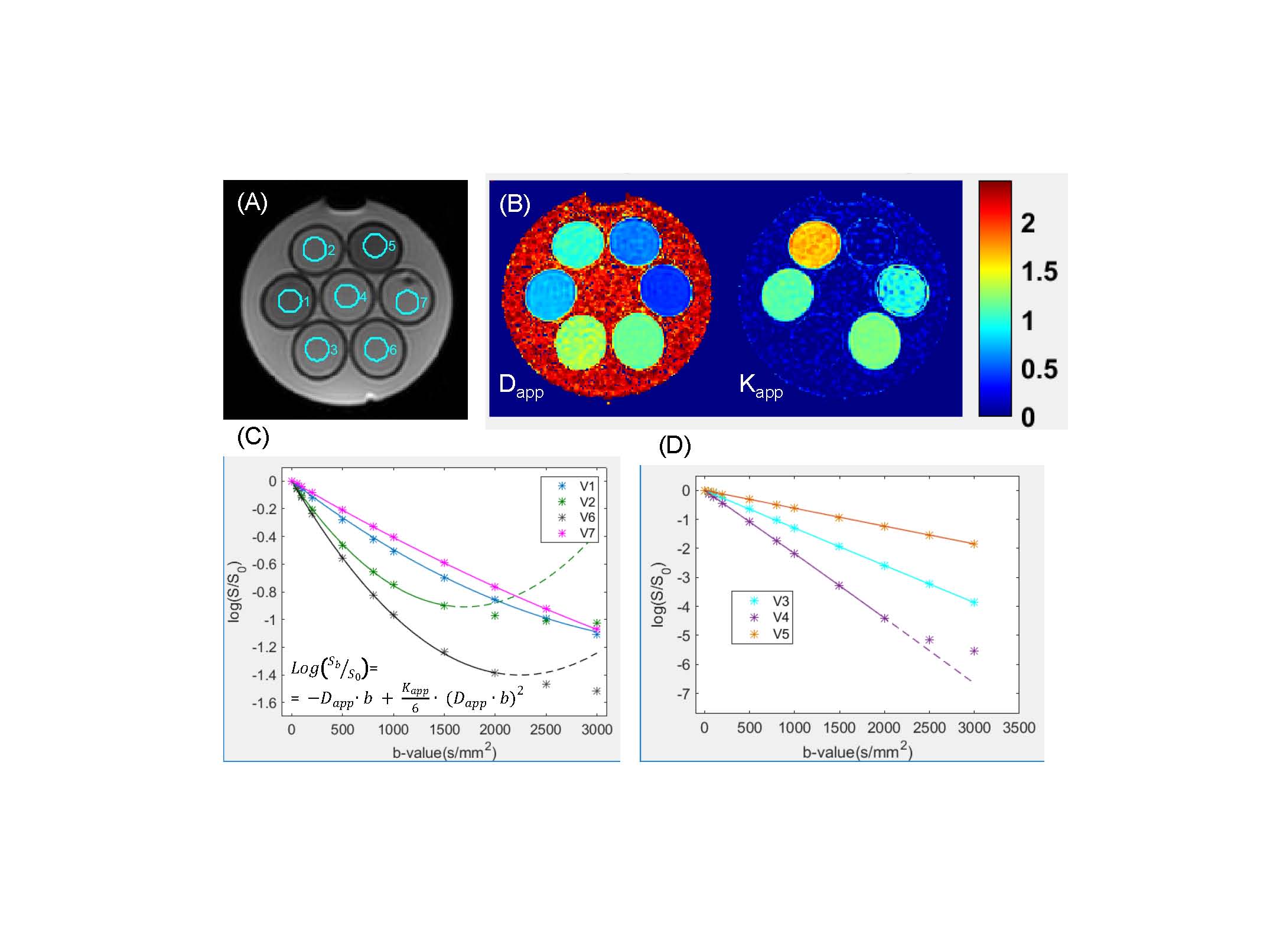
The developed prototype iDKI phantom consisted of seven sample vials immersed in water bath. Four restricted diffusion materials were tested based on water solutions of cetyltrimethylammonium bromide (CTAB) or behentriammonium chloride (BTAC) and cetearyl (CA) or decyl (DEC) alcohols, as well as prolipid 161 (PL161). Three negative control, mono-exponential diffusion samples included polyvinylpirrolidone (PVP) solutions in water at 0, 20% and 40%. The constructed iDKI phantoms were scanned at three imaging centers on four MRI scanners (two at 1.5T and 3T each) at ambient temperature over a period of six months. Shared multi-b DKI scan protocol included 11 b-values (b = 0, 50, 100, 200, 500, 800, 1000, 1500, 2000, 2500, 3000s/mm2), repeated two times. All image and statistical analysis was automated using MATLAB R2015b (Mathworks, Natick MA).
Seven circular ROIs (12mm diameter, 155 pixels) were defined separately for the test-retest runs on DWI(b=0) for phantom tubes (Figure 1A) avoiding air-bubble susceptibility artifacts. The parametric maps of apparent diffusion, Dapp , and kurtosis, Kapp, coefficients (Fig.1B) were derived using linear-least-squares fit of voxel DWI log-signal to quadratic dependence on b-value4 (Fig.1C). To satisfy iDKI model4 convergence and ensure SNRbmax>2, maximum fit b-values were constrained to bmax=1500 s/mm2 for CA-BTAC (V2), and bmax=2000 s/mm2 for water (V4) and PL-161 (V6) vials (Fig.1C, D). The apparent scan temperature (Ta) was self-calibrated using water (V4) ADC(bmax=1000 s/mm2) based on Speedy-Angell relation8, and ranged between 19.5 and 25.5 (±1oC).
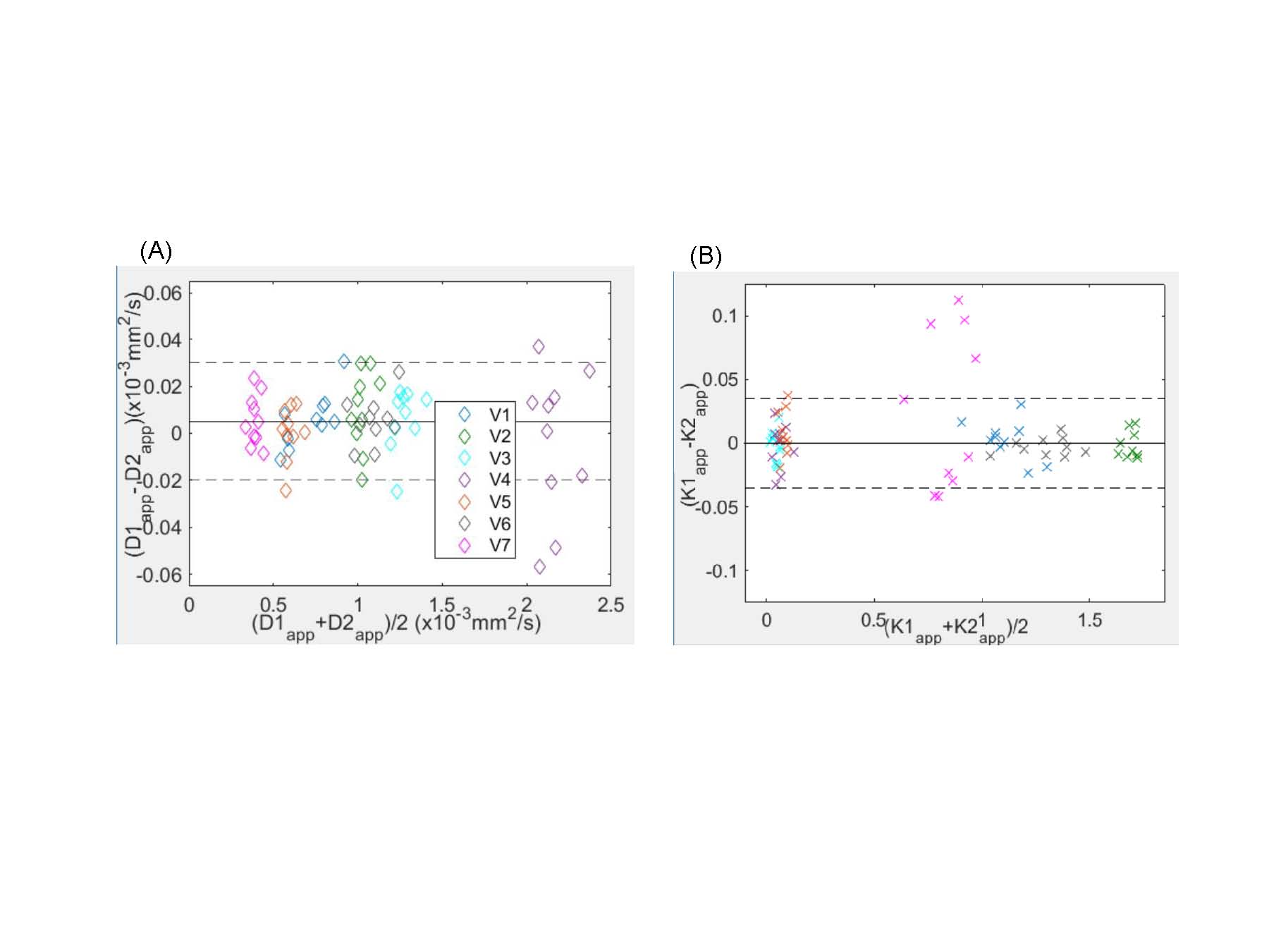
Bland-Altman (BA) repeatability analysis was performed
for Dapp and Kapp across all
test-retest sample scans (pool of 70), excluding outliers outside 1.5 interquartile ranges above/below the upper/lower
quartiles. Sample-specific coefficient of variance (wCV) and
corresponding 95% confidence interval (CI) were assessed1 (including BA-outliers) for all test-retest
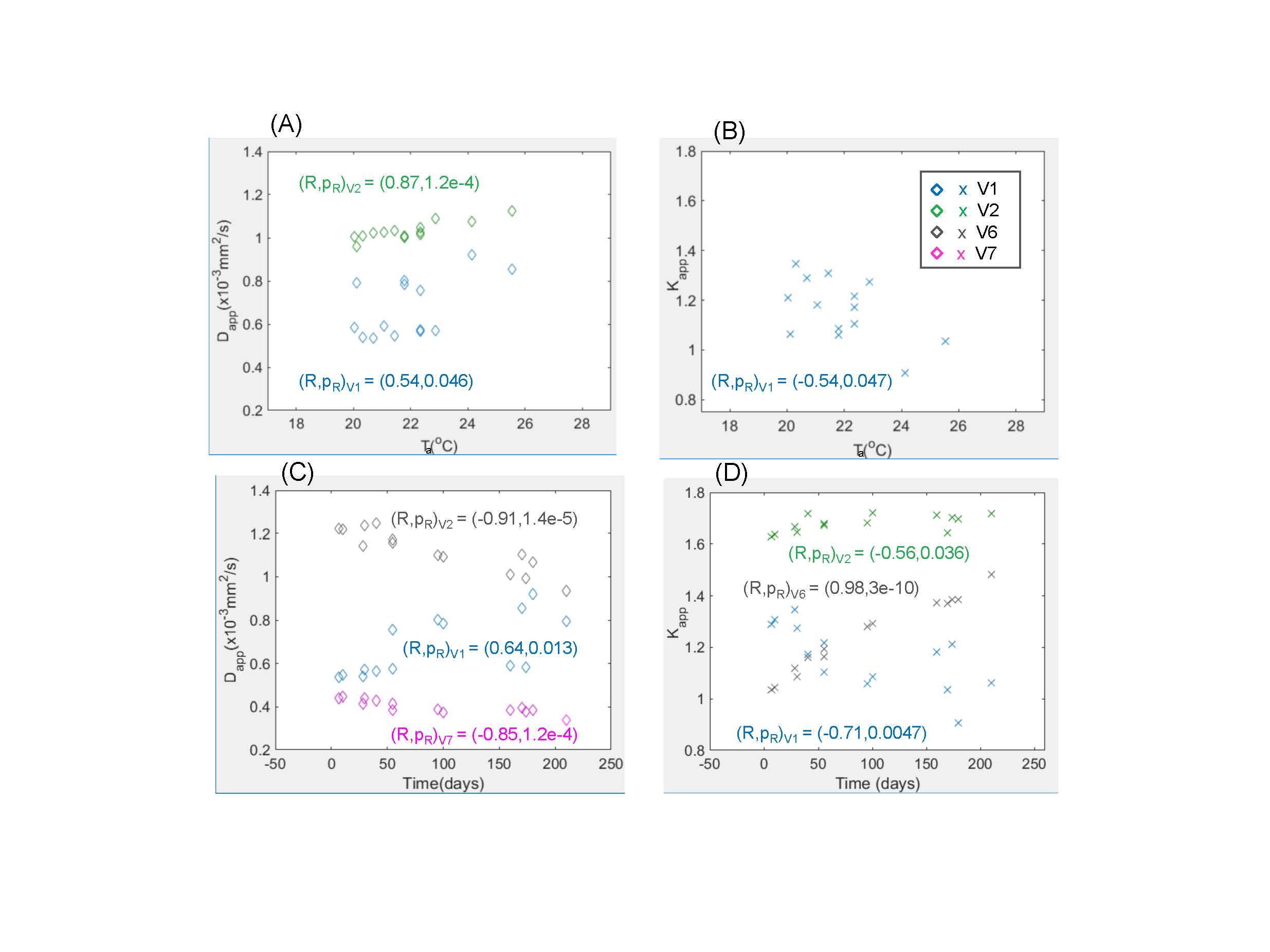
parameter values. Thermal and temporal phantom
stability was evaluated by inter-scan Pearson correlation, R, for the derived kurtosis parameters versus Ta and days from phantom manufacturing (using R-significance threshold of pR<0.05). Temperature and scan date were independent covariates (R=0.13, pR=0.66).
Results and Discussion
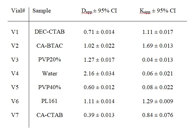
Four different chemical designs tested for iDKI phantom materials in V1, V2, V6, and V7 (Figure 1) exhibited restricted diffusion at high b-values (>1000 s/mm2), with DWI signals sustained above 20% of S0 (Fig.1C) and Kapp exceeding negative control noise-induced bias (Fig.1B, Kapp<0.1). All four tested iDKI materials allowed physiologically-relevant Kapp ranges 0.4-1.7(Table 1). Except for V7 (Kapp) outlier, the achieved intra-scan iDKI parameter precision (95% CI) was 1%-3.5% (Figure 2, Table 1) and independent of magnetic field (SNR).
The inter-scan variability of Dapp for the negative control samples (not shown) was fully explained by dependence on scanner ambient temperature (R>0.96, pR<1e-7). Observed kurtosis phantom Dapp sensitivity to temperature (2-3%/ oC) was higher than that of Kapp (Figure 3A,B). All iDKI phantom materials underwent initial parameter stabilization period of 3-4 weeks following preparation (Fig.3C,D), coincidental with evident sample degassing, when parameters changed by 6-22%. The parameter values for vesicular phase materials remained temporally stable post initial stabilization period (Fig.3C,D).
The candidate materials based on more-viscous multi-lamellar vesicle phase, exhibited either poor temporal stability (V6:PL161) or notable dependence on site storage and thermal equilibrium conditions (V1:DEC-CTAB). The kurtosis parameter values of CA-CTAB vesicular material had limited Kapp precision (9%, Table 1) and large (22%) initial Dapp stabilization change (Fig.3C(V7), due to apparent formation of in-volume gas micro-bubbles), but provided good thermal stability (no significant Ta dependence). CA-BTAC (V2) phantom has shown best intra-scan repeatability and inter-scan stability with the lowest 6% change in Dapp during stabilization stage and moderate thermal dependence (2%/ oC; R=0.87, pR=1.2e-4).
Conclusion
The chemical designs that were evaluated for the prototype iDKI phantom provided repeatable quantitative diffusion characteristics, within physiologically-relevant parameter ranges observed for tumors, and allowed sufficient SNR to avoid noise-bias or noise-limited precision. The most promising iDKI phantom design recommended for multi-site trials is based on CA-BTAC vesicular suspension that allowed easy preparation, temporal stability and independence of storage. iDKI material preparation should include degassing to improve initial parameter stabilization and precision. Temperature control or monitoring is desired for inter-scan Dapp reproducibility.Acknowledgements
This research was funded by National Institutes of Health Grants: U01CA166104, U01 CA211205, R01CA190299, P01CA085878 and P30 CA008748
Disclosure: S.D.Swanson, D.I.Malyarenko and T.L.Chenevert are co-inventors on intellectual property assigned to and managed by the University of Michigan for the technology underlying the manufacturing of the quantitative diffusion kurtosis phantoms utilized in this manuscript.
References
1Raunig DL, McShane LM, Pennello G, Gatsonis C, Carson PL, Voyvodic JT, et al. Quantitative imaging biomarkers: A review of statistical methods for technical performance assessment. Statistical methods in medical research. 2014.
2Jansen JF, Stambuk HE, Koutcher JA, Shukla-Dave A. Non-gaussian analysis of diffusion-weighted MR imaging in head and neck squamous cell carcinoma: A feasibility study. AJNR American journal of neuroradiology. 2010;31(4):741-8.
3Rosenkrantz AB, Padhani AR, Chenevert TL, Koh DM, De Keyzer F, Taouli B, et al. Body diffusion kurtosis imaging: Basic principles, applications, and considerations for clinical practice. Journal of magnetic resonance imaging : JMRI. 2015;42(5):1190-202.
4Jensen JH, Helpern JA, Ramani A, Lu H, Kaczynski K. Diffusional kurtosis imaging: the quantification of non-gaussian water diffusion by means of magnetic resonance imaging. Magn. Reson. Med. 2005;53(6):1432-40.
5Fieremans E, Pires A, Jensen JH. A simple isotropic phantom for diffusional kurtosis imaging. Magnetic resonance in medicine : official journal of the Society of Magnetic Resonance in Medicine / Society of Magnetic Resonance in Medicine. 2012;68(2):537-42.
6Phillips J, Charles-Edwards GD. A simple and robust test object for the assessment of isotropic diffusion kurtosis. Magnetic resonance in medicine : official journal of the Society of Magnetic Resonance in Medicine / Society of Magnetic Resonance in Medicine. 2015;73(5):1844-51.
7Portakal ZG, Shermer S, Jenkins C, Spezi E, Perrett T, Tuncel N, et al. Design and characterization of tissue-mimicking gel phantoms for diffusion kurtosis imaging. Medical physics. 2018;45(6):2476-85.
8Holz M, Heil SR, Sacco A. Temperature-dependent self-diffusion coefficients of water and six selected molecular liquids for calibration in accurate H-1 NMR PFG measurements. Phys Chem Chem Phys. 2000;2(20):4740-2.
Figures